Menu

Prenatal exposure to endocrine disruptors and behavioral problems in children

© Fotolia

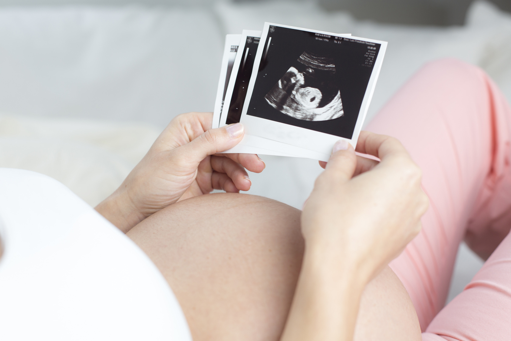
An epidemiological study carried out by Inserm on families from the EDEN cohort (500 boys born between 2003 and 2006 and their mothers) shows that exposure during pregnancy to certain phenols and phthalates is associated with behavioral problems in boys between 3 and 5 years of age. The most worrying compounds in this respect are bisphenol A, triclosan and dibutyl phthalate (DBP). The results of this study have recently been published in Environmental Health Perspectives.

In France, bisphenol A has been banned from use in all food containers since January 2015, occurring after this study was performed. Triclosan is an antibacterial agent found in certain toothpastes and soaps. DBP is used as a plasticizer in PVC plastics, certain glues, nail varnishes and hair sprays. According to the regulations, triclosan and DBP are permitted in limited quantities in certain product families but banned in others (in the EU for example, DBP is forbidden in cosmetics and triclosan is forbidden in clothing). Toxicological in vitro and animal studies have revealed that these compounds are endocrine disruptors and could interact with hormone systems involved in the normal development of the central nervous system. The specific mechanisms that could explain an effect of endocrine disruptors on neurodevelopment and behavior could involve the impaired functioning of thyroid hormones, of steroid hormones such as estrogen, or of other hormones such as oxytocin or vasopressin which are secreted by the hypothalamus.

In light of these initial findings in animals, the researchers wanted to study the association between exposure to endocrine disruptors during pregnancy and the subsequent behavior of the children.

The study concerned 529 young boys from the EDEN Mother-Child Study Group, set up by Inserm. The pregnant women participating in this cohort were recruited between 2003 and 2006 in the university hospitals of Nancy and Poitiers. At their children’s third and fifth birthdays, they completed a standardized questionnaire called the “Strengths and Difficulties Questionnaire” (SDQ), which evaluated certain aspects of their children’s behavior, such as hyperactivity, emotional problems and relationship problems. This questionnaire, which has been in use for 20 years, establishes scores for various aspects of behavior, such as emotional symptoms, peer relationship problems, conduct problems, hyperactivity and inattention problems. A urine sample taken during pregnancy was used to screen for the biomarkers characteristic of exposure to phenols and phthalates at the Environmental Health Laboratory of the CDC in Atlanta, which is responsible for biomonitoring campaigns in the US.

Between 70 and 100% of women from the Eden cohort, recruited during their pregnancy between 2003 and 2006, had been exposed to detectable levels of various substances. Levels in urine ranged from 1 to 3 µg per liter for bisphenol A, from 10 to 100 µg per liter for triclosan, and from 50 to 200 for methylparaben. The results suggest that maternal exposure to certain phenols and phthalates is linked with behavior problems in young boys.

Exposure to bisphenol A was associated with increased relationship problems at 3 years and hyperactivity problems at 5 years,  thus confirming that the effects of bisphenol A on the behavior observed in laboratory animals also occur in humans at low levels of exposure, levels that are probably lower than those recommended by the European Food Safety Authority (EFSA).

The metabolite of DBP was linked to increased emotional and relationship problems, including internalizing behavior, at 3 years of age. However, this was not the case for emotional problems at 5 years of age. Links between these compounds and behavior had already been revealed in previous studies in young boys and animals. In one such study on mother-child pairs from New York, an increase in internalizing behaviors in children of 3 years of age had been reported with an increase in levels of the DBP metabolite in 2012.

The results of this study have also shown a link between triclosan and increased emotional problems at 3 and 5 years of age. This is the first study to evaluate the effects of this compound on behavior, for which the environmental epidemiology team from Grenoble had already revealed a reduction in head circumference at birth, in the same population. At the molecular level, triclosan can interact with the thyroid axis which, during pregnancy, is involved in fetal brain development.

The sample size of this study, which is one of the largest on the subject, did not enable direct study of the onset of neurodevelopmental disorders, such as those of the autism spectrum, which would require the study of tens of thousands of children.

The research teams will now endeavor to replicate these results within the SEPAGES mother-child cohort, a study which is currently taking place in the Grenoble region, coordinated by Inserm and with the support of the European Research Council. In this study, a number of urine samples are collected from each participant, during pregnancy and the first years of the child’s life. This approach will make it possible to minimize errors in the measurement of exposure, as well as to identify potential periods of sensitivity to phenols and phthalates in relation to various health events, such as growth, behavior or respiratory health. It will also mean that the potential effect of these substances can be studied in young girls, a population that it was not possible to take into consideration here. It is possible that their sensitivity to endocrine disruptors differs from that of boys.

How do arteries protect themselves from atherosclerosis?

Associated with an aging population and the development of metabolic syndrome, atherosclerosis is a leading cause of death worldwide. Researchers from Inserm Unit 970, the “Paris Cardiovascular Research Center” (Inserm/Université Paris Descartes), have succeeded in revealing the mechanisms underlying the formation of atherosclerotic plaques. In particular, they have discovered the protective role of autophagy, a mechanism for cleaning and recycling cell components, in the cells that line the inner artery wall. These results, published in PNAS on September 25, 2017, provide us with an improved understanding of the initial stages of plaque development, and open up the possibility of developing a preventive treatment.

Atherosclerosis is a cardiovascular disease characterized by the build-up of plaque primarily made up of lipids (atheroma) on the inner artery wall. While some of these plaques remain stable, others erode or break apart with dramatic consequences for the patient, including heart attack or stroke.

The risk factors for cardiovascular disease are multiple and include diabetes, obesity, smoking, and hypertension. Although these factors are general, atherosclerotic plaques primarily develop in very specific areas of the circulatory system: the arterial bifurcations and curvatures. These areas are subject to low frictional forces from the flowing blood. In contrast, areas of the arteries exposed to higher frictional forces are protected from atherosclerosis. The mechanisms involved in this protective role of the frictional forces on the development of atherosclerosis remain poorly understood.

A recent study led by Chantal Boulanger and Pierre-Emmanuel Rautou from Inserm Unit 970, the “Paris Cardiovascular Research Center”, has filled this gap in our knowledge of atherosclerosis. Their research has shown the key role played by endothelial autophagy, i.e. the ability of the cells lining the inner wall of the arteries to break down and recycle their own components.

Autophagy is an intracellular process through which the cell breaks down part of its cytoplasm (the contents of the cell between the membrane and the nucleus) in response to stress or a lack of nutrients. The research team observed that the friction exerted by the blood strongly stimulates autophagy on the surface of the arterial wall. This phenomenon enables the endothelial cells to maintain a healthy physiological state, and prevents the development of atherosclerosis by protecting them from cell death, senescence, and inflammation.

The researchers then blocked the mechanism of autophagy on the surface of the arterial wall and observed increased plaque formation in the arterial areas usually protected from the development of atherosclerosis. Endothelial autophagy thus represents the previously missing link between atherosclerosis and the forces exerted by the flowing blood.

These results prove that inhibiting autophagy is not beneficial in the case of atherosclerosis. Specific stimulation of autophagy, on the other hand, could enable us to prevent the formation of atheroma and thus reduce the risk of heart attack and stroke, which are major public health challenges, ” concludes Chantal Boulanger.

The Yin and Yang of Low-dose Irradiation on Hematopoiesis

A team of researchers from CEA, Inserm, and the Paris-Sud and Paris Diderot universities has shown that exposure to low doses of irradiation (0.02 Gy) leads of a loss of hematopoietic stem cell[1] (HSC) function. The team has also shown that irradiation at this low dose facilitates efficient bone marrow transplantation without myeloablation[2]. These results, published in Cell Reports on September 26, 2017, show both the positive and negative aspects of low doses of irradiation.

What are the consequences of exposure to low doses of ionizing radiation, for example during medical examinations using X-rays? Previous epidemiological studies associated exposure to low doses of irradiation (<0.1 Gy) with an increased frequency of hematologic disease onset. However, no biological link between exposure to low doses of irradiation and hematopoietic cell abnormalities had been shown. The results obtained by the researchers from CEA, Inserm, and the Paris-Sud and Paris Diderot universities show that low-dose irradiation of HSCs, cells at the origin of all blood cells, leads to a decrease in HSC number and function. These effects on the stem cells can also be observed in vivo in inflammation and may lead to a deficiency in blood cell production and the risk of aplastic anemia[3] or leukemic transformation.

This team has used this property to test a new protocol to facilitate efficient bone marrow transplantation without the need for myeloablation. Indeed, the protocol, currently used during autologous bone marrow transplantation[4] involves the use of drug treatment to destroy the patient’s bone marrow prior to the transplant (myeloablation), a procedure which is unfortunately associated with a number of side effects. On the basis of their observations, the researchers have shown that a very low dose of irradiation, a dose used in medical imaging, preceded by a treatment that is currently used in the clinical setting and which removes the HSCs from the bone marrow, would facilitate efficient bone marrow transplantation without myeloablation.


These results point to the need for careful management when performing medical imaging, particularly in patients presenting signs of inflammation. It could also provide major therapeutic benefit to patients who are candidates for autologous bone marrow transplantation, particularly when a gene therapy protocol is used.


Hematopoietic stem cells (HSCs), non-irradiated (left) and irradiated (right) at a dose of 0.02 Gy. In blue the nucleus and in red the activated and nuclear Nrf2 protein, indicator of HSC response to oxidative stress caused by irradiation at a dose of 0.02 Gy.


Irradiation à faibles dosesLow-dose irradiation
YangYang
YinYin
Greffe de moelle osseusse sans myéloablationBone marrow transplant without myeloablation
Perte de fonction des cellules souches hématopoiétiquesLoss of hematopoietic stem cell function

[1] Bone marrow stem cells that produce blood cells: red cells, white cells and platelets.

[2] Drug treatment to destroy the patient’s own bone marrow prior to transplant.

[3] Insufficiency of the bone marrow to produce the various blood cell lines caused by more or less lasting HSC rarefaction.

[4] In gene therapy, autologous transplant consists of taking a patient’s HSCs and reconstituting his hematopoiesis with his own, genetically modified stem cells.

Maternal mortality: deaths from hemorrhage reduced but inequalities remain

© Fotolia

Between 2010 and 2012, 256 women died in France from causes linked to pregnancy, labor, or following childbirth, amounting to 85 such cases a year. Although inequalities remain, improvements have been observed in the provision of labor care, with the death rate from hemorrhage halving. These epidemiological results have been made public by the triennial report of the Confidential Inquiry into Maternal Deaths (Enquête Confidentielle sur les Morts Maternelles, ENCMM) in regard to the period 2010-2012, which was led by Inserm’s EPOPé – “Obstetrical, Perinatal, and Pediatric Epidemiology Team” – at the Center of Research in Epidemiology and Statistics Sorbonne Paris Cité (CRESS, Unit 1153).

Considered internationally as a reflection of the overall quality of a country’s health care system, maternal mortality rate constitutes a key indicator of public health. Co-ordinated by the Inserm EPOPé team since 1996 and led by Catherine Deneux-Tharaux, the Confidential Inquiry into Maternal Deaths (ENCMM) facilitates comprehensive identification of the causes of deaths in women occurring before, during, or following labor. This enables all the information to be collated in order to understand the chain of events leading to a death, and the lessons that can be learned for the future. This analysis, led by the National Expert Committee on Maternal Mortality (part of Santé Publique France, the French public health agency, since 2014), allows for any failings in the health care system to be brought to light, and is also responsible for considering non-fatal complications, which are more common but more difficult to study.

For the period 2010 to 2012 under review, 256 maternal deaths were identified: a total of 85 women dying each year in France. This figure corresponds to around 10 maternal deaths per 100,000 live births. It remains stable in comparison to the previous period (2007-2009) and is in line with the European average. However, 56% of these deaths were considered “avoidable” or “potentially avoidable”, and in 59% of cases optimal care was not provided, demonstrating clear room for improvement.

Reduction in direct mortality (during labor)

A major result of this report is the reduction by a third compared to 10 years ago of deaths directly related to obstetrical complications such as hemorrhage and eclampsia. For the first time, a statistically significant reduction in obstetrical hemorrhage has been observed, with the rate halving over the last 10 years. These results demonstrate an overall improvement in the quality of obstetrical care during the period under review.

However, almost all the deaths from hemorrhage were judged to be avoidable, and it remains the leading cause of maternal mortality in France (11% of deaths), although it has become rare in other countries. The researchers state therefore that: “we cannot drop our guard, and the results of this report enable us to identify new areas for focus in order to further reduce deaths from hemorrhage.”

Regional and social inequalities persist

Some troubling inequalities in maternal mortality persist. These are regional disparities: 1 out of every 7 maternal deaths occurs in the French overseas departments (DOMs), and the number of maternal deaths reported per live births in the DOMs is 4 times higher than in Metropolitan France (40 compared to 9 deaths/100,000 live births).

And social disparities: mortality among migrant women remains 2.5 times higher than among women born in France. This excess death rate is particularly marked in women born in Sub-Saharan Africa, among whom it is 3.5 times higher than that of women born in France. Investigating the care pathway of these women suggests that language barriers, among other factors, may sometimes play a role in the chain of events leading to their death.

Overall stability masking other trends

The overall stability of maternal mortality since 2007 can be explained by two trends:

  • the changing characteristics of the population of pregnant women, which place them at higher risk of maternal death (maternal age continues to rise along with the number of overweight and obese patients)
  • The increase in causes of death not directly linked to labor complications: These include maternal deaths due to infection (9%), including those linked to flu in unvaccinated women; sudden maternal deaths (9%); and maternal suicides (4%).

 “Going beyond the figures, the members of the National Expert Committee on Maternal Mortality (CNEMM) have drawn out 22 key messages from this analysis for clinicians and the public authorities (ed: report in full). According to the general principle of the inquiry, ‘better understanding for better prevention’, we have targeted specific elements to be improved within the provision and organization of health care in order to avoid patient deaths, but also in all likelihood serious maternal complications, which do not lead to death but result from the same failings” concludes Catherine Deneux-Tharaux, who led the inquiry.

“Humanized” Mouse Model Developed to Study Hepatitis B Infection

©Fotolia

The ANRS consortium “Humanized Mouse Models for Viral Hepatitis”1, made up of 6 teams of researchers, has developed a mouse model for studying the interaction between the immune system and the liver following infection by the hepatitis B virus. This research, coordinated by Dr. Hélène Strick-Marchand (Inserm joint unit 1223, “Physiopathology of the Immune System”, Institut Pasteur), responds to a real lack of animal models for studying this disease and thus opens up the possibilities for evaluating new therapeutic strategies. These results have been published in the journal Gastroenterology.

Over 250 million people around the world are chronic carriers of the hepatitis B virus (HBV), and this figure is growing despite the existence of a highly effective preventive vaccine. Once it has developed, chronic hepatitis can progress to liver fibrosis, cirrhosis, and hepatocellular carcinoma (liver cancer). There are treatments to prevent the disease from progressing by keeping the virus under control, but they must be taken for life as they do not eliminate it completely. In order to better understand the consequences of HBV infection, interactions between the infected hepatocytes (liver cells) and the immune system response, and to be able to test new therapeutic strategies, researchers required an animal model with a physiology similar to that of humans. For a number of years, the ANRS consortium “Humanized Mouse Models for Viral Hepatitis”1, which is made up of 6 teams of researchers and coordinated by Dr. Hélène Strick-Marchand (Inserm joint unit 1223, “Physiopathology of the Immune System”, Institut Pasteur), has therefore been working to develop so-called “humanized” mice. This research has led to development of the HIS-HUHEP model, for which promising results have been published in the journal Gastroenterology.

With the aim of modeling interactions between human hepatocytes (the target of HBV infection) and the human immune system, the HIS-HUHEP model is doubly engrafted with a humanized immune system, and human hepatocytes in its liver. Researchers analyzed the physiological responses of these mice to HBV infection, which proved to be comparable to those observed in humans. In addition, giving the infected HIS-HUHEP mice entecavir (an antiviral used to treat HBV infection) reduced their viral load and liver inflammation.

The doubly “humanized” HIS-HUHEP mice represent an important animal model for studying the interactions that take place between the immune system and the liver in liver disease. This new model fills a real gap. It opens the way to better understanding of the immune response developed to HBV, and to testing new therapeutic strategies with the final goal of eliminating the virus completely from the bodies of infected individuals.


1 For further information on the Humanized Mouse Models for Viral Hepatitis consortium, see:       www.anrs.fr/fr/actualites/285/consortium-anrs-modeles-murins-humanises-pour-letude-des-virus-hepatites

Usher syndrome: gene therapy restores hearing and balance

The cochlear sensory epithelium (organ of Corti), and the vestibular sensory epithelia, ampullar cristas (inset) of the inner ear ijntected with the AAV8 virus producing GFP and the SANS protein. The cochlear and vestibular sensory cells were immunostained for GFP and myosin VI and analyzed with a confocal microscope. The green and orange cells produce the protein of the therapeutic gene.
© Institut Pasteur

Scientists from the Institut Pasteur, Inserm, the CNRS, Collège de France, University Pierre et Marie Curie, and University Clermont Auvergne*, have recently restored hearing and balance in a mouse model of Usher syndrome type 1G (USH1G) characterized by profound congenital deafness and vestibular disorders caused by severe dysmorphogenesis of the mechanoelectrical transduction apparatus of the inner ear’s sensory cells. By locally injecting the USH1G gene, critical for the formation and maintenance of the hair bundle, the mechanosensory antenna of these cells, the scientists successfully restored the function and the structure of this apparatus, resulting in the first recovery of hearing and balance by gene therapy for this mouse model. These findings, published in the journal PNAS, open up new possibilities for the development of gene therapy treatments for hereditary forms of deafness.

Hearing loss, sometimes associated with other disorders such as balance defects, is the most common sensory deficit, affecting more than 280 million people worldwide, according to WHO. In France, one child in 700 is born with severe or profound hearing loss, and one in every 1,000 will lose their sense of hearing before adulthood.

Over the past 20 years, scientists have made remarkable progress in deciphering the genetic origins of congenital hereditary hearing loss, which is usually caused by inner ear dysfunction. The inner ear comprises the hearing organ or cochlea, together with the five balance organs (the saccule, utricle and three semicircular canals), which contain the sensory cells, or hair cells, that detect mechanical vibrations and convert them into electrical signals. To date, mutations in more than 100 genes have been associated with inner ear defects, and it is estimated that mutations in more than 100 genes can cause genetic forms of deafness.

The various hereditary forms of hearing loss include Usher syndrome type 1 (USH1), a particularly severe clinical form of deaf-blindness, and specifically the USH1G genetic form. USH1G patients are profoundly deaf and have no balance function at birth, and they subsequently suffer from prepubertal-onset sight loss leading to blindness. USH1G syndrome is due to mutations in the gene encoding the scaffold protein sans, which is essential for the cohesion of the hair bundle of the inner ear hair cells.

Patients with hearing loss and balance dysfunction are currently fitted with auditory prostheses and may be given balance rehabilitation therapy, but the outcomes are variable. One possible alternative for treating such hereditary inner ear defects is gene therapy. This approach entails transferring a healthy (non-mutant) copy of the defective gene to restore the expression of the missing protein. So far, gene therapy attempts have only resulted in partial improvements of hearing in mouse models of specific human deafness forms that did not include severe anomalies in hair cell structure.

In this context, scientists from the Institut Pasteur, Inserm, the CNRS, Collège de France, University Pierre et Marie Curie, and University Clermont Auvergne*, have now succeeded in restoring hearing and balance in a mouse model of USH1G syndrome using gene therapy. With a single local injection of the USH1G gene just after birth, the scientists observed a restoration of the structure and mechanosensory function of the inner ear hair bundles – profoundly damaged before birth –, resulting in a long-term partial recovery of hearing, and complete recovery of vestibular function in these mice. These results unexpectedly establish that inner ear defects due to major morphogenetic abnormalities of the hair bundle can be reversed even after birth, with durable efficacy, by gene therapy.

Hair bundles of vestibular sensory cells analyzed using scanning electron microscopy. The image shows a normal hair bundle with its characteristic “staircase” pattern (in yellow), a defective Usher type 1G hair bundle (in pink) and a treated Usher type 1G hair bundle (in green), whose normal/characteristic form was restored with gene therapy.
© Institut Pasteur

The scientists injected the USH1G gene into the inner ear using the innocuous AAV8 virus, which enabled them to specifically target the hair cells. The expression of the therapeutic gene was detected 48 hours after injection. The team demonstrated that a single injection to restore the production and localization of the missing protein in hair cells successfully improved hearing and balance functions in the young mice. These findings suggest that the therapeutic protein was able to interact normally with its binding partners among the USH1 molecular complex (the proteins cadherin 23, protocadherin 15, myosin VIIA and harmonin), as required for the mechanoelectrical transduction apparatus of the hair bundle to function correctly.

As Saaïd Safieddine, CNRS Director of Research at the Institut Pasteur and co-senior author of the study with Prof. Christine Petit, explains, “we have just shown that it is possible to partially correct a specific form of hereditary hearing loss accompanied by balance problems using local gene therapy performed after the embryogenesis of the ear, which is primarily affected by the mutation responsible for the disorder. This suggests that the time window for effectively treating USH1 syndrome using gene therapy may be larger than initially thought.

This study represents a significant step towards the development of clinical trials in gene therapy for the curative treatment of hereditary deafness and balance loss in humans.


*From the Genetics & Physiology of Hearing Laboratory (Institut Pasteur/Inserm/UPMC), the Genes, Synapses and Cognition Laboratory (CNRS/Institut Pasteur, the Center for Neurophysics, Physiology and Pathology (CNRS/Paris-Descartes University), and the Sensory Biophysics Laboratory (University Clermont Auvergne).

Researchers Warn About Mixtures of Endocrine Disruptors During Pregnancy

© Fotolia

A new study led by Inserm researchers from Irset, the Research Institute for Environmental and Occupational Health,[1] shows for the first time in humans that simultaneous exposure to endocrine disruptors exacerbates the effects observed from exposure to each chemical independently. This study focused principally on the human fetal testes and the potential consequences of these mixtures on development of the reproductive system, as the selected chemicals inhibited testosterone production. These results were published in Environmental Health Perspectives.


The increased use of new materials, products, and industrial/agricultural processes characteristic of a “modern” way of life has led to environmental contamination (including domestic, professional, and food-related environments) with multiple chemicals. Many of them have been identified as having endocrine-disrupting effects, and in particular as antiandrogens (=antitestosterone). It now appears clear that continuing to focus research on these “individual” chemical products would underestimate the risk linked to their simultaneous exposure, particularly in pregnant women. 


Experimental data, notably across a range of different animal species and on cell lines in culture, supports the idea of a “mixture” or “cocktail” effect. Paradoxically, however, in view of the stakes for human health, it has not yet been proven that these “cocktail effects” are present in humans. The authors of this new article have developed mathematical models to predict these combined effects based on the individual toxicology profiles of the chemicals. These mathematical models are the first stage in evaluating the risk of exposure to mixtures of endocrine disruptors in humans, and in particular in pregnant women. The study had two objectives:

  • to expand the known list of endocrine-disrupting chemicals in humans; and
  • to verify that experimental data are in line with mathematical predictions for the mixtures.


The Irset researchers – with the support of colleagues from the Rennes University Hospital, and Professor Andréas Kortenkamp and Dr. Martin Scholze from Brunel University in London – undertook an entirely new experimental approach, screening 27 chemicals, including 7 drugs, 14 industrial chemicals (pesticides) and 6 so-called sociocultural chemicals (alcohol, caffeine, etc.). Eleven endocrine-disrupting chemicals were identified in this way, some for the very first time in humans. 


From these 11 chemicals, four mixtures were developed and tested on human fetal testes. The experimental results for these mixtures corroborated the mathematical predictions, for a number of components greater than 3. This demonstrates that the model developed by the authors of the article is capable of illustrating cocktail effects, for the first time in a human organ, and that the combined effects observed can be predicted mathematically.


Finally, the authors of this article were able to measure the exacerbation of the individual effects of each of the chemicals in the mixture. In other words, they were able to provide a response to the question “how much more powerful is the chemical in a mixture than alone?” by showing that this exacerbated potency varies from a factor of 10 to 1,000, depending on the chemical in question. 


According to Bernard Jégou, Irset director, Inserm researcher, research director of the EHESP-School of Public Health and the lead on this study, Pierre Gaudriault, pharmacist and doctor at the Université de Rennes 1, and Inserm researcher Séverine Mazaud-Guittot, the conclusions of this work, which was supported by the French Agency for Food, Environmental and Occupational Health & Safety (ANSES), must be taken seriously: “there is a very precise critical window during the 1st trimester of fetal development during which simultaneous exposure to weak doses of multiple endocrine disruptors may represent a risk to the development of the child’s genitals and reproductive system. It is particularly concerning as the individual potency of these chemicals can be exacerbated by up to a factor of 1,000. All the experimental data from different models lead us toward the same conclusions. This experimental proof of concept study shows that intensifying research into identifying the real mixtures to which individuals are exposed, and testing the effects on appropriate models, is a necessity.”

[1] Research Institute for Environmental and Occupational Health; Inserm; EHESP-School of Public Health, Université de Rennes 1.

Regulating or Stimulating Our Antibodies Based on Our Immunological Needs Will Soon Be Possible

©Fotolia


Clinician-researchers from the Université Pierre et Marie Curie (UPMC) and Inserm, in collaboration with the Biological Therapy Department of the Pitié-Salpêtrière Hospital (AP-HP), have revealed a new mechanism for regulating antibody production. The results of this study were published in Science Immunology on Friday, September 8, 2017.


The immune system protects the body from pathogenic agents, in particular infectious ones, that cause damage or disease in humans. Two types of defense work in parallel: cell-mediated immunity, which destroys the infected cells, and humoral immunity, which produces antibodies. These antibodies neutralize the pathogens in a targeted manner.

“As with all immune responses, the humoral immune response must be controlled. Too weak a response would be ineffective, but too strong a response against our own tissues could lead to autoimmune disease,” explains Professor David Klatzmann, clinician-researcher at UPMC and the Pierre et Marie Curie Faculty of Medicine, director of the Immunology – Immunopathology – Immunotherapy laboratory (UPMC/Inserm) and head of the Biological Therapy Unit at the Pitié-Salpêtrière Hospital (AP-HP).


Professor Klatzmann’s team is interested in cells that control the intensity of the humoral response: follicular helper T cells (Tfh), which stimulate the production of antibodies, and follicular regulatory T cells (Tfr), which reduce it. Tfr cells were discovered in 2011. Their numbers are limited, and little is currently known about their mechanisms of action.

The research group first redefined the characteristics for identifying Tfr cells. From this basis, the researchers were able to identify a new mechanism for regulating antibody production

by showing the key role of interleukin-1 (IL-1), a soluble mediator, in triggering these responses. Tfh cells receive IL-1, which activates them and enables an increased antibody response; conversely, Tfr cells reduce the antibody response by neutralizing IL-1 and depriving the Tfh cells of this stimulation.


IL-1 therefore appears to play a major role in the stimulation of humoral immunity via the Tfh cells, and this axis appears to be regulated by the Tfr cells. “We aim to stimulate the antibody response with vaccinations, and reduce it in autoimmune disease,” explains Professor Klatzmann. “This discovery therefore means that we have a new method for regulating antibody immune response.” 


The full results can be found in Science Immunology, published on Friday, September 8.

A New Avenue to Explore in the Fight Against Sepsis

©Fotolia

Inserm researchers have succeeded in producing a human protein in laboratory conditions and to use it against bacterial infections and for the treatment of sepsis. Sepsis is a systemic inflammatory response by the body to a serious infection. Should the inflammation reach a critical stage (septic shock), the condition becomes life-threatening. Sepsis kills one person every 3 to 4 seconds worldwide. This research, published in Scientific Reports, therefore represents a serious avenue to explore in the fight against a condition that currently remains a medical emergency.


In developed countries, there are 377 cases of sepsis per 100,000 inhabitants. Each year, it is responsible for the deaths of 6 million infants. In France, the mortality rate for sepsis is 27%, which rises to 50% for the most severe form of the infection. The number of cases is expected to double in 50 years’ time, mainly as a result of population aging (source:  Institut Pasteur). On the global scale, the situation is so serious that WHO delegates decided to recognize sepsis as a major public health concern at the World Health Assembly held in Geneva in May 2017. The next World Sepsis Day, an initiative of the Global Sepsis Alliance, will take place on September 13, 2017.

In the majority of cases, sepsis involves an infection caused by Gram-negative bacteria naturally present in the body (generally in the intestine) that become toxic in susceptible individuals. The toxic part of the bacterium is found on its outer membrane in the form of a lipopolysaccharide complex, known as an endotoxin.

The specific chemical aspect of these endotoxins formed the starting point of the study conducted by the Inserm researchers.  Indeed, previous studies have shown that plasma phospholipid transfer protein (PLTP) is able to bind to endotoxins located on the outer membranes of bacteria and even transport them to the liver. When infection is present, this protein therefore appears to play a role in eliminating the endotoxins.

The research team, with the help of an American team, studied a genetically engineered mouse model which had the particularity of no longer expressing the PLTP gene to verify this hypothesis. When these mice were injected with bacterial endotoxins, the researchers observed that they died without being able to fight the infection generated. Hence their hypothesis that PLTP presents previously unknown and potentially major importance in the domain of innate immunity.

The challenge was then to obtain sufficient quantities of human PLTP in order to conduct therapeutic tests to demonstrate its ability to counteract the effects of these endotoxins. For this, the researchers approached the “Development and Reproduction Biology” joint research unit at Inra, which is able to produce this protein in the milk of transgenic female rabbits.

Once obtained, the researchers tested the ability of PLTP to fight the inflammatory response in mice with sepsis. Quite small quantities of PLTP were all it took to obtain considerable improvements in the health of these mice. “However, our ultimate objective was to understand how it all works,” summarizes Laurent Lagrost.

Continuing their work, the researchers demonstrated that PLTP can block the proliferation of bacteria by weakening their outer membranes. They also observed that PLTP, in addition to being able to neutralize the activity of these endotoxins, can also disintegrate them before transferring them to lipoproteins. These lipoproteins, usually simple cholesterol transporters, have the ability to transform into emergency vehicles to escort the endotoxins to the liver for elimination via the biliary route.

“Unless we endogenously neutralize, within the body of the individual patient, the bacterial endotoxins that will be responsible for the inflammatory response and the entire resultant cascade of harmful effects, we cannot definitively resolve the problem. However, it appears that PLTP is able to neutralize these endotoxins and detoxify the blood, at least in mice,” conclude the researchers.

This approach is part of an original concept of biomimetics in which “the more we copy nature, the closer we get to the truth,” they add.

Sunday, September 10, World Suicide Prevention Day

©Fotolia 

Suicide is the cause of over 800,000 deaths around the world each year according to estimates from the World Health Organization, amounting to one death every 40 seconds. It affects all age groups and all parts of the world[1]. In France, the emergency services handle 220,000 suicide attempts every year[2], and 10,500 individuals take their own lives.

Several risk factors appear to increase suicidal ideation. These include problems with alcohol, drugs, and sexual identity, strong impulsivity, social isolation, and advanced age. Depression is the cause of suicide in 70% of cases. Antoine Pelissolo, an Inserm researcher from Unit 955, “Mondor Institute of Biomedical Research” (IMRB), sought to understand this link between depression and suicide in his book Dépression: s’enfermer ou s’enfuir [Depression: sinking or coping], which was published by Le Muscadier in 2015.

© OMS

Among 15-29 year-olds, suicide is the second leading cause of mortality. A large-scale survey, coordinated by Inserm Unit 1178, “Mental Health and Public Health”, and the Fondation Vallée’s University Research Center, which was carried out in 2013 among 15,235 students aged 13 to 18, also showed that suicide attempts appear to be more common now than in the past: 7.8% of young people have already tried to commit suicide once, and 3.7% more than once. These alarming figures highlight the need for more work on preventing suicidal behavior.

Read the report “Le nouveau visage de nos adolescents [The new face of our adolescents]

Catherine Jousselme, an Inserm researcher from the “Mental Health and Public Health” Unit, explains that social networks can have both incredibly positive and negative effects: they enable greater self-expression, but also lead to isolation. When adolescents feel down, feeling like they exist and are important help them do better. The depiction of suicide on social networks gives them the impression of “dying a hero”.  Certain measures are now in place to prevent suicide (including censorship on social networks, flagging inappropriate content, and the presence of prevention services on social networks), but educating young people and teaching them the right actions to take remains the best way to combat this plague.

For more information on adolescence and suicide, see the book Ados & Suicide: en parler et se parler [Teens & Suicide: talking to them and to each other] in the Choc Santé [Health Shock] series from Inserm and Le Muscadier.

Unemployment can also be a cause of suicide. This was shown by a study from January 8, 2015, led by the Inserm Epidemiology Center on Medical Causes of Death (CépiDc) and the Paris public hospital system (AP-HP). For further data on deaths due to suicide in France, please contact Grégoire Rey, director of the CépiDc-Inserm.

In the context of suicide prevention, joint research unit 1123–Clinical Epidemiology and Economic Evaluation Applied to Vulnerable Populations (ECEVE) has developed the STOPBLUES application, an interventional and evaluative research program. STOPBLUES enables users to access information (videos of professionals, people with depression, and their friends and family) and complete anonymous self-evaluation questionnaires. A map feature also enables them to find the doctors, specialist mental health care centers, and supportive organizations closest to them. It is a kind of virtual companion, designed to support, inform, and reassure users in difficulty. For further information, please contact Karine Chevreul, the deputy director of ECEVE, or Kathleen Turmaine, Inserm researcher and lead on the STOPBLUES project. The application will be available in November 2017, and a website will also be available.

© STOPBLUES

[1] For more information, see the Inserm collection on this topic

[2] “Dépression: s’enfermer ou s’en sortir” [Depression: sinking or coping], Antoine Pelissolo, Le Muscadier, 2015.

fermer