Menu

Filming the brain to shed light on sleep

©Adobe Stock

What happens inside our brains when we sleep? To answer this question, French researchers have, for the first time, filmed the entire brain in sleeping rats, thanks to innovative ultrasound imaging technology. They were thus able to closely observe brain function in rodents, particularly during the REM sleep phase. These results were obtained in shared Inserm, ESPCI Paris, CNRS, and Sorbonne University laboratories. Published in Nature Communications, these findings allow this period to be redefined as a brain hypersynchronization phase, characterized by massive peaks in blood flow, particularly in the hippocampus. These new data, which question the role currently ascribed to REM sleep, have yet to be confirmed in humans.

REM sleep is a specific sleeping state in which brain activity is similar to the waking state, while being associated with inhibition of muscle activity. It is notably characterized by rapid eye movements, and has long been considered to coincide only with dreaming and emotional processes. However, recent studies showed that it also played a major role in neuronal plasticity in the hippocampus, i.e., the ability of neurons to reconfigure their connections.

To have a better understanding of brain function during REM sleep, researchers from Inserm unit 979 “Wave Physics for Medicine” managed by Mickaël Tanter at Institut Langevin (ESPCI Paris/CNRS), and recently accredited as a “Biomedical ultrasound” technology research accelerator in partnership with the Paris-Seine Neuroscience Laboratory (Sorbonne University/CNRS/Inserm), studied brain activity in sleeping rats. To do so, they combined electroencephalography (EEG), which records neuronal electrical activity, with an ultrarapid ultrasound imaging technique known as fUS (functional ultrasound). This innovative technique, developed by the team led by Mickaël Tanter, allows highly precise visualization of variations in blood flow related to neuronal activity throughout the brain in alert, moving rats.

The research team observed that REM sleep in rats is associated with substantially increased blood flow in the brain, in the form of waves which first reach the subcortical regions and then move along the hippocampus, followed by the cortex. In comparison, non-REM sleep and waking phases in inactive rats show relatively low cerebral blood volumes.

This vascular hyperactivity during REM sleep is characterized by two phases: one, similar to the findings observed when recording active rats, and the other, unknown until now, consisting of sudden elevations in blood flow which the researchers describe as “vascular surges”. Although lasting 5 to 30 seconds on average, these can persist for 1 minute in the cortical regions, and are particularly strong in the hippocampus.

The researchers successfully identified an electrical signal in the hippocampus (a crucial zone for memory), characteristic of these elevated blood flow peaks. This signal – high-frequency gamma oscillations – is ordinarily observed in alert rats. Their intensity during REM sleep is directly correlated with vascular surge intensity, which suggests that these local oscillations could control vascular flow throughout the brain. “This information is crucial,” points out Antoine Bergel, co-author of the study, “as it allows us to target very precise regions of the brain potentially involved in the genesis of these intense vascular events.”

The scientists also noted that, during REM sleep, a vascular synchronization phenomenon exists between distant areas of the brain (cortex, hippocampus, and thalamus), which is much more extensive compared to any other sleeping or waking states.

These studies reveal the very first films of the entire brain during REM sleep and confirm the benefit of neurofunctional ultrasound to fundamental neuroscience research. At present, the fUS technique is still difficult to apply in adult humans. However, these results can now be confirmed in neonates, although caution should be exercised in terms of extrapolation to human physiology. These results nonetheless represent major progress in understanding the connection between electrical and vascular activity (a phenomenon involved in numerous human disorders, such as stroke or epilepsy), and challenge our understanding of REM sleep, the function of which has yet to be elucidated.

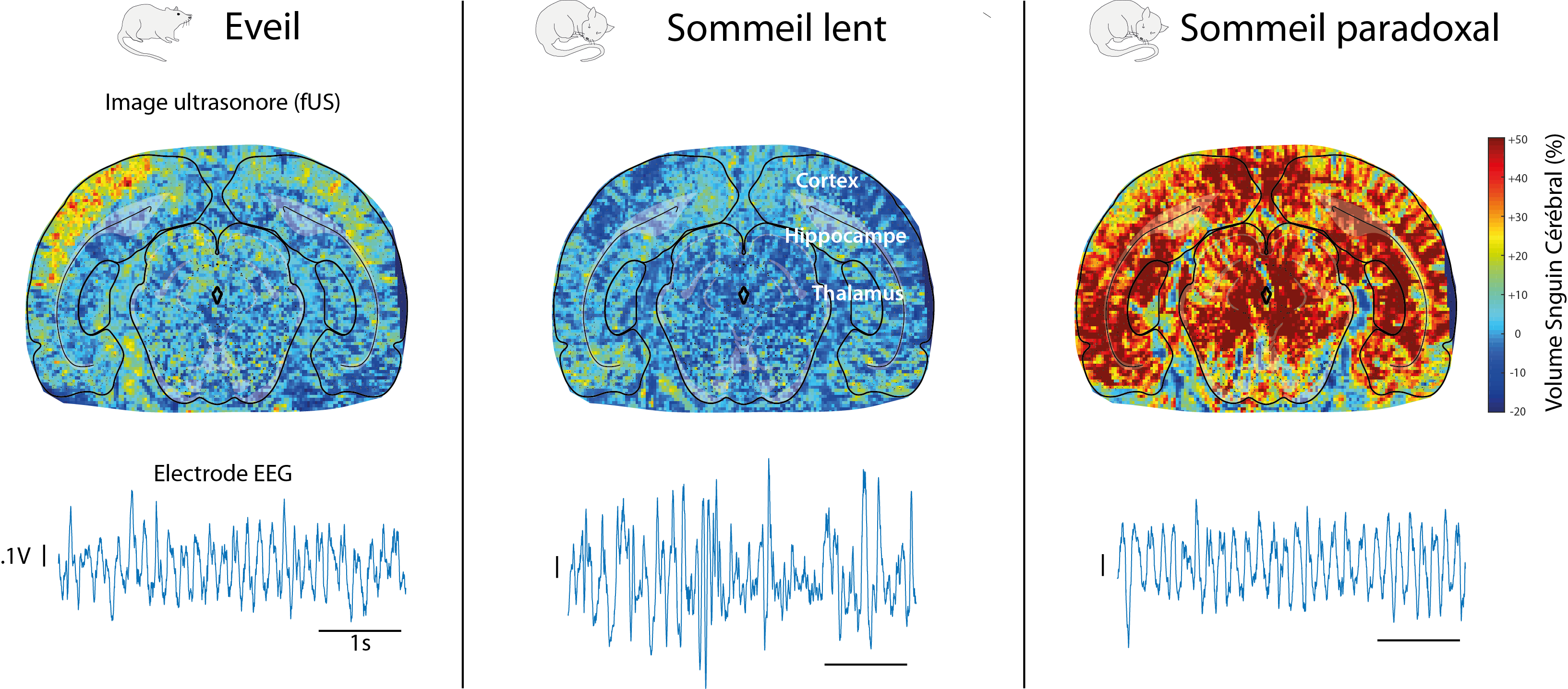
Images obtained using the fUS technique and EEG signals generated in a rat brain during the waking, deep sleep and REM sleep phases. In contrast to the highly similar electrical signals between waking and REM sleep, vascular brain activity is much more intense and more “synchronized” than during the waking state. The brain structures are identified by superimposing a brain atlas onto the vascular network images.

Discovery of novel mechanisms that cause migraines

©Photo by Anh Nguyen on Unsplash

Researchers at CNRS, Université Côte d’Azur and Inserm have demonstrated a new mechanism related to the onset of migraine. In fact, they found how a mutation, causes dysfunction in a protein which inhibits neuronal electrical activity, induces migraines. These results, published in Neuron on December 17, 2018, open a new path for the development of anti-migraine medicines.

Even though 15% of the adult population worldwide suffers from migraines, no long-term, effective, curative treatment has been marketed to date. Migraine episodes are related, among other factors, to electric hyperexcitability in sensory neurons. Their electrical activity is controlled by proteins that generate current called ion channels, specifically by the TRESK channel, which inhibits electrical activity. The researchers have shown that a mutation in the gene encoding for this protein causes a split between two dysfunctional proteins: one is inactive and the other targets other ion channels (K2P2.1) inducing a great stimulation of the neuronal electrical activity causing migraines.

Though researchers had already shown the hereditary nature of migraines, they did not know the mechanism underlying migraine. By demonstrating that the TRESK split induces hyperexcitability in sensory neurons leading to migraine, this work, carried out at the Institut de Biologie Valrose (CNRS/Inserm/Université Côte d’Azur), opens new research path for the development of anti-migraine medicines. A patent application has been filed1: the scope is targeting K2P2.1 channels to reduce the electrical activity of neurons and prevent migraines from being triggered.

What is more, the researchers propose that this new genetical mechanism, causing the formation of two proteins instead of just one, has now to be considered for the study of other genetic diseases and for diagnosing them.


1 Patent PCT/EP2018/067581 “Methods and compositions for treating migraine”

MON 810 and NK603 GM Maize: No Effects Detected on Rat Health or Metabolism

Photo by Charles Deluvio ???? on Unsplash


A diet based on MON 810 or NK603 transgenic maize does not affect the health or metabolism of rats, under the conditions of the GMO 90+1 project1. This unprecedented study performed by a research consortium led by Inra brought together a number of partners2, including Inserm. The research was performed as part of the Risk’OGM program funded by the French Ministry of Ecological and Inclusive Transition. For six months, rats were fed a diet containing either GM maize (MON 810 or NK603) or non-GM maize, in varying concentrations. The researchers, using high-throughput biology techniques, did not identify any significant biological markers related to the transgenic maize diet. Neither did anatomic pathology examination reveal any alteration of the liver, kidneys or reproductive system of the rats fed diets containing these GMOs. This research, published on December 10, 2018 in Toxicological Sciences, does not reveal any harmful effects related to the consumption of these two types of GM maize in the rat even after lengthy periods of exposure.

The researchers used two well-known types of GM maize: MON 810, which produces the protein Bt rendering it resistant to certain insects, and NK603 in which the modification of a gene renders it resistant to glyphosate. For 6 months, the rats were fed a diet containing either transgenic maize or non-GM control maize.  This time period, which is double that of the test required by European regulations, is equivalent to one third of the average lifespan of rats.

The objective of the researchers was to test for early biomarkers of biological function alterations in rats fed GM maize over periods of 3 and 6 months. For that, they used two ultra-sensitive high-throughput techniques: transcriptomics (gene expression) and metabolomics (study of the compounds derived from the body’s functioning). These techniques were used to identify and measure metabolites (amino acids, sugars and other small molecules) and to characterize the expression of messenger RNA and cellular microRNA. These methods are capable of detecting a broad spectrum of metabolic variations. The researchers identified markers able to differentiate the MON810 and NK603 diets. However, following the six-month period of the experiment, no significant differences were identified between the GM and non-GM diets, from the biological point of view.

In addition, in the rats fed the GM diets, anatomic pathology techniques (macro- and microscopic study of the tissues to detect potential abnormalities) revealed no alteration of the organs, particularly the liver, kidneys and reproductive system.

As such, the researchers did not detect any harmful effects of the MON810 and NK603 maize diets on the health and metabolism of the rodents, even after a lengthy exposure period.


1 The GMO 90+ project In 2010, the French Ministry of Ecology launched the Risk’OGM program, in the context of the 2008 law on genetically modified organisms for the establishment of a new legal and regulatory framework based notably on the principle of a triple evaluation of the impact of GMOs – from the health, environmental and socioeconomic standpoints. To set this dynamic in motion and fulfil public authority requirements in terms of expertise, guidance and completed research on GMOs, two calls for research proposals were held, in 2010 and 2013, respectively. The GMO 90+ project was selected during the 2013 call for proposals, with the following scope: to test for biomarkers predictive of biological effects in the study of the subchronic toxicity (3 and 6 months) of GMOs in the rat. Driven by a consortium pooling the various scientific competences, the purpose of this research was to determine whether the feeding of rats with genetically-modified maize led to metabolic changes which could be linked to early effect biomarkers (measurable biological characteristic). The objective was to provide key data which could be used in risk evaluation processes. http://recherche-riskogm.fr/fr/page/gmo90plus  
2 List of project partners: 1-Toxalim (Research Centre in Food Toxicology), Université de Toulouse, INRA, ENVT, INP-Purpan, UPS, Toulouse, France. 2-INSERM UMR-S1124, Toxicologie Pharmacologie et Signalisation cellulaire, Université Paris Descartes, USPC, Paris, France 3- Centre de Recherche sur l’Inflammation (CRI), INSERM UMRS 1149, Paris, France. 4- Laberca, ONIRIS, UMR INRA 1329, Nantes, France 5- Université de Rennes, Inserm, EHESP, Irset (Institut de recherche en santé, environnement et travail) – UMR_S 1085, Rennes, France. 6- Methodomics, France. 7- Institut de Mathématiques de Toulouse, UMR5219 – Université de Toulouse, CNRS – UPS IMT, Toulouse, France. 8- Anses, Maisons-Alfort, France. 9- Profilomic, Saclay/Gif sur Yvette, France 10- UMR1332 Biologie du Fruit et Pathologie, INRA, Université de Bordeaux, Villenave d’Ornon, France. 11- UR 1264, MycSA, INRA, Villenave d’Ornon, France. 12- Laboratoire Reproduction et Développement des Plantes, University Lyon, ENS de Lyon, UCB Lyon 1, CNRS, INRA, Lyon, France 13- CRO CitoxLAB, Evreux.

Type 2 Diabetes: A Therapeutic Avenue is Emerging

Physical contact between HSL and ChREBP in human adipose cells. Each red dot represents an interaction between the two proteins. The cell nucleus is stained in blue. (Credit: I2MC).

Restoring the action of insulin is one of the keys to fighting type 2 diabetes. Researchers from Inserm led by Dominique Langin at the Institute of Cardiovascular and Metabolic Diseases (Inserm/Université de Toulouse) are developing a therapeutic strategy that uses the properties of an enzyme (hormone-sensitive lipase) which, when stimulating fatty-acid synthesis in the fat cells, has a beneficial effect on insulin action. This research has been published in Nature Metabolism.

Diabetes is a disease in which blood sugar levels are high over a prolonged period (hyperglycemia). In the case of type 2 diabetes, this phenomenon which is caused by a disruption of the glucose metabolism develops progressively and insidiously. In France, the prevalence of diabetes is estimated at over 5 % of the 2015 population, with type 2 accounting for 90 % of cases. These figures do not include those who are unaware of their condition, particularly among the overweight or obese.

Hormone-sensitive lipase (HSL) is an enzyme which converts fats into fatty acids and releases them into the bloodstream. In obese patients, these fatty acids trigger the gradual insulin resistance at the origin of type 2 diabetes. Previous research by the Inserm team of Dominique Langin had shown that a decrease in HSL expression in the adipocytes led to a better response to insulin, a sign of good health for these cells.

Surprisingly, the researchers observed that the beneficial effect of a reduction in HSL was not actually due to the reduced release of fatty acid. It was explained by the increased synthesis of oleic acid, the principal fatty acid component of olive oil.

This initial observation gave a glimpse of an interesting avenue for treating obese patients who are at greater risk of developing type 2 diabetes.

To envisage a therapeutic strategy, it therefore had to be elucidated how reducing HSL exerted this beneficial effect on the action of insulin. The group of Prof. Langin discovered the existence of a physical interaction between HSL and a transcription factor responsible for the synthesis of fatty acids, ChREBP. HSL, when binding to ChREBP, blocks its activity. As such, a decrease in HSL leads to the release of this factor in the nucleus, promoting its activity, oleic acid synthesis and sensitivity to insulin.

Preliminary results indicate that a known inhibitor of HSL blocks the interaction with ChREBP. These data therefore pave the way for the development of molecules which target this interaction. In collaboration with global biopharmaceutical company AstraZeneca, the researchers in Toulouse are currently testing different approaches to block the interaction between HSL and ChREBP. Ultimately this project could lead to the development of new drugs to treat the increasing global epidemic of type 2 diabetes.

fermer