Menu

Microdystrophin restores muscle strength in Duchenne muscular dystrophy

©fotolia

Researchers from Généthon, the AFM-Téléthon laboratory, Inserm (UMR 1089, Nantes) and the University of London (Royal Holloway) demonstrated the efficacy of an innovative gene therapy in the treatment of Duchenne muscular dystrophy. Indeed, after injecting microdystrophin (a “shortened” version of the dystrophin gene) via a drug vector, the researchers managed to restore muscle strength and stabilise the clinical symptoms in dogs naturally affected by Duchenne muscular dystrophy. A first. This work, published today in Nature Communications, has been achieved thanks to donations from the French Téléthon.

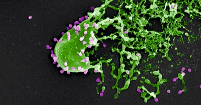
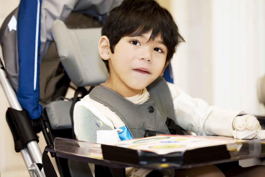
Duchenne muscular dystrophy is a rare progressive genetic disorder involving all the muscles of the body, and affects 1 in 5,000 boys. It is the most common neuromuscular disorder in children. It is associated with abnormalities in the DMD gene, which encodes dystrophin, a protein that is essential for proper muscle function. This gene is one of the largest in our genome (2.3 million base pairs, of which over 11,000 are coding). Because of this size, it is technically impossible to insert the entire DNA for dystrophin into a viral vector (or even the 11,000 coding base pairs alone), as is usually done for gene therapy.

To meet this challenge, teams at Genethon developed, in collaboration with a team at Royal Holloway University of London led by Pr. Dickson, and produced, a gene therapy drug combining an AAV-type viral vector with a shortened version of the dystrophin gene (approximately 4,000 base pairs), allowing the production of a functional protein. Dr Le Guiner’s team tested this innovative treatment in 12 dogs naturally affected by Duchenne muscular dystrophy.


By injecting this microdystrophin intravenously, and hence into the whole body of the dogs, the researchers observed that dystrophin expression returned to a high level, and muscle function was significantly restored, with stabilisation of the clinical symptoms observed for over 2 years following injection of the drug (see video). No immunosuppressive treatment was administered beforehand, and no side-effects were observed.

Some Golden Retrievers develop Duchenne muscular dystrophy naturally. The successful treatment of these dogs, which show the same clinical symptoms as children with this disease, and are of a similar weight, is a decisive step toward developing the same treatment in children.

“This preclinical study demonstrates the safety and efficacy of microdystrophin, and makes it possible to consider developing a clinical trial in patients. Indeed, this is the first time that it has been possible to treat the whole body of a large-sized animal with this protein. Moreover, this innovative approach allows treatment of all patients with Duchenne muscular dystrophy, regardless of the genetic mutation responsible,” says Caroline Le Guiner, the main author of this study.

“This is tremendously exciting progress towards a gene therapy for DMD. The studies in GRMD dogs have been spectacular and exceeded our expectations. My team has worked for many years to optimise a gene therapy medicine for DMD, and now the quite outstanding work of colleagues in France, in Genethon, in Nantes and in Paris has taken us close to clinical trials in DMD patients. I pay thanks also to the amazing and steadfast support of this research by AFM-Telethon and MDUK (Muscular Dystrophy UK) which has been essential to this achievement.” commented George Dickson.

For Frédéric Revah, Chief Executive Officer of Généthon: “For the first time, researchers obtained a systemic therapeutic effect on a neuromuscular disease in dogs using microdystrophin, and without immunosuppressive treatment. This highly complex cutting edge technology has been developed as part of an exceptional collaborative effort between Genethon and academic teams from Britain and France. Now our bioproduction experts have the task of producing a sufficient quantity of these new drug vectors, under GMP conditions, for the clinical trial.”
“This new evidence of the efficacy of gene therapy in Duchenne muscular dystrophy strengthens the therapeutic arsenal developed (exon skipping, CRISPR Cas-9, pharmacogenetics, etc.), and the first results are there. We need to forge ahead to complete the final phase and transform these scientific advances into drugs for children,” emphasises Serge Braun, Scientific Director of AFM-Téléthon.

Gene therapy: first results in children with Sanfilippo B syndrome

©Fotolia

On July 13, 2017, the journal Lancet Neurology published the results of a gene therapy trial conducted in four children with Sanfilippo type B syndrome (also known as MPS IIIB). This trial is the achievement of a two-decade partnership with financial support of AFM-Téléthon and the cooperation of the charity “Vaincre les Maladies Lysosomales” (VML). After monitoring of the treated children for 30 months, Dr. Jean-Michel Heard, from the Institut Pasteur and Inserm, and Professors Marc Tardieu and Michel Zérah, from the Paris public hospital administration (AP-HP) and the Paris-Sud and Paris Descartes Universities, conclude that the treatment was well tolerated and associated with neurocognitive benefits for the patients.

Sanfilippo syndrome is a rare genetic disease which affects approximately one in every 100,000 children. It alters brain development after birth and leads to brain degeneration several years later. The first symptoms of the disease are hyperactivity and delayed cognitive acquisition, which are usually noticed when children are around two-years old. A genetic anomaly prevents the production of an enzyme needed to breaking down mucopolysaccharides. Mucopolysaccharides are large macromolecules that help the neurons develop effective connections in young children during learning. Incomplete degradation and accumulation are toxic for brain cells. The disease progressively leads to a state of severe and multiple impairments and to premature death within periods of 5 to 10 years.

The challenge to treat Sanfilippo syndrome lies in the design of a method to supply the missing enzyme to the brain as early as possible after birth.  The therapeutic trial conducted by the Institut Pasteur at the Bicêtre Hospital (AP-HP) used gene therapy for that purpose. A gene therapy vector (AAV2/5) capable of inducing the production of the missing enzyme by brain cells was injected at several sites in the brain and cerebellum of affected children. The specific aim of this phase I/II trial was the assessment of tolerance to the surgical procedure and to the candidate drug delivered by gene therapy.

The clinical study initiated in 2013 was preceded by more than ten years of preclinical studies in animals naturally affected by the disease. The researchers administered the treatment for the first time to four children aged between 1.5 and 4 years (20, 26, 30, and 53 months). No particular clinical, radiological or biological side effects associated with the treatment were observed within 30 months of administration, indicating that it was well tolerated.

Within one month of treatment and throughout the 30 months of the trial, researchers detected the previously missing enzyme in the cerebrospinal fluid of the four treated children. Moreover, very careful and regular neurocognitive monitoring revealed positive impact on cognitive acquisition and behavioral development, which were more pronounced in the youngest treated child.

The encouraging findings of this phase I/II clinical trial suggest that treatment could be proposed to patients with Sanfilippo syndrome in the future.  To reach that goal, the next step would consist in a large and multicentric phase III clinical trial, if an appropriate partner is found.

9th IAS Conference on HIV

© Fotolia

From July 23 to 26, 2017, the Ninth IAS Conference on HIV Science, of which Inserm is a partner, will be held at the Palais des Congrès in Paris. For this edition, the International AIDS Society (IAS) has teamed up with ANRS, the autonomous agency of Inserm, to coordinate and fund research on HIV/AIDS and hepatitis.

The program will cover innovative approaches to HIV prevention, treatment and care. Various speakers, including Inserm researchers, will highlight the various advances in vaccines, new therapeutic approaches and prevention of HIV and hepatitis.

Yves Lévy, Inserm Chairman and CEO, will speak at the inaugural conference on Monday, July 24 on “Exploiting the immune system to prevent and control HIV infection”.

The scientific results of several Inserm researchers, including Dominique Costagliola, Patrizia Carrieri, Guillemette Antoni and Marie Jauffret-Roustide, will be presented at this conference.

Access the program for IAS 2017

Hereditary hearing loss: the ear and auditory brain are both affected

Brain slice showing neurons migrating to the auditory cortex. The nuclei are shown in blue. In green, molecules from the “molecular code” that sends the neurons to this brain region.

©Institut Pasteur

Scientists from the Institut Pasteur, Inserm, the Collège de France and Pierre & Marie Curie University have recently demonstrated that mutations in three genes responsible for Usher syndrome – a hereditary condition that affects both hearing and sight – influence not only the workings of the ear, specifically the function of sensory cells in the cochlea, but also the development of the auditory cortex. Their discovery could explain why some patients, even after being fitted with a cochlear implant (an electro-acoustic device that bypasses the defective cochlea), still have difficulties understanding speech. The findings are reported this week in a paper in the Proceedings of the National Academy of Sciences of the USA.

In most cases of hereditary hearing loss in humans, damage to the auditory sensory organ, the cochlea, is sufficient to account for patients’ hearing impairment. Many forms of hereditary hearing loss affect the hair bundle that acts as the sound receptor for auditory sensory cells. Cochlear implants stimulate the auditory nerve directly, bypassing the need for the sound signal to be processed by the cochlea and restoring a good level of hearing. In some cases, however, patients still have difficulties in understanding speech.

The team led by Prof. Christine Petit[1] from the Genetics & Physiology of Hearing Unit (Institut Pasteur/Inserm/UPMC) – with research by Baptiste Libé-Philippot supervised by group leader Dr. Nicolas Michalski –, working in cooperation with Dr. Christine Métin (Inserm/UPMC), recently identified three genes in mice which, when damaged, affect not only the cochlea but also the auditory cortex, the brain region responsible for analyzing auditory information. These three genes are among the nine currently recognized as causing Usher syndrome (type I and II), the leading hereditary cause of hearing and sight loss. Since damage to the cochlea in these patients prevents the auditory brain from receiving some or all of the acoustic information it normally receives, the cerebral damage had previously gone unnoticed.

The scientists demonstrated that, during embryogenesis, the proteins coded by these three genes are involved in the migration and maturation of some cells destined to become “inhibitory” neurons, which specifically colonize the auditory cortex. These neurons produce parvalbumin and are closely involved in cortical plasticity, which determines the structure and function of neural networks in the cortex based on auditory experience. They also play an important role in the temporal precision of sound detection that is required to understand speech. In mice, a single mutation in one of these three genes prevented this population of neural precursors from entering the developing cortex and reaching the auditory cortex.

The scientists also revealed that these neurons synthesize molecules that act as molecular markers. “These molecules serve as labels that instruct neurons to be sent from their birthplace in the subpallium, in the center of the brain, to the cortical area, their final destination. For the first time, we suggest that inhibitory neuron precursors have a “molecular mailing code”, explains Prof. Christine Petit.

These findings therefore demonstrate that hearing loss genes, previously thought to influence just the cochlea, also have another independent role in the development and formation of neural networks in the auditory cortex. In embryogenesis, this role is performed prior to the one played by the same genes in the cochlea.

Prof. Christine Petit’s team is at the forefront of pioneering research into hereditary hearing loss. It previously demonstrated that the proteins coded by the genes that cause type I and type II Usher syndrome are also responsible for the development and workings of the hair bundle, and went on to unravel the related molecular networks. The scientists hope to use these new findings to develop innovative aural rehabilitation methods designed specifically for patients with damage to the auditory cortex.

This research was funded by the French National Research Agency (LIGHT4DEAF [ANR-15-RHUS-0001] and LIFESENSES LabEx [ANR-10-LABX-65]), the European Commission (ERC-2011-ADG_294570), the BNP Paribas Foundation, the FAUN Stiftung (Suchert Foundation) and the LHW-Stiftung (CP), and the “scientific discovery” award and SEIZEAR grant from the “Agir Pour l’Audition” foundation (NM).

[1] Prof. Christine Petit is a Professor at the Collège de France and at the Institut Pasteur.

Vision restoration by optogenetic therapy within easy reach?

©fotolia

The Vision Institute (Inserm, Université Pierre et Marie Curie (UPMC), National Center for Scientific Research (CNRS)) via the Fondation Voir & Entendre has signed a contract with the United States Defense Advanced Research Projects Agency (DARPA), which could ultimately represent $ 25 million. With the help of an international consortium, researchers from Inserm, CNRS and UPMC, working within the Vision Institute, want to develop a system capable of restoring vision by optogenetic stimulation of the visual cortex. This project is called CorticalSight.

The consortium is coordinated by Professor José-Alain Sahel (Vision Institute and University of Pittsburgh School of Medicine). It is composed of academic partners: Stanford University, Friedrich Miescher Institute for Biomedical Research, the French Alternative Energies and Atomic Energy Commission – Leti and companies GenSight Biologics, Chronocam and Inscopix. Serge Picaud, Inserm research director, will coordinate the research activities at the Vision Institute.

The retinal ganglion cells are neurons which integrate the visual information of the environment in the eye’s photoreceptors and transmit it to the higher visual centers. Impairment of these cells deprives the centers of any visual information coming from the outside, thus causing complete blindness.

The degeneration of retinal ganglion cells is one of the leading causes of blindness in the Western world. It can be the result of various pathological conditions, including ocular trauma, retinal disorders such as glaucoma, diabetic retinopathy or optic neuropathies.

In animals, the restoration of sight after photoreceptor degeneration works thanks to the development of a very recent technique: optogenetic therapy. By this method it becomes possible to optically take control over the activity of very precise areas of the brain to induce behavior in the animal. In this specific case, the visual areas would be directly activated to induce visual perception even though the photoreceptors have not been activated. This first step in animals paves the way for the transfer of this technology to humans.

The CorticalSight project, financed by this contract, thus aims to restore visual perception in people who have become blind, by acting directly on the higher centers of the brain. To do this the researchers will use an intelligent image capture device combined with optogenetic stimulation.

In detail, the system as a whole will consist of several devices operating in series. On the face, a first device attached to glasses will consist of a camera filming the live environment of the patient in high resolution. A second device in the brain will transform the visual information, through complex algorithms, into light signals that the brain can interpret.

And this is where otogenetics comes into play. Using this technique, neurons specific to the visual cortex will be made sensitive to light by the expression within them of a microbial opsin (this algae protein transforms light energy into electrical activity).

It is then sufficient to couple the two external and internal devices so that the light signals coming from the outside are transformed into optical stimulation capable of activating the neurons of the visual cortex.

The human brain then does the rest of the work, as it knows how to, by translating the visual perception into a mental image representing the environment: a face, a tree, etc.

The Consortium

The CorticalSight project is coordinated by the Vision Institute (Inserm/CNRS/UPMC) and brings together international researchers in the field of vision whose individual expertise will be needed at each stage of scientific development.

Phage therapy : synergy between bacteriophages and the immune system is essential

Bacteria (in green) assaulted and killed by bacteriophages (in purple). Electron microscopy image provided courtesy of M. Rohde and C. Rohde (Helmholtz Centre for Infection Research, Braunschweig/Leibniz Institute DSMZ, Braunschweig, Germany) and colorized by Dwayne Roach (Institut Pasteur).
© M. Rohde and C. Rohde

Phage therapy involves the use of bacteriophages, or phages, for treating bacterial infections. Phages are viruses that specifically attack bacteria and are harmless to humans. A significant decline in the use of this therapeutic strategy introduced 100 years ago was seen in the West following the development of antibiotics. However, there is now new interest in phage therapy, especially in Europe, given the alarming increase in the number of antibiotic-resistant bacterial infections.

Until now, there has been insufficient scientific data to understand how phage therapy works in vivo. While most in vitro studies have proven that phages specifically target and kill bacteria, none of these studies took account of the importance of the host’s response to this activity.

Two Institut Pasteur teams (Laurent Debarbieux’s Bacteriophage-Bacteria Interactions in Animals Group and the Innate Immunity Unit led by James di Santo (Inserm U1223)) in partnership with Joshua Weitz’s team at the Georgia Institute of Technology (Atlanta, U.S.), recently showed the importance of patients’ immune status in terms of the chances of phage therapy success. This finding is the result of an original dual approach combining an animal model and mathematical modeling.

In order to evaluate the efficacy of treatment with a single phage species, the researchers focused on the bacterium Pseudomonas aeruginosa, which is involved in respiratory infections such as pneumonia. This bacterium, which is resistant to carbapenems, or ‘antibiotics of last resort’, was ranked by WHO as one of the four biggest global threats in terms of antibiotic resistance.

The researchers demonstrated that phage therapy is effective in animals with a healthy immune system (known as ‘immunocompetent’). The innate immune system can be triggered quickly and phages initially act in tandem with it to fight off infection. Then, after 24 to 48 hours, some bacteria naturally develop resistance to the phages which consequently cease to function. The innate immune system then takes over to destroy the bacteria. Of all the immune cells involved, neutrophils (white blood cells originating in the bone marrow) play a predominant role.

In parallel, in silico simulations have shown that the innate response needs to destroy 20-50% of the bacteria in order for phage therapy to be effective, regardless of whether phage resistance is observed. Thus, in the model studied, the researchers proved that there are no circumstances under which phages are capable of eradicating a P. aeruginosa infection alone.

These findings are particularly significant since they suggest that patients’ immune status should be considered when undertaking phage therapy. Laurent Debarbieux explains: “In terms of clinical consequences, one could reconsider the selection of patients likely to benefit from phage therapy. It may not be appropriate or recommended for people with severe immunodeficiency”.

The researchers are now planning to decipher the exact immune processes involved and the underlying mechanisms. At the same time, clinical trials are ongoing, notably including the Phagoburn trial on skin infections in burn patients funded by the European Union’s 7th Framework Programme.

What life expectancy in good health?

Copyright Fotolia

Life expectancy has been on the increase for several decades in Western countries. But what about life expectancy in good health?

It is clear that the increase in life expectancy is a new source of inequality between men and women in terms of disability.

The work of Jean-Marie Robine (Inserm unit 1198 “Molecular mechanisms in neurodegenerative dementias”) and Claudine Berr (Inserm unit 1061 “Neuropsychiatry: epidemiological and clinical research” in Montpellier) shows that from 2004 to 2015, disability-free life expectancy (DFLE) had increased by 1.1 years for men, going from 61.5 years in 2004 to 62.6 years in 2015. For women, DFLE had virtually stagnated over this period, going from 64.2 years in 2004 to 64.4 in 2015.

These are the findings of a study published in the latest Weekly Epidemiological Bulletin (BEH) published on July 11, which draw on the work of Inserm researchers on aging.

Mobilized on the subject, Inserm researchers are available to answer your questions.

An antidiabetic drug moves a step forward

In humans, apelin is able to regulate blood sugar levels and increase the sensitivity of cells to insulin. These two observations have paved the way for a clinical trial led by Inserm researchers from Toulouse, and represent a promising step forward for the development of a new treatment for diabetes, in particular type 2 diabetes.

This work has been published in the journal Diabetes, Obesity and Metabolism


 It is a journey that began over 10 years ago. A classic research story that demonstrates the long road between discovering a therapeutic molecule and its possible use in humans. The potential use of apelin was demonstrated in 2008 by university professor Philippe Valet and his Inserm team. This ubiquitous molecule (it is found throughout the body) can, if necessary, regulate the body’s blood sugar level instead and in place of insulin. However, this rescue pathway is only activated if the main pathway does not function properly.

Normally, sugar from food is stored in the liver, muscle, and adipose tissue, and is released as and when the body requires. This process is however dependent on the action of insulin, which “captures” sugar for storage. If insulin does not function properly, it leads to diabetes (increased blood sugar levels). Either it is not produced by the body at all: this is type 1 diabetes. Or the insulin receptors located on the surface of the liver, muscle, and adipose tissue cells become desensitized: this is type 2 diabetes. This results in two problems: the levels of circulating glucose are too high, and in time this becomes harmful to the body.

After discovering this alternative pathway, which enables another way of absorbing sugar, the researchers soon had the idea of stimulating this natural pathway and producing synthetic apelin.

Today, the researchers report the positive results of a clinical trial in 16 patients that was carried out within the Diabetology Department headed by Professor Pierre Gourdy. Healthy but overweight men were recruited to take part in a study that aimed to prove the efficacy and tolerability of two different doses of intravenously-administered apelin. The first group received a dose equivalent to 9 nmol/kg, and the second group received 30 nmol/kg. The patients’ glycemia was measured before and after the injection.

The results show that the injection of the smallest dose led to better absorption of circulating blood glucose, while administration of the highest dose also led to a demonstrable increase in cell insulin sensitivity. No side effects were observed.

“This is what we call a ‘proof of concept’ study”, explains Philippe Valet. “Although the sample is a small one, the results that we have just obtained encourage us to move on to larger studies in order to confirm them on a larger scale and be able to consider proper marketing authorization.”

This work could notably contribute to research into the treatment of diabetes, which affects over 400 million people around the world.

Physical activity does not protect against the onset of dementia

 
© fotolia

Physical activity in adulthood is not associated with a reduced risk of developing dementia. However, a decline in this activity is observed during the decade preceding its diagnosis. If this decline in activity cannot be considered as an early sign of dementia, it could be one sign – among others – to be taken into account by the attending physician. This is demonstrated by a study conducted by an Inserm research team of the Center for Research in Epidemiology and Public Health (Unit 1018 Inserm / Université Paris-Saclay). The results were published in the British Medical Journal on June 22, 2017.

Over the past forty years in France, life expectancy in good health has increased by 10 years, from 72 years in 1970 to 82 years in 2010. This increase in life expectancy can be explained by the improvement in quality of life but above all by the progress made in the field of medicine. However, living longer also leads to a higher probability of developing dementia (reduction of cognitive abilities leading to difficulties in everyday activities). The World Health Organization (WHO) estimated in 2015 that 47 million people worldwide were suffering from this disease and this number is expected to triple by 2050.

Until very recently, many studies suggested that physical activity could be a neuroprotective factor, thus delaying the onset of cerebral pathologies. In order to study this hypothesis, a longitudinal study conducted by a team of Inserm researchers from the Center for Research in Epidemiology and Public Health (CESP), in collaboration with University College London, followed more than 10,300 people aged 35 to 55 at the time of inclusion, between 1985 and 2012 (i.e. 27 years). Every 4 years the researchers measured the physical activity of each subject and made them take numerous cognitive tests.

This study showed that physical activity in adulthood would not have a protective effect on the risk of dementia. Indeed, it could not be demonstrated that there was any association between the practice of physical activity (whether mild or moderate to intense) and the onset or not of dementia. Moreover, people following public health recommendations in terms of physical activity, i.e. more than 2 hours 30 min of moderate to vigorous physical activity, showed a decline in cognitive functions, such as memory or thinking skills, similar to people not following these recommendations. As stressed by Séverine Sabia, Inserm researcher heading this study, “These results are corroborated by two recent intervention trials1,2 and an expert report published on June 22, 2017 that concludes there is a lack of sufficient evidence of a protective effect of increased physical activity on the risk of dementia”.

Nevertheless, researchers observed a decrease in physical activity (up to 2 hours / week less) in the 9 years preceding diagnosis in subjects with dementia. These findings suggest that the decline in physical activity in these individuals may be part of the changes that occur during the preclinical phase of dementia such as Alzheimer’s disease. Even today, it is very complicated to diagnose this disease before certain clinical signs such as memory problems or loss of autonomy appear. It therefore remains to be seen whether maintaining a good level of physical activity during this preclinical phase of dementia could slow down the disease process.

While physical activity appears to have no protective effect on the onset of dementia, it is important to remember that practicing a sport is beneficial for the cardiovascular system and the prevention of obesity and type 2 diabetes. It is therefore necessary to maintain good regular physical activity, even of moderate intensity, as it is a major determinant of a person’s state of health at all ages of life.

Gut bacteria can help to predict how the body will respond to fatty foods

Scientists have found that certain compounds, produced by microbes in the guts of mice, could be used to show which animals are at greater risk of becoming obese, or developing health conditions such as diabetes or cardiovascular disease.

The group, led by scientists at Imperial College London and INSERM UMRS 1138 in Paris, tested the urine of mice for a number of these microbial compounds, finding that certain key chemical signatures could accurately predict how the animals would respond to a high-fat diet before they received it.

High-fat diets are a major driver of obesity and related health conditions, such as diabetes and cardiovascular disease. However, evidence from previous studies suggests different people eating the same high-fat diet may have different outcomes, making it hard to define a one-size-fits-all ‘healthy diet’.

Previous research has shown that the hundreds of species of bacteria and other microbes which inhabit our gut work with our own cells to carry out a number of roles, and that this microbial garden can be shaped by what we eat or medicines we take, such as antibiotics.

In the latest study, published in Cell Reports, researchers used genetically similar mice to highlight the role that gut bacteria played in how the body responds to changes in diet and the impacts on health.

Before animals switched diets, their urine was screened for compounds produced by their gut bacteria using magnetic resonance spectroscopy, giving the mice a profile of chemical signatures, generated by metabolites from their microbiomes.

The team found that once the mice were switched to the same high-fat diet, they had a range of outcomes, with some animals gaining more weight than others, or becoming less tolerant to glucose – one of the early warning signs of diabetes.

Analysis revealed that key chemical signatures in their urine were predictive of some outcomes, such as changes in behaviour, weight gain and tolerance to glucose. One compound in particular, trimethylamine-N-oxide (TMAO), was shown to be predictive of glucose tolerance.

“We know that our environment and genetics can influence our risk of obesity and disease, but the effects of these communities of bacteria living inside us are less well understood,” said Dr Marc-Emmanuel Dumas, from the Department of Surgery & Cancer at Imperial, who led the research. “By using a group of mice with the same genetic makeup, we were able to zoom in on the variability in animals switched to a high-fat diet.”

“This study shows that value of a diet is determined not only by your genes, but also the genes of your gut microbes. This work has implications in lots of different areas, which is why it’s so exciting.” Senior investigator on the study, Dr Dominique Gauguier, from INSERM-Paris and a visiting professor at Imperial, said: “Our results illustrate the strong capacity of an organism’s gut microbiome to drive the adaptation to environmental challenges regardless of genetic variation and underline the need of deeper physiological and molecular phenotyping of individuals in large scale genetic studies.”

The findings will be explored further as part of an ongoing large clinical trial of 2,000 patients, where details of their lifestyle, diet, medication and other factors, as well as their microbiomes being characterised. Pulling together all of these data, and building on previous findings, they will be able to reveal how people react to different diets, and how their microbiomes influence the outcome.

According to the researchers, the hope is that in future, a patient’s profile could be generated from urine and blood samples and used to predict which diet they will respond to best.

“Our findings reveal that measuring metabolites in urine before the diet switch, we can predict which animals will get fat and become intolerant to glucose and which ones won’t,” added Dr Gauguier. “These findings open up really strong perspectives into designing personalised diets and harnessing our gut bacteria to promote health.”

fermer