Menu

The nature of the Ebola virus responsible for the 9th epidemic currently raging in the DRC characterized


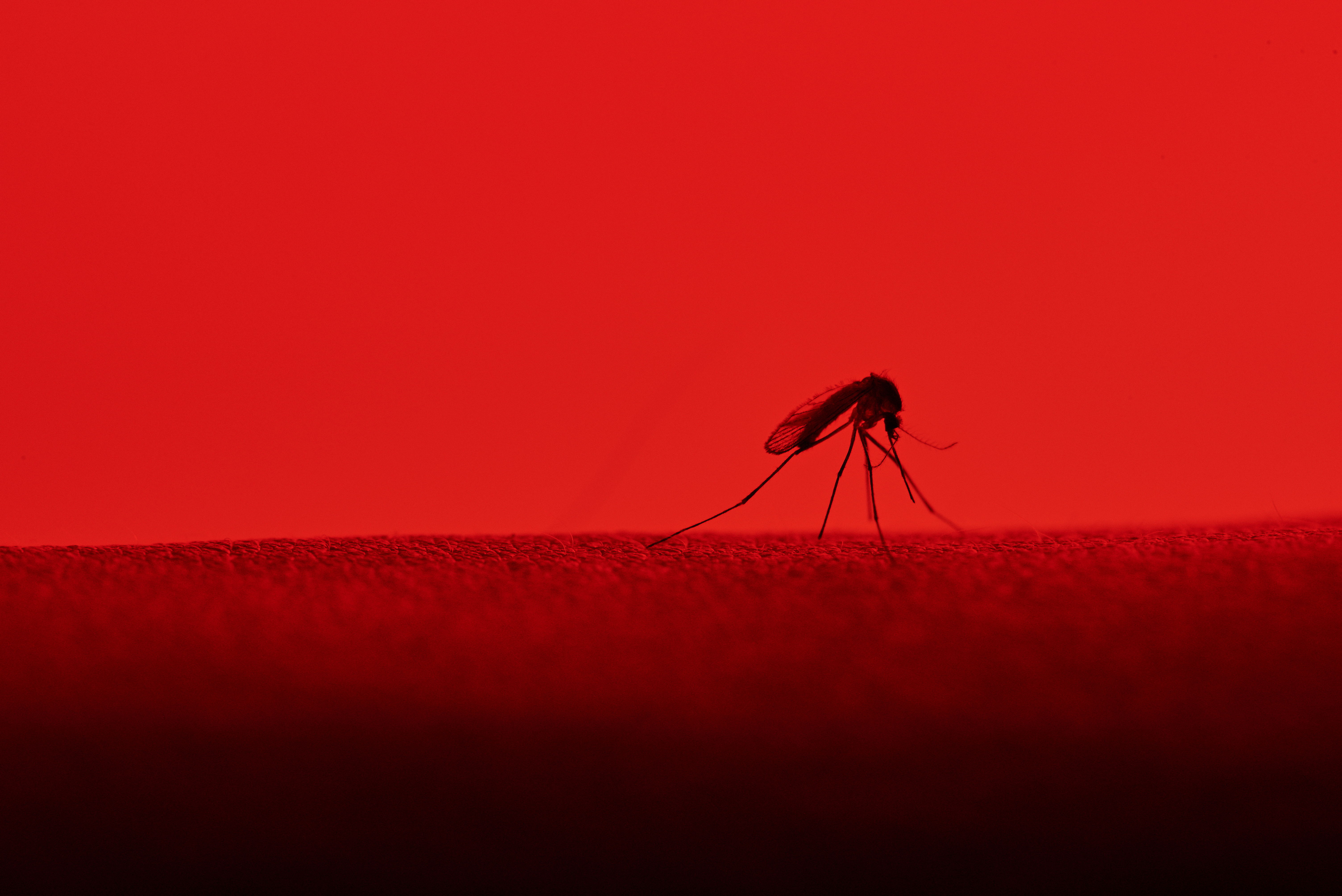
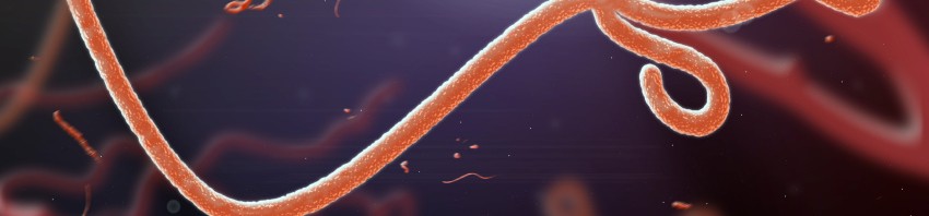
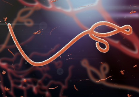
©Adobestock Microscopic view of the ebola virus

The National Institute for Biomedical Research (INRB) of Kinshasa and Inserm have characterized the nature of the Ebola virus responsible for the 9th epidemic currently raging in the Democratic Republic of Congo (DRC). The strain identified is the so-called Ebola Zaire strain.

According to a latest report published by the WHO (19/5/2018), a total of 46 cases (of which 21 confirmed), due to the current epidemic have been reported in the Democratic Republic of Congo (DRC). The risk for public health can be considered as high because of the possible extension in urban areas and neighboring countries.

Since the outbreak in West Africa in 2014, we have learned that the responsiveness of national and international health authorities is fundamental to accelerate the management and deployment of vaccines and possible treatments such as antivirals, or neutralizing antibodies. The deployment of these strategies, currently under discussion between the DRC authorities and the WHO, depends on the characterization of the virus responsible for the epidemic. Five Ebola strains are known today.

The collaboration established between INRB and INSERM (UMR INSERM / IRD / University of Montpellier), the technology transfer and the exchange of researchers between the two institutes allowed the genetic characterization of the circulating virus in the DRC, responsible of the current epidemic. The strain identified is the so-called Ebola Zaire strain. 

The approach used by the researchers used standard on-site techniques and new generation techniques that did not require the isolation of the live virus. The partnership between the two institutes as part of Inserm’s Reacting platform and its program to monitor the reservoir of the Ebola virus in Africa, supported by the french ministry for education and research with the help of the IRD and of the University of Montpellier, allowed a rapid deployment of these technologies. Reacting, a platform coordinated by Inserm, with its partners in the alliance Aviesan (alliance for life sciences and health), is a multidisciplinary consortium bringing together teams and laboratories of excellence, to prepare and coordinate research to deal with sanitary crises.

“This collaboration has allowed us to respond quickly to a health emergency, to ensure that the nature of the Ebola virus responsible for the epidemic is identified by DRC researchers and to alert the health authorities to rapidly deploy treatment on sites, “says Professor JJ Muyembe, Director General of INRB.

“I congratulate the INRB for this breakthrough under difficult conditions in its mission of monitoring and diagnosis and the excellence of our collaboration,” said Professor Eric Delaporte.

“In this context of health crisis, our experience in Guinea during the Ebola outbreak in 2014, the structuring of the Reacting platform conducted under the responsibility of Prof. Yazdanpanah, and the North / South collaborations put in place allowed us to better prepare and respond quickly. I would like to congratulate Professor Delaporte and Professor Muyembe for this breakthrough. This opens the way to the care of the sick people”, says Pr Yves Lévy, CEO of Inserm. 

The organization of the intestinal microbiota governed by antibodies

Credits: Fotolia

In a paper published in the journal Science Translational Medicine , Guy and his team Gorochov research center CIMI (Inserm / Université Sorbonne) and Immunology Department at the Pitié-Salpêtrière Hospital, AP-HP, reveal that our IgA act as conductor of the intestinal microbiota.They effectively prevent intestinal colonization by the oral flora and promote the presence of certain bacteria, totally innocent of infectious standpoint, but playing a beneficial role.

Our pact with microbes, otherwise known as symbiosis, we make them indispensable to normal life. Obesity, cancer, autoimmunity, accompanied unlike dysbiosis, that is to say, a disturbance of the bacterial ecosystem in favor of the action of “bad” bacteria. Until recently, the IgA antibodies that we secrete heavily in our digestive tract (66 mg / kg / day) was considered a defense to prevent the passage of potentially harmful bacteria through the intestinal barrier so that its effects potential microbial ecology sheltered by man remained unclear. This is precisely what the researchers wanted to understand.

It is not possible to inactivate a gene in humans to elucidate its function, as is done in mice. To assess the impact of IgA on the microbiota, the authors have taken advantage of a clinical situation of immune deficiency resulting in the almost complete absence of IgA in blood and secretions. Typical bacterial targets of IgA in the general population were also determined by purifying the part of the fecal microbiota naturally covered with IgA in healthy subjects, a unique approach developed by Martin Larsen in the laboratory. Then, total or fractionated microbiota were analyzed in a so-called metagenomic approach comprising sequence simultaneously all bacterial genomes present in a sample. Finally,

The work published today reveals that IgA plays an organizing role of the intestinal microbiota. IgA prevent intestinal colonization by the oral flora while promoting the presence of certain commensal, totally innocent of an infectious standpoint, but play a beneficial role.

This work has also helped to break an old mystery by explaining why the IgA deficiency (affecting about 1 in 500 Caucasian) is not accompanied more often fatal infections. The study shows that IgM, another type of antibody, can partly compensate IgA in its functions of interaction with the microbiota. A compensation however incomplete because patients with IgA deficiency suffer from respiratory infections, but also autoimmunity and atopy. These symptoms although point out the specific roles and not strictly anti-infectives, played by IgA.

These findings were obtained with the help of 21 deficient patients IgA, followed in hospitals of the AP-HP. Besides the fundamental breakthrough in understanding the role of IgA in the establishment of a physiological balance essential to health, the article opens up new therapeutic prospects oral supplementation these IgA deficient patients.

Finally, this study illustrates how anti-microbiota analysis of the antibody response can be a convenient way to study the interface between the host and its microbiota own, and thus the immune footprint of that scale the entire body. The study of anti-microbiota individual serological signatures representing a new biomarker for studying microbiota associations / disease that currently show the open, especially in the field of cancer.

Hepatitis C: a novel point-of-care assay

Darragh Duffy and Alba Libre, Immunobiology of Dendritic Cells Unit, Institut Pasteur / Inserm, using the Genedrive HCV assay. ©Institut Pasteur

One of the major challenges identified by the WHO in efforts to eradicate the hepatitis C virus (HCV) is the diagnosis of chronic cases that are generally asymptomatic. Major progress is required for new diagnostic techniques that can be “decentralized”, in other words accessed by populations and countries with limited resources. Scientists from the Institut Pasteur and Inserm, in collaboration with the company genedrive, have developed and validated a rapid, reliable, point-of-care HCV assay. This new screening assay means that patients can begin treatment for the disease as soon as they are diagnosed. The results have been published in the journal Gut on April 4th, 2018.

Hepatitis C is a liver disease caused by the hepatitis C virus (HCV). The virus can result in chronic infection, which may lead to severe complications such as cirrhosis and liver cancer many years later. Chronic infection with the hepatitis C virus affects approximately 1% of the global population (71 million people) and claims 400,000 lives every year when it develops into severe disease.

New direct-acting antivirals can successfully treat more than 95% of patients with chronic HCV infection if they are taken in time. In 2016, the WHO therefore published a plan to eliminate this major threat for public health by 2030. But the main challenge in meeting this ambitious target remains the diagnosis of asymptomatic patients, especially in low- or middle-income countries, where access to traditional screening assays is limited.

The current method for HCV diagnosis involves two stages. The first is to screen for specific HCV antibodies, but this does not reveal whether patients were infected in the past (and experienced spontaneous HCV clearance) or are still chronically infected. So the second stage requires a PCR1 assay to detect HCV RNA in the blood to confirm or rule out chronic infection.

There are rapid serological assays for HCV antibodies, but PCR screening requires dedicated infrastructure and qualified staff. In countries with limited resources, this type of assay is only available in centralized laboratories, which means that less than 1% of infected individuals in these regions actually know that they are infected. PCR screening may also involve several visits, and the time required between each result increases the risk of losing patients before the final diagnosis. To improve patient care from diagnosis to treatment, a screening assay for HCV RNA that can be “decentralized” and used in rural or low-income areas is urgently needed.

The team of scientists led by Darragh Duffy (Immunobiology of Dendritic Cells Unit, Institut Pasteur / Inserm) developed an assay in collaboration with the company genedrive that detects HCV RNA as reliably as existing assays but is faster and can be utilized at the point of care. PCR can be performed with the miniaturized device that enables the necessary succession of 40 reaction cycles to be carried out more quickly than in a conventional platform. The analysis can be performed in approximately an hour. This type of device is ultimately less costly than the current assays, which require significant laboratory infrastructure and maintenance.

The scientists began by clinically validating the assay on cohorts from the Institut Pasteur in France and the National Health Service in Nottingham, UK, then with data from Johannesburg-based Lancet Laboratories using samples from South Africa, Kenya, Ghana, Nigeria and Uganda.

The study demonstrated that the assay had a specificity of 100% – in other words there were no false positives – and a sensitivity of 98.6%, thereby meeting WHO requirements for this type of assay.

The kit has obtained CE certification for distribution in Europe and will be available for sale in the Middle East, Africa, South-East Asia and India once local regulatory clearance is obtained.

This study was funded by the organizations listed above and by the EU FP7 project POC-HCV.

Genedrive HCV assay. ©Institut Pasteur


1 PCR: Polymerase chain reaction, an enzyme reaction used to select then amplify an RNA fragment in large quantities. PCR consists of a series of repeated cycles (20 to 40 on average), each involving three temperature steps.

How allergens trigger asthma attacks

©Adobe Stock

A veritable sensor: a team of Inserm and CNRS researchers at the Institute of Pharmacology and Structural Biology (IPBS, CNRS/Paul Sabatier University – Toulouse III) has identified a protein that is able to detect various allergens in the respiratory tract, which are responsible for asthma attacks. This study, co-led by Corinne Cayrol and Jean-Philippe Girard, was published in Nature Immunology on March 19, 2018. It offers hope for breakthroughs in the treatment of allergic illnesses.  

What do mold, pollen and cockroaches have in common? Although they belong to three distinct kingdoms of the living world, they can all trigger asthma attacks in people sensitive to them. And all of them, despite their very different compositions, contain enzymes called proteases.   

The IPBS team has identified a human protein that reacts to a large number of environmental allergens: interleukin-33 (IL-33). When they enter the human respiratory tract, the allergens release their proteases. These split the IL-33 into extremely reactive fragments which trigger the chain reactions behind allergy symptoms.

This appears to be a general mechanism responsible for triggering allergic reactions. Indeed, IL-33 was shown to detect each of the 14 allergens tested, some of which are present in ambient air (several types of pollen, house dust mites, fungal spores) and others implicated in occupational asthma (such as subtilisin, found in detergents). 

These findings are all the more important because they establish a direct link between genetics and the environment. Indeed, the gene that codes for IL-33 is recognized as being one of the principal genes of asthma predisposition in humans.

Furthermore, clinical trials now under way are focusing on this protein. And they are on the right track, given this recent discovery of a single mechanism for detection of airborne allergens by IL-33. Inhibiting the production of reactive IL-33 fragments following allergen exposure could, for example, limit severe allergic reactions in asthmatic patients. 

This study was funded by the French National Research Agency (ANR). 


Mucous production in the lung following inhalation of an allergen : IL-33 sensor present (left), IL-33 sensor deactivated (right) (lung sections, mucous shown in magenta).   

Excessive mucous production is characteristic of allergic asthma. Protein IL-33, a major factor predisposing humans to asthma, detects allergen protease activity. Activated by proteases, IL-33 sets off a cascade of reactions, including mucus production, that are associated with asthma and other allergic illnesses. When IL-33 activation is inhibited (on the right), these reactions are not triggered.

© Corinne Cayrol and Jean-Philippe Girard / IPBS / CNRS-Paul Sabatier University – Toulouse III


Zika: an accurate estimation of the neurological risks in unborn children

Thanks to a study conducted in pregnant women and their unborn children during the Zika epidemic in the French territories in the Americas, researchers from Inserm, Institut Pasteur and the University Hospital of Guadeloupe have been able to accurately estimate the risk of severe neurological complications in babies. They have also determined that the first trimester of pregnancy is the period which presents the highest risk. While the overall risk is 7%, this rises to 12.7% – i.e. more than 1 in 10 children – if infection occurs during the first 3 months of pregnancy. This research has been published in the New England Journal of Medicine (NEJM).

In February 2016, faced with a drastic increase in the number of Zika infections and in order to establish a link between the virus and neurological complications, the World Health Organization (WHO) declared a “Public Health Emergency of International Concern (PHEIC)”. In March 2016, with the aid of the REACting consortium, Inserm took charge of the establishment, sponsorship and scientific follow-up of a cohort of pregnant women exposed to Zika in the French territories in the Americas, monitored by the French Antilles-French Guiana Clinical Investigation Center (Inserm CIC 1424). The objective? To study the fetal and neonatal complications associated with Zika virus infection in an epidemic situation. This cohort was funded by the French Ministry of Health and Solidarity (Exceptional Support of Research and Innovation) and included in the European ZIKAlliance[1] program.

Several thousand women who were pregnant during the Zika epidemic in the French territories in the Americas were enrolled in this cohort between March 2016 and August 2017. The article published in NEJM addresses those women from the cohort who presented with Zika virus infection confirmed by laboratory testing between March 2016 and November 2016. These women were then monitored every month until the end of their pregnancy. All complications and treatments received were recorded and if fetal abnormality was detected during an ultrasound, an additional examination by magnetic resonance imaging was performed.

The results obtained by the researchers show a 7 % rate of congenital neurological abnormalities observed in the fetuses and neonates of the cohort, which is a lot lower than that initially observed in Brazil, and close to what was observed in the US registry.

The study confirms an especially high risk in the event of infection occurring during the first trimester.

When broken down, the results show that the frequency of neurological complications is:
12.7% when the mother is infected during the 1st trimester
3.6% when the mother is infected during the 2nd trimester
5.3% when the mother is infected during the 3rd trimester

Likewise, the percentage of severe microcephaly (head circumference < -3SD) is 1.6% overall, and:
3.7% when the mother is infected during the 1st trimester
0.8% when the mother is infected during the 2nd trimester
0 when the mother is infected during the 3rd trimester

“These are the initial findings of the analyses of this cohort, given that the babies are still very young. It will be essential to monitor all the children in order to identify any later complications,” explains Bruno Hoen, physician researcher at Inserm and University Hospital of Guadeloupe and principal investigator of the study.

“Even if these complication rates are low in relation to other viral infections in pregnant women, they remain worrying given that the Zika virus can infect over 50 % of a given population in the epidemic phase”, comments Arnaud Fontanet, who heads the Emerging Diseases Epidemiology Unit at Institut Pasteur, and who is co-investigator of the study.

REACTing (REsearch and ACTion targeting emerging infectious diseases)

Inserm and its Aviesan partners have created REACTing, a multidisciplinary consortium that brings together research groups and laboratories of excellence, in order to prepare for and coordinate research to combat the health crises linked to emerging infectious diseases. Since its creation, REACTing has also set up programs centered around the Chikungunya, Ebola and Zika epidemics.

Clinical research at Inserm

The Clinical Research Unit manages sponsor activities for the clinical trials of which Inserm is a sponsor and, together with the Directorate of Health Care Supply (DGOS), jointly supervises the Clinical Investigation Centers (CIC). In 2017, he was in charge of 238 studies, including 15 European and/or international projects.


[1] ZIKAlliance is a 3-year project funded by the European Union’s Horizon 2020 Research and Innovation Programme under Grant Agreement No. 734548.

Tattoos: are they really indelible?

©AdobeStock

Researchers from Inserm, CNRS and Aix Marseille University at the Center of Immunology Marseille-Luminy (CIML) have discovered that while a tattoo may be forever, the skin cells that carry the tattoo pigment are not. These cells transmit this pigment to new cells when they die. Acting on this process could improve current laser removal techniques. This study was published on March 6, 2018 in Journal of Experimental Medicine.

For many years, it was thought that tattoos worked by staining fibroblast cells in the dermal layer of the skin. More recently, however, researchers suggested that skin macrophages (specialized immune cells that reside in the dermis) “gobble up” the tattoo pigment, as they would normally engulf an invading pathogen or piece of a dying cell. In either case, it was presumed that the pigment-carrying cell lived forever, allowing the tattoo to be more or less permanent.

A hypothesis which has been called into question by a team of researchers from Inserm and CNRS, led by Sandrine Henri and Bernard Malissen of the Center of Immunology Marseille-Luminy which, with the help of the Marseille Center for Immunophenomics, has developed a genetically-engineered mouse capable of killing the macrophages residing in its dermis. Over the weeks that followed, the researchers observed that the thus destroyed cells had been replaced by new macrophages derived from precursor cells present in the blood and originating from the bone marrow, known as monocytes.

They found that the dermal macrophages were the only cell type to take up the pigment when they tattooed the mice’s tails. Despite the programmed death of these macrophages, the appearance of the tattoo did not change. As such, the team concluded that the dead macrophages released the pigment into their surroundings where, over the following weeks, it was taken up by new macrophages. 

©Baranska et al., 2018

The appearance of a tattoo appears to be the same before (left) and after (right) the dermal macrophages were killed.

This cycle of pigment capture, release and re-capture occurs continuously in tattooed skin, even when macrophages are not killed off in one go. The researchers transferred a piece of tattooed skin from one mouse to another and found that, after six weeks, most of the pigment-carrying macrophages were derived from the recipient rather than the donor animal.

We think that, when tattoo pigment-laden macrophages die during the course of adult life, neighboring macrophages recapture the released pigments and insure in a dynamic manner the stable appearance and long-term persistence of tattoos,” explains Sandrine Henri, Inserm researcher and co-leader of the research project.

©Baranska et al., 2018

The green pigment of the tattoo is taken up by the macrophages (left). The pigment is released when these cells are killed (center) but, 90 days later, it is taken back up into new macrophages that have replaced the old ones (right).

Tattoos can be removed by laser pulses that cause the death of the skin cells and the release and fragmentation of their pigment. The latter can then be transported away from the skin via lymphatic vessels that drain the skin. “Tattoo removal using this laser technique can be likely improved by the temporary elimination of the macrophages present in the tattoo area”, declare the researchers. “As a result, the fragmented pigment particles generated using laser pulses will not be immediately recaptured, a condition increasing the probability of having them drained away via the lymphatic vessels. “

Type 1 diabetes: the role of the thymus is not what we thought!

©nerthuz – adobestock 

A small revolution has taken place in the world of type 1 diabetes research. A study conducted by an Inserm team led by Roberto Mallone at the Cochin Institute (Inserm, CNRS, Paris Descartes University) is calling into question the role long attributed to the thymus in selecting and eliminating white blood cells associated with type 1 diabetes and reveals that we are all auto-immune. Discoveries which change our understanding of the mechanisms of type 1 diabetes and point to new therapeutic strategies in fighting this disease. 

This research has been published in Science Immunology. 

In the large family of white blood cells, lymphocytes are in charge of immune response during infection. Among them, the T-cells are responsible for the recognition and specific destruction of pathogens. The “T” in T-cells is derived from the thymus, an organ to which the T-cells migrate from their place of birth, the bone marrow, prior to entering the blood circulation. Up until now, it was thought that the thymus was a place for the maturation and selection of T-cells, particularly that of CD8+ T-cells – a rare subset implicated in type 1 diabetes (T1D). So rare in fact that 10 mL of blood contains only 5 to 10 of these cells! These auto-immune lymphocytes become active when they encounter certain characteristic proteins for the first time, such as those of the β cells of the pancreas, subsequently leading them to consider them as undesirable and destroy them.

Up until now, it was accepted that the thymus “presented” to the CD8+ T-cells protein fragments characteristic of pancreatic β cells in order to pre-activate, detect and eliminate them. In T1D, it was thought that the selection by the thymus was altered – that if a healthy thymus filtered virtually all CD8+ T-cells, then that of a person with diabetes allowed many more to pass into the blood circulation. However, by comparing blood samples from healthy subjects and those with T1D, the Inserm researchers observed that not only did the blood of the healthy subjects present CD8+ T-cells but also that it contained as many as that of the diabetic subjects. These unexpected results call into question the role of the thymus in the selection of T-cells: since its presentation of β fragments to the CD8+ T-cells does not lead to their elimination, its selection turns out to be incomplete and inefficient.

The astonishing part of this discovery is that we are all auto-immune. This is the price we pay for being well-protected against the threat of infection, because the CD8+ T-cells spared by the thymus are also capable of recognizing microbial protein fragments that are similar to those of the β cells (a phenomenon known as “cross-recognition”).

But if we are all auto-immune, why then are we not all diabetic? According to Roberto Mallone, Inserm researcher at the Cochin Institute who led this study: “The next challenge is to better understand the ingredients that transform the “benign” auto-immunity of Mr. Average into T1D. Identifying these ingredients will allow us to diagnose T1D earlier and develop therapies to revert the auto-immunity to its benign state.”

 Two principal hypotheses are under investigation: the first one is that non-diabetic individuals may be capable of keeping their CD8+ T-cells under control, either due to a “policing” role played by other regulatory T cells or thanks to low CD8+ T-cell activation. The second is based on potential β cell vulnerability in T1D individuals, leading either to their abnormal recognition by the CD8+ T-cells or to their self-destruction.

Diabetes is a common disease in which complex genetic factors are involved. That is why it is the focus of the French Plan for Genomic Medicine 2025, supported by Aviesan and Inserm. As of 2019, a pilot experiment on diabetes as a common-disease model will be conducted in France to determine how the access to genetic sequencing could lead to earlier and more refined diagnosis than at present and the implementation of suitable treatments. New programs to screen the relatives of T1D patients are also being set up with the studies TRAKR and INNODIA, in order to achieve early diagnosis and the subsequent launch of clinical prevention trials.

Clever bacteria put human ingenuity to the test

How do bacteria manage to infect our body? What tactics do they use to slip through the cracks in our immune system? This is what the team of Thomas Henry, Inserm researcher, and his coworkers from the CNRS of Claude Bernard Lyon 1 University and the École Normale Supérieure de Lyon grouped within the International Center for Infectiology Research (CIRI) are trying to elucidate.  In a paper published in Nature Communication, the researchers studied a key component in the escape mechanism of bacteria and found, in humans, the major player involved in its detection.

Detecting the presence of the enemy is the first essential step in implementing a combat response. For our bodies, that role is played by the immune system,  which is confronted with various types of pathogens – particularly bacteria that employ every possible strategy to thwart its surveillance.

Normally, when invading the body, the bacteria betray their presence. The culprit, which is known as LPS and located in the bacterial cell wall, enables the human cells to recognize them and trigger an immune response. However, certain bacteria that are equipped with a more discreet form of LPS have a greater ability to pass under the immune system’s radar and increase their chances of infecting the body.

To understand the body’s defense mechanisms against bacteria, the researchers studied Francisella novicida, a pathogen model equipped with this discreet LPS.  This bacterium has the capacity to escape from within the innate immune cells (macrophages) which are supposed to destroy them.

Organizing the bacterial escape

Normally, the arrival of LPS within the cytoplasm of the macrophage is detected and the inflammatory response triggered, with the death of the cell halting the propagation of the pathogen. In reality, there is a constant race between bacterial multiplication and the detection systems of the host cell. Among the macrophage’s many alarm systems, Aim2 has been identified as being that which – in the mouse – is able to detect the arrival of these bacteria within the cytoplasm. However, it is impossible to reproduce the same result in humans. So, the challenge was then to understand how this counterattack takes place in humans.

Organizing the counterattack: at the right time, together!

This discovery also helps to explain why humans are more susceptible than mice to septic shock, which occurs when bacteria invade the blood or certain organs. Given that caspase-4 is particularly sensitive, the large quantities of LPS circulating in the blood provoke an immune system surge with irreversible life-threatening consequences. Despite everything, the diversity of the detection mechanisms and their partial redundancy help ensure that, following encounters with bacteria, humans often emerge the winner.

Identification of the functioning of caspase 4 and its cofactors represents a step towards the implementation of anti-inflammatory treatments in septic shock.

Illustration: Human macrophage (nucleus shown in blue) infected with Francisella novicida (red). The pathogen has escaped from the phagolysosomal compartment (shown in white) (one of the first defense mechanisms of the macrophage) but another defense protein, GBP2 (green), detects certain bacteria and enables caspase-4 to reveal the LPS of Francisella and implement antibacterial responses. Credit: Thomas Henry/Inserm

New Antiviral Targets Identified to Combat Dengue

©PixieMe – stock.adobe.com

The dengue virus – like all other viruses – hijacks many of the host cell’s functions to accomplish its infectious cycle. For the very first time, researchers from Inserm, CNRS and Université Paris Diderot have recently identified all of the cellular factors that interact with the virus as it replicates. By providing proof of concept that some of these molecules can be inhibited, these scientists are paving the way for new antiviral therapies for dengue and also for other viruses in the same family, such as the Zika and West Nile viruses.

This research has been published in Cell Reports.

The dengue virus is a major public health problem that affects millions of people throughout the world, and for which no antiviral treatment is available. The only vaccine currently available is recommended by WHO only in highly endemic (national or regional) geographical contexts, and for people who have already been infected at least once. The virus causes the body to develop disorders, often harmless, ranging from mild to moderate fever, but may also cause hemorrhagic fever which can be fatal, especially in children.

The dengue virus genome is an RNA molecule that codes for three structural proteins forming the viral particle, and for seven non-structural (NS) proteins. The NS proteins are responsible for viral replication in the host organism and also control the host’s antiviral immune response. These two functions are essential to the survival of the virus in the infected organism.

During the infectious cycle, NS proteins assemble and recruit cellular factors, which are still relatively unknown, to form a replication complex essential to amplifying the viral genome. Understanding this crucial step in the life of the virus is paramount if researchers seek to discover strategies to curb infection.

By using miniature modified dengue virus genomes, the team led by Ali Amara from the “Pathology and Molecular Virology” laboratory (Inserm, CNRS, Université Paris Diderot), in collaboration with Dr. Pierre-Olivier Vidalain from the Pharmacological and Toxicological Chemistry and Biochemistry Laboratory (Université Paris Descartes, CNRS), has succeeded in purifying and analyzing the protein composition of the dengue virus replication complex. Researchers were thus able to identify a whole network of cellular factors which interact with NS proteins during the infectious cycle. Some act as virus restriction factors while others are essential to viral replication.

The researchers have also provided proof of concept that these interactions between the virus and host cell are potential targets for new antiviral therapies. To do so, they first demonstrated that the OST cellular complex, normally responsible for the transfer of sugar motifs on cellular proteins, is also hijacked by the virus for some of its own proteins. The scientists then explained that an inhibitor of OST complex activity, NGI-1, prevents glycosylation of certain viral proteins and strongly inhibits dengue virus replication, along with the secretion of virotoxin NS1, an early marker for severe forms of the disease. They have also shown that these results can be extrapolated to other pathogenic flaviviruses, such as the Zika and West Nile viruses.

How Zika virus induces congenital microcephaly

©Fotolia

A team of researchers from ZIKAlliance discovers a specific mechanism of the infection

(Liège-Paris, December 11, 2017) –  Epidemiological studies show that in utero fetal infection with the Zika virus (ZIKV) may lead to microcephaly, an irreversible congenital malformation of the brain characterized by an incomplete development of the cerebral cortex. However, the mecha- nism of Zika virus-associated microcephaly remains unclear. An international team of researchers within the European consortium ZIKAlliance (coordinated by Inserm in France) has identified a specific mechanism leading to this microcephaly. Their findings are published this week in Nature Neuroscience.

To understand this mechanism, the scientific team led by Dr. Laurent Nguyen (frs-F.N.R.S., GIGA Neuroscience, University of Liège) and Prof. Marc Lecuit (Institut Pasteur, Inserm, University Paris Descartes, Necker Children’s Hospital, AP-HP) combined analysis of human fetuses infected with Zika virus, cultures of human neuronal stem cells and mice embryos. They showed that ZIKV infec- tion of cortical progenitors (stem cells for cortical neurons) controlling neurogenesis triggers a stress in the endoplasmic reticulum (where some of the cellular proteins and lipids are synthe- tized) in the embryonic brain, inducing signals in response to incorrect protein conformation (re- ferred to as “unfolded protein response”).

When it reaches the brain, Zika virus infects neuronal stem cells, which will generate fewer neu- rons, and by inducing chronic stress in the endoplasmic reticulum, it promotes apoptosis, i.e. the early death of these neuronal cells. These two combined mechanisms explain why the cerebral cortex of infected fetuses becomes deficient in neurons and is therefore smaller in size.

“These discoveries demonstrate a hypothesis that we had made following a basic research study we had just carried out in our laboratory, and thus confirm the physiological importance of the unfolded protein response in the control of neurogenesis,”  says Laurent Nguyen.

Researchers continued their studies on mice by administering inhibitors  of protein-folding  re- sponse in cortical progenitors and found that this inhibited the development of microcephaly in mice embryos infected with Zika virus.

Furthermore, the defects observed are specific to an infection by ZIKV, as other neurotropical vi- ruses of the flavivirus family (West Nile virus, yellow fever,…) did not cause microcephaly, in con- trast to Zika virus.

According to Prof. Marc Lecuit, “these results illustrate how studying fundamental biological pro- cesses is an essential step in understanding the mechanisms of infections, and lead to novel thera- peutic strategies.”

fermer