Menu

Ebola survivors: life afterwards

photo-postebogui

© Livia Saavadra REA / For Waha International: ophthalmic consultation as part of the PostEbogui programme in Guinea.

The long-term clinical and social sequelae following survival of Ebola infection are unknown. In November 2014, i.e. less than a year after the start of the Ebola epidemic in West Africa, Inserm, in partnership with the French National Institute for Sustainable Development (IRD) and the Department of Infectious Diseases at Donka University Hospital, Conakry in Guinea, organised medical follow-up of people who survived infection by the virus as part of a large research cohort. 802 individuals (adults and children) were included in the PostEbogui cohort[1] from March 2015.

Results of follow-up show that on average one year after their initial period in hospital, 3 in 4 survivors still report health problems. 40% suffer from tiredness or fever, but also from muscular (38%) and abdominal pain (22%), vision disorders that are sometimes serious and can lead to blindness (18%), and depression (17%). One quarter of survivors also claim to be victims of stigmatisation. As regards persistence of the virus in the semen, this is true: it can be found up to 18 months after the acute phase. This study has led the researchers to define what they now call the post-Ebola syndrome. This work is published in the journal The Lancet Infectious Diseases.

There are few data regarding people who have survived Ebola epidemics in the past. The reason: the too-small number of survivors and the inadequate structures for performing research in a state of emergency. The last epidemic of Ebola in West Africa was so large that it led both to an unprecedented number of deaths, and a number of survivors that had never been attained in the past (17,000 survivors). Given this unprecedented situation, basic medical and research questions emerged. What long-term complications could be caused by the virus? What might be the psychosocial consequences for the survivors? Is there a risk of delayed reactivation of the virus? How long does it persist in the body and what is the possibility of sexual transmission?

In order to answer all these questions, Inserm joined forces with the Guinean health authorities to organise follow-up of those who survived the infection. Researchers at the International Joint Unit “Translational Research on HIV and Infectious Diseases” (TransVIHMI; Inserm/IRD) established a follow-up cohort of people who had survived Ebola virus infection in Guinea, the PostEbogui cohort, in March 2015. 802 individuals (of the 1,270 reported survivors of the epidemic in Guinea) entered this multidisciplinary study, an average of one year after their initial infection.

They received biological, psychological and sociological follow-up, and their viral load was measured 1, 3, 6, 9 and 12 months after their inclusion in the cohort. Datas of this study refer to a follow-up until July 2016. Follow-up of immunological responses will be performed on a segment of the people, in partnership with the researchers of the Vaccine Research Institute (Inserm/ANRS).

45% of the participants are men. The median age is 28 years (range 1-79 years). One in five patients is a child under 18 years.


One year after their initial period in hospital, three quarters of survivors continue to report clinical symptoms.

Clinical data

40% of patients in the cohort suffer from so-called symptoms of a general nature (tiredness/fever/anorexia). Vision disorders affect 18% of patients (conjunctivitis; visual deficits [up to blindness] and eye pain). 38% of patients suffer from musculoskeletal pain (joint pain and muscular weakness), 35% complain of headaches, 2% of deafness and 22% of abdominal pain.

“Fortunately, these symptoms tend to become less frequent over time, and they tend to become less severe the longer the period after the acute phase of the infection,” explains Eric Delaporte, director of the International Joint Unit “Translational Research on HIV and Infectious Diseases” (TransVIHMI).

Compared with adults, children have more episodes of fever in the long term, but less musculoskeletal pain and fewer eye problems than adults.

Biological and virological data

26% of survivors suffer from anaemia. Ebola virus is still present in the semen of 5% of men 1-18 months after infection. These results were published in greater detail in The Journal of Infectious Diseases in May 2016.

Psychological and sociological data

The risk of depression is increased in survivors. Moreover, 26% of patients claim to be stigmatised after contracting the disease.

“The results of this first large cohort allow us to better characterise what we now call the post-Ebola syndrome. Medical complications persist or appear after the acute phase of the infection, and are not negligible. They justify medical follow-up of patients with Ebola for at least 18 months following infection,” concludes Eric Delaporte.

[1] This operational research programme, “[Re]vivre après Ebola en Guinée” (Living after Ebola in Guinea), led by Inserm, is developed by the International Joint Unit TransVIHMI (UMI 233 IRD / U1175 Inserm), in partnership in Guinea with several departments of Donka University Hospital, INSP, Macenta, N’Zérékoré and Forécariah Hospitals, the ALIMA association and the Socio-Anthropological Analysis Laboratory of Guinea (LASAG) of the University of Sonfonia, and in France with the Biology of Viral Emerging Infections Unit at Institut Pasteur Lyon, the Inserm U955 Immunomonitoring Platform, and the Inserm U980 Laboratory of Human Genetics of Infectious Diseases.

Identification of one of the keys allowing entry of Zika virus into brain cells

PhotoCP Zika

A team led by Ali Amara, Inserm Research Director at Unit 944, “Pathology and Molecular Virology” (Inserm/CNRS/Paris Diderot University) describes, in an article published in Cell Reports, the mechanisms that allow Zika virus to infect the cells of the nervous system.

 The ZIKAlliance project, coordinated by Inserm and funded under the Horizon 2020 programme of the European Commission’s Directorate-General for Research and Innovation, is aimed at characterising the fundamental and clinical aspects of infection by Zika virus, an emerging pathogen in America. The infection is generally mild, but the virus can also cause severe neurological diseases and congenital microcephaly in the foetus.

The researchers have shown that the Axl protein, which is expressed in numerous glial cells, facilitates entry of Zika virus into the brain. Entry of the virus into these cells requires a second protein, Gas6. The latter constitutes a mediator between the viral particles and glial cells.

The researchers also discovered that activation of the Axl protein lowers the immune response to Zika virus, thus promoting infection.

This study improves the state of knowledge of the molecular interactions that take place at the moment the virus enters the glial cells. These results represent a major step in understanding the neurological complications of the infection. Furthermore, they demonstrate in vitro that inhibition of the Axl pathway could represent a potential therapeutic target, although any side-effects associated with blocking it have yet to be identified.

Hepatitis C virus observed under a microscope for the first time

BO56374 éluats Fraisjc AR3A VLDL-0026b

(c) BMJ / British Society of Gastroenterology 2016

Scientists have finally observed the hepatitis C virus (or HCV) using an electron microscope. This is the first time since the virus became known in 1990. Inserm researchers at Tours (Inserm unit 966, “HIV and Hepatitis Viruses: Morphogenesis and Antigenicity”) have taken other scientists by surprise, including an American team believed to have accomplished this feat in 2013. The latter had in fact misunderstood the nature of the particles observed.

This research is published in the journal Gut.


The scientific community has waited twenty-five years for this. One of the greatest challenges of our era is to observe the hepatitis C virus, or HCV, under a microscope. It is responsible for 130 to 150 million cases of hepatitis C worldwide and about 700,000 deaths each year. This goal has become a reality thanks to the work by Inserm.

Viruses are normally discovered and described by observing them. However, HCV is an exception. All available data on this virus since 1990 were obtained by molecular biology as no one could view it under a microscope. This is due to the ability of the virus to hijack the machinery of the liver to give the appearance of a single lipid particle. This strategy allows the virus to easily penetrate cells and circumvent the immune system, also making it visually undetectable. “It resembles a single, small blank sphere amidst other blank lipospheres in the blood” explains Jean-Christophe Meunier, Inserm Research Fellow who is leading this study. “The virus takes advantage of the synthesis pathway for lipoproteins, particles that transport fat within an organism, so that it can replicate and closely associate with lipoprotein components.”

The virus effectively positions and attaches itself to the formation route of new lipoproteins and their components (phospholipids and their proteins).

Under its “disguise”, the virus becomes a genuine lipoviroparticle. This phenomenon was known for a long time, which makes it impossible to directly observe in the bloodstream of patients.

By contrast, lipoproteins sometimes inadvertently integrate viral proteins during formation, so it is possible to think that one is dealing with a virus when it is a single lipid particle. “This is precisely what happened in 2013 when an American team believed that they had observed the HCV virus”, explains Jean-Christophe Meunier.


Well-Structured Lipoviroparticles

Except this time, researchers are sure of their findings. They have used several viral protein-specific antibodies and were finally able to distinguish these famous lipoviroparticles from single lipoproteins circulating in the serum of individuals.

This work was possible through the electron microscopy platform at the University of Toulouse backed by the Inserm unit.

image VHC

(c) BMJ / British Society of Gastroenterology 2016

And as scientists observed, these chimeric particles ultimately have a similar structure to [lipoproteins]. They take the form of a sandwich lipid composed of a viral RNA centre, core antigen and enveloped by a monolayer of phospholipids. This is surrounded by a mixture of fatty acids and cholesterol, which are further enveloped by a second monolayer of phospholipids. Finally, the size of the virus varies according to the number of lipid layers it contains. “This structure is completely consistent with previous molecular biology work that predicted this formation. These observations validate twenty-five years of work for the scientific community.” says Jean-Christophe Meunier.

image VHC 2

(c) BMJ / British Society of Gastroenterology 2016

Apart from the satisfaction of accomplishing this technical feat, researchers stress the relevancy of this work. “Effective treatments are now available for hepatitis C, but a vaccine has yet to be found. Now, knowing the exact structure and organisation of these lipoviroparticles will be highly beneficial for those working in the field.”, states Jean-Christophe Meunier.

Combating Ebola: Encouraging Results for the PREVAIL Clinical Trial

DSC_0094

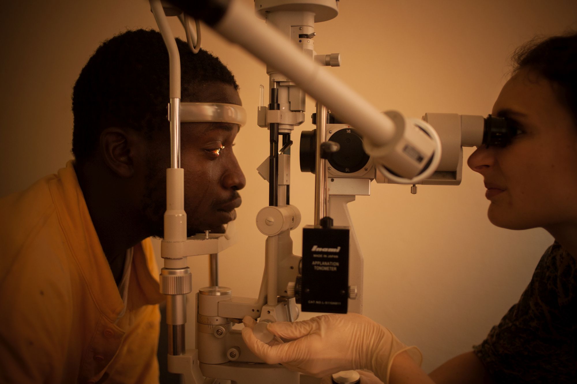
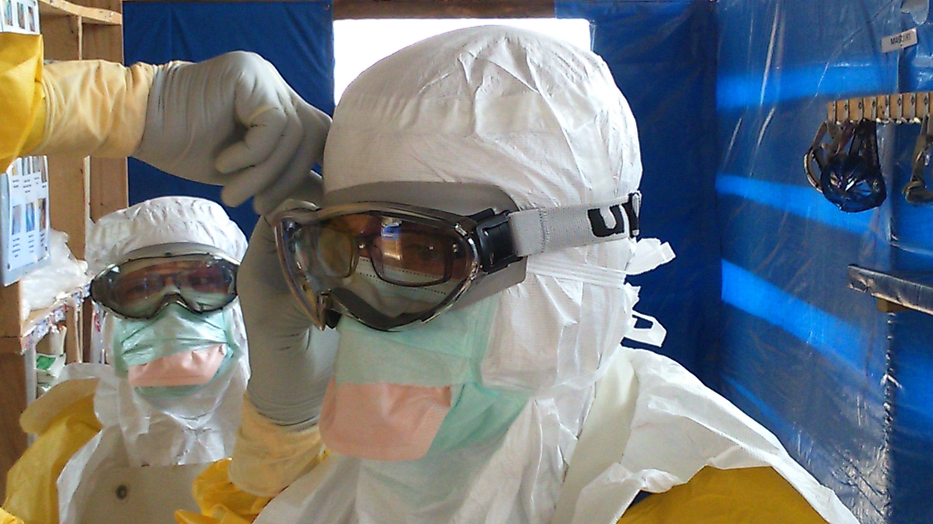
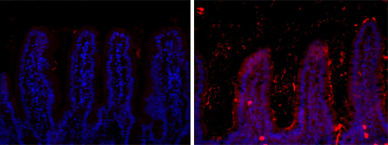
(c) Inserm/Anglaret, Xavier/Sissoko, Dadoua

An international research team involving Inserm and the National Institutes of Health (NIH) has just published the results of the PREVAIL study on treatment for the Ebola virus. While not definitive, the results show that ZMapp may benefit patients with the Ebola virus disease.

These results are published in The New England Journal of Medicine.


In this trial conducted in the context of the Ebola epidemic in West Africa, the effectiveness of the experimental drug ZMapp[1] was compared with the best supportive[2] care given at treatment sites. Used in combination with current care, ZMapp was well-tolerated and is leading to a higher survival rate than supportive care alone. However, the decreasing epidemic trend in West Africa at the time of the trial prevented the enrolment of a sufficient number of participants to prove this point definitively.

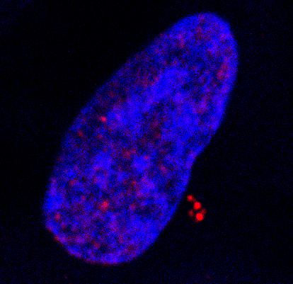
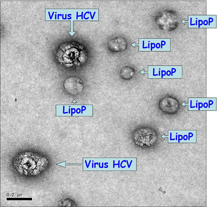
ZMapp is a combination of three antibodies that target a protein on the surface of the Ebola virus. By binding to the virus in an infected individual, they block its ability to infect human cells. It is believed that this blockade provided by the drug and administered as early as possible reduces the growth of the infectious agent and allows the patient’s immune system to mount an effective response in sufficient time.

The study opened in March 2015. All patients (except the first patient) were enrolled in West Africa where the epidemic is ongoing: 5 in Liberia, 12 in Guinea, and 54 in Sierra Leone. Patients of all ages with Ebola infections were eligible. The average age of the participants was 24 years, and slightly more than half were women. Although the team planned to enrol up to 200 patients, the study closed in late January 2016 once it became clear that the epidemic was curtailed in this region.

This was a randomised, controlled trial. Each patient was assigned by lot into two study groups with different treatment strategies. One group only received the supportive care available at their treatment sites. In Guinea, this supportive care also included Favipiravir[3], an antiviral drug whose potential benefit against the Ebola disease was previously demonstrated by Inserm[4]. Meanwhile, the second group received supportive care plus three consecutive infusions of ZMapp over the course of one week including, where applicable, Favipiravir.

A total of 21 patients, nearly one-third, died during the study. To assess whether ZMapp was effective, the team compared the number of deaths in each group after 28 days.

8 patients out of 36 (22%) died in the group that received ZMapp, compared to 13 patients out of 35 (37%) in the other group. This translates as a 40% lower risk of death among those who received the drug.

Given the lack of scope due to enrolment difficulties and insufficient staffing, this difference did not reach the statistical significance required to offer a firm conclusion. However, based on these results, the administration of ZMapp is approved should new Ebola cases arise in these 3 countries. This authorisation is issued under an expanded access protocol[5] (EAP, expanded access protocol).

According to Denis Malvy, Inserm researcher and co-investigator in this trial in Guinea: “The results from this trial mark a prominent step in treating the Ebola virus disease. I am also pleased that these results were obtained thanks to the unique partnership between researchers, authorities in Guinea and ONGs. I have no doubt that these collaborations will continue to bear fruit in the months to come.”

Inserm’s involvement in this clinical trial was made possible through the REACTing network (REsearch and ACTion targeting emerging infectious diseases) designed to:
• improve research planning during periods between crises: governance, preparation of research tools, identification of research priorities, application for funding, and ethical and legal aspects;

• fund and establish research projects during periods of epidemic crisis: coordination, strategic priorities, methodological assistance, and informing the authorities and general public.

This network is organised around a steering committee of some fifteen human health specialists, and relies on an 8-member scientific committee and methodological centres located in the North (F. Mentré, Bichat University Hospital) and in the South (A. Fontanet, Institut Pasteur). REACTing does not target any particular disease, and can get involved in all emerging infectious diseases. Its scope of action is broad, from fundamental research to human and social sciences, promoting a cross-disciplinary approach.

Guinea’s involvement (also called JIKIMapp) in the PREVAIL trial was led by the JIKI team at the treatment site in Forécariah, Coastal Guinea, directed by the French Red Cross. This study is part of the ongoing pioneering trial to assess the antiviral drug Favipiravir in Guinée-Forestière between December 2014 and April 2015.

Financial or logistic support for the trial was provided by NIAID/NIH; the French Institute national de la santé et de la recherché médicale (INSERM); the Republic of Sierra Leone Armed Forces; the Ministries of Health and U.S. Embassy staff in Liberia, Sierra Leone, and Guinea; the U.S. Centers for Disease Control and Prevention (CDC) and the CDC Foundation; the U.S. Biomedical Advanced Research and Development Authority; and the U.S. Defense Threat Agency. The trial was conducted in partnership with other academic, governmental and non-governmental agencies, including Bordeaux University Hospital, in the Franco-Guinean area.

[1] Made by Mapp BioPharmaceutical Inc. of San Diego, CA, USA

[2] Among other things, this optimised supportive care includes administering fluids, balancing electrolytes, maintaining physiological functions, reinstituting nutrition and treating other infections if they occur.

[3] Made by laboratories in Toyama/Fujifilm, Japan

[4] https://presse.inserm.fr/resultats-definitif-de-lessai-jiki/22789/

[5] An EAP is a U.S. regulatory mechanism that enables an unlicensed drug to be made available to treat a serious or life-threatening disease for which no approved therapy is available. The U.S. Food and Drug Administration has approved the EAP for use in the United States.

Zika virus found inside spermatozoa

Zika spermaozoides (c) Suberbielle

Spermatozoa infected by Zika virus (green; arrowhead) 

© Elsa Suberbielle, CPTP / Inserm

Recent work has shown that Zika virus persists in semen for up to 6 months after infection[1]. In a correspondence published in The Lancet Infectious Diseases, the researchers, in addition to confirming its long persistence in semen (in this case for more than 130 days, i.e., over 4 months), reveal the presence of the virus even within spermatozoa. This work results from collaboration between researchers from Inserm, CNRS, and academic practitioners from University Toulouse III – Paul Sabatier and Toulouse University Hospital.


In this letter, the scientists report the case of a 32-year-old man returning from French Guyana with symptoms suggestive of Zika virus infection, namely, moderate fever, rash, and muscle and joint pain. Zika virus was detected in the patient’s plasma and urine 2 days after the onset of these symptoms. Samples of semen (11 samples), blood (10) and urine (5) were taken and analysed over a total period of 141 days.

Upon analysis, Zika virus was found to be present in all samples up to the 37th day. Beyond that point, the virus was found only in the semen, where it persisted for over 130 days, while the patient was in good health. This result was confirmed in two other patients, in whom the virus persisted for 69 and 115 days in the semen. For the moment, the factors responsible for this variation in persistence amongst individuals are unknown. From the time of diagnosis, however, these patients were advised to use protection during sexual relations.


The research team subsequently analysed the semen from the patient and examined the spermatozoa it contains using various microscopy techniques.

“We detected the presence of Zika virus inside approximately 3.5% of this patient’s spermatozoa,explains Guillaume Martin-Blondel, Inserm researcher at the Toulouse Purpan Pathophysiology Centre (Inserm/CNRS/University of Toulouse III – Paul Sabatier) and physician at the Infectious and Tropical Diseases Department of Toulouse University Hospital.

The researchers explain that for other sexually-transmitted viruses, such as HIV, the virus remains “stuck” at the surface of spermatozoa. For purposes of in vitro fertilization, it is therefore possible to “wash” the spermatozoa from HIV-infected patients, whereas this seems to be excluded for patients positive for Zika virus. However, the “active” nature of the Zika virus present in spermatozoa remains to be determined, as well as the ability of these spermatozoa to transmit infection (since the virus is also present outside the spermatozoa, in the seminal fluid).


In conclusion, analysis of this case has important implications for the prevention of sexual transmission of this virus, by means that remain unknown at present. These observations also raise many questions on the need to include screening for Zika virus in the testing of sperm donations in fertility centres.

[1] https://www.eurosurveillance.org/ViewArticle.aspx?ArticleId=22556

A new way to combat viruses

The team “Physiopathology and therapeutics of chronic viral hepatitis and related cancers” of the Mondor Institute of Biomedical Research (Inserm/UPEC), located on the premises of Henri Mondor Hospital AP-HP, in collaboration with the researchers of the Centre for Structural Biochemistry (CNRS/Inserm/Montpellier University), with the support of ANRS, has created a whole new family of molecules that inhibit cyclophilins – proteins indispensable to cell metabolism – and have strong therapeutic potential as broad-spectrum antiviral drugs. This discovery, published in Nature Communications on 22 September 2016, also opens the possibility of using these new inhibitors to protect cells in the context of ischaemia-reperfusion (organ transplants, recovery after ischaemic accidents, or neurodegenerative diseases).


Cyclophilins are cellular proteins that play complex and indispensable roles in cell metabolism. They are very often used by viruses to help complete their life cycle. Cyclophilins therefore represent a potential target for broad-spectrum antiviral drugs, i.e. drugs that are effective against many virus families. Cyclophilin inhibitors also have cell protection properties associated with inhibiting the opening of the mitochondrial pore. Unfortunately, the existing inhibitors, all derived from cyclosporin A, present considerable problems that impede their clinical development.


The teams led by Professor Jean-Michel Pawlotsky (Mondor Institute of Biomedical Research [IMR], Henri Mondor Hospital AP-HP, Inserm/University Paris-Est Créteil [UPEC]) and Dr Jean-François Guichou (Centre for Structural Biochemistry [CBS], Montpellier University/CNRS/Inserm), have joined forces and used innovative techniques to create and optimise a whole new family of chemicals that directly target cyclophilins. This work was supported by the French National Agency for Research on AIDS and Viral Hepatitis (ANRS). The new inhibitors thus specifically block the action of cyclophilins by inhibiting the multiplication of viruses from different viral families such as hepatitis C virus, HIV, hepatitis B virus and several coronavirus strains.

This work therefore made it possible to create a whole new family of cyclophilin inhibitors with strong therapeutic potential as broad-spectrum antiviral agents, i.e. active against many viral families for which there is currently no treatment. This discovery also provides an opportunity to use new inhibitors to protect cells in the context of ischaemia-reperfusion (organ transplants, recovery after ischaemic disease, or neurodegenerative diseases).


This work was carried out as part of a close collaboration between Dr Abdelhakim AHMED-BELKACEM and Prof. Jean-Michel PAWLOTSKY for the team “Physiopathology and therapeutics of chronic viral hepatitis and related cancers” (Mondor Institute of Biomedical Research, Inserm/UPEC). It also involves Dr Lionel COLLIANDRE and Dr Jean-François GUICHOU from the Centre for Structural Biochemistry (CBS; Montpellier University/Inserm/CNRS) and researchers from the Institute of Analytical Sciences (ISA; CNRS/ENS Lyon/Claude Bernard Lyon 1 University).

The team “Physiopathology and therapeutics of chronic viral hepatitis and related cancers” at the Mondor Institute of Biomedical Research is developing research on two main themes: the development of broad-spectrum antiviral approaches (building on its experience in the development of new treatments for hepatitis C), and the role of inflammation and the liver microenvironment in the occurrence and progression of primary liver cancers.

Meanwhile, the team “Structures and screening of therapeutic and environmental targets” at the Centre for Structural Biochemistry is developing an integrated approach to spur and rationalise the development of new molecules with potential therapeutic applications, using X-ray crystallography and structural bioinformatics.

Too much fat rapidly unbalances the intestinal flora

Visuel sansonneti microbiote

Localisation of the bacteria the ileum of a mouse fed a standard diet (left) or a high-fat diet (right).

© Institut Pasteur

A disruptive element such as a change in diet, and the whole intestinal flora becomes disrupted, with possible repercussions for health. An international research study conducted by the Molecular Microbial Pathogenesis Unit (Institut Pasteur/Inserm), directed by Philippe Sansonetti, has just demonstrated, in mice, the direct influence of a diet too high in fat on the intestinal flora and its environment. In response to this new diet, the bacterial communities reorganise themselves and the small intestine itself is transformed. And this happens in the first month. These results were published in the journal PNAS on 16 September.


The billions of bacteria that populate our intestine – known as the microbiota – play a central role in digestion, but also have a role in some diseases such as type 2 diabetes or obesity. These diseases have often been associated with an imbalance of the intestinal flora, with some bacteria becoming clearly predominant, and a permeable intestine, likely to release inflammatory substances into the bloodstream. But although many studies have been conducted on the state of the microbiota once the disease has become established, few have focused on the development of this intestinal imbalance, e.g. when a high-fat diet is introduced. For this reason, an international research team has addressed the question. “We wanted to see, from an early stage, how the intestinal bacteria behave in the presence of a high-fat diet,” emphasises Thierry Pédron, a research engineer in the Molecular Microbial Pathogenesis Unit (Institut Pasteur/Inserm). “And we soon focused our research on the small intestine, because that was where we saw the most obvious variations!”


Some mice in the study were therefore fed an ordinary diet, whereas others were fed a diet containing 70% lipid. Using genomic techniques, the researchers were able to identify the different bacterial species contained in faecal samples, and monitor the changes in composition of the microbiota in time. They also localised and accurately identified the bacteria within the small intestine.

“Only one month after starting this new high-fat diet, we observed changes in the composition of the microbiota,” says Thierry Pédron. “Some bacterial species proliferated, whereas others declined, and the species Candidatus arthromitus actually disappeared completely. Furthermore, and as never seen before, we observed a massive concentration of bacteria between the intestinal villi.”


Ordinarily, bacteria cannot come close to or even cross the intestinal wall because the epithelium releases antimicrobial peptides and is lined with a protective mucus. The researchers then focused on these intestinal wall defences: they found that production of antimicrobial peptides fell following massive ingestion of fat, and that the mucus layer became thinner. In other words, not only does the microbiota become reorganised under the influence of lipids, but the intestine itself undergoes transformation. And the modifications do not end there. Additional measurements made it possible to demonstrate an increase in the permeability of the small intestine, and a reduction in PPAR-γ activity. “PPAR-γ is a molecule with many functions. It plays an important role in fatty acid metabolism, as well as in inflammation and embryonic development,” explains Thierry Pédron. “This drop seems closely related to the drop in antimicrobial peptide level.” And although the connections between all these results and their potential involvement in some dietary imbalances have not yet been established, it is reassuring to note that when the mice are put back on a balanced diet, everything returns to normal within a month!

An antibody-based drug for multiple sclerosis

Inserm Unit U919, directed by Prof. Denis Vivien (“Serine Proteases and Physiopathology of the Neurovascular Unit”) has developed an antibody with potential therapeutic effects against multiple sclerosis. The study, directed by Fabian Docagne and published in Brain, paves the way for a new strategy to control the disease.

Multiple sclerosis is a disease that affects the central nervous system, particularly the brain and spinal cord. It is the most common cause of neurological disability in young adults.

The disease is considered autoimmune since the immune system, which is there to protect the body from external assault, attacks its own constituents. The cells of the immune system, particularly the lymphocytes, bring about the destruction of the myelin sheath that surrounds and protects the extensions (axons) of the neurons. This demyelination, which marks the beginning of axon degeneration, disrupts the transmission of nerve impulses. Lesions in the form of “plaques” are dispersed over the brain and spinal cord. They cause symptoms that vary greatly from one individual to another.

Usually, the disease is characterised by exacerbations, with the appearance of motor, sensory and cognitive disorders, followed by remission a few weeks later. But with the passage of years, these symptoms can progress to irreversible disability. Current treatments reduce the exacerbations and improve the quality of life of patients, but do not control the progression of the disease.

sclérose en plaq

(C) Inserm/Fumat, Carole

In order for the cells of the immune system circulating in the bloodstream to reach the central nervous system, they must penetrate the blood-brain barrier (haematoencephalic barrier) and blood-spinal cord barrier (haematomedullary barrier).

During previous work on a mouse model of stroke, the team from Inserm Unit 919 studied a factor involved in opening the blood-brain barrier, the NMDA receptor. In particular, they observed that blocking the interaction of this receptor with tPA (a member of the serine protease family) has beneficial effects associated with maintaining the integrity of the barrier.

In this study, the researchers developed a strategy for blocking the interaction of tPA with the receptor, in multiple sclerosis. In the laboratory, they developed a monoclonal antibody (Glunomab®) directed against the specific site on the NMDA receptor to which tPA binds.

schéma docagne english

(C) Fabian Docagne, Inserm/Servier Medical Art

In cellular models of the human blood-brain and blood-spinal cord barriers, the use of this antibody prevented opening of the barrier under inflammatory conditions, limiting the entry of lymphocytes. The team then tested the therapeutic effects of the antibody in an experimental mouse model of multiple sclerosis. After intravenous injection of Glunomab, the progression of motor disorders (partial or total paralysis of the limbs), as assessed by a clinical score, was blocked. In these treated mice, this effect was associated with reduced infiltration of lymphocytes into the nervous tissue, and reduced demyelination.

By thus preventing myelin destruction by the cells of the immune system, this strategy might represent a promising therapy for the control of multiple sclerosis.

A patent application has been filed on this work.

Malaria: a genetically attenuated parasite induces an effective, long-lasting immune response

With nearly 3.2 billion people currently at risk of contracting malaria, scientists from the Institut Pasteur, the CNRS and Inserm have experimentally developed a live, genetically attenuated vaccine for Plasmodium, the parasite responsible for the disease. By identifying and deleting one of the parasite’s genes, the scientists enabled it to induce an effective, long-lasting immune response in a mouse model. These findings were published in the Journal of Experimental Medicine on July 18, 2016.

Anopheles stephensi infectée par Plasmodium berghei

Anopheles stephensi infectée par Plasmodium berghei. © Institut Pasteur

Despite increased prevention and eradication efforts over the past fifteen years, especially targeting mosquito vectors, malaria remains the parasitic disease that poses the biggest threat for the world’s population. Approximately 214 million cases and 438,000 deaths from malaria were recorded in 2015[1], mostly children under the age of five and pregnant women. An effective vaccine is needed to combat this disease, but the complex biological make-up of Plasmodium and the many strategies the parasite has evolved to outmaneuver the host immune response mean that developing a malaria vaccine is a difficult task. One notable feature of patients infected by the malaria parasite is the difficulty in mounting a long-lasting protective immune response. Premunition, or relative immunity, is only acquired after several years of exposure. An important feature during malaria infection is that the parasite prevents the establishment of immunological memory.

The team led by Salaheddine Mécheri in the Biology of Host-Parasite Interactions Unit (a CNRS / Inserm unit at the Institut Pasteur), working in cooperation with Robert Ménard from the Institut Pasteur’s Malaria Infection & Immunity Unit, decided to take an original approach to attenuate parasite virulence for effective vaccine development. The scientists genetically modified strains of the Plasmodium parasite by deleting the gene that codes for the HRF (histamine-releasing factor) protein.

The resulting mutants, which no longer expressed HRF, proved to be highly effective in triggering a potent immune response. The absence of HRF boosted the production of the IL-6 cytokine, known for its ability to stimulate the immune response, in the liver and the spleen. This conferred mice with protection from any potential reintroduction of the Plasmodium parasite, including highly virulent strains. This protection was long lasting as it was maintained for more than a year, suggesting that a long-term immunological memory had been established. The protection was also effective against all stages of the parasite’s life cycle. Finally, unlike the standard wild-type Plasmodium berghei strain, which does not induce a cellular or humoral response, this vaccine strain not only induced a cellular response (CD4 and CD8 T cells) but also triggered high levels of specific antibodies that recognized parasite antigens known to be vaccine targets.

The HRF mutants obtained in this study are the first genetically modified parasites whose mutation has a direct impact on the host’s immune response. Use of this target gene, or a similar strategy to stimulate immunity, could lead to the development of effective, long-lasting live vaccines for malaria.

“In recent years, the vaccine strategy of choice using live, genetically attenuated parasites to combat malaria has received renewed interest. The HRF mutant is a promising prototype in this respect, offering a rapid, long-lasting and wide-ranging protective effect,” commented Salaheddine Mécheri.

[1] WHO figures.

Stroke study prompts call for revised treatment guidelines

Doctors should rethink how they treat patients who have suffered the deadliest form of stroke, a clinical trial suggests.The study recommends a change in guidelines for treating any patients who have had a stroke caused by bleeding into the brain – known as intracerebral haemorrhage (ICH) – while regularly taking aspirin.

AVCAccident vasculaire cérébral ischémique (AVC)

(c) Inserm/Koulikoff Frédérique


Prof. Yvo Roos (Academic Medical Center Amsterdam) who initiated the study said: “Until now, a treatment known as platelet transfusion has been used by some doctors in the hope that it will aid recovery. It had been thought that platelets – blood cell fragments that play a vital role in blood clotting – could block ruptured blood vessels and prevent further bleeding into the brain in patients who are using anti-platelet therapy”.

However, the team from the Netherlands, France, UK has found that giving platelet transfusions to patients who had suffered an ICH while taking aspirin reduced their chances of recovery.


Prof. Charlotte Cordonnier (Inserm Unit 1171, Univ. Lille, CHU Lille) adds :”Each year, about two million adults worldwide suffer stroke caused by ICH, which accounts for half of all stroke deaths. Two out of five people die within one month, and a further two out of five become dependent on carers. One quarter of patients are taking aspirin before they suffer an ICH.”

Stroke patients who participated in the clinical trial received standard stroke care, but they were also randomly assigned either the platelet transfusion or no extra treatment. Researchers found that platelet transfusions increased the risk of death and long-term disability compared with usual care.

It is unclear why patients given platelet transfusions were worse off than those who were not, researchers say. They suggest that the treatment may cause blood clots to form or trigger inflammation in the brain, worsening bleeding.

Prof. Rustam Al-Shahi Salman, of the University of Edinburgh’s School of Clinical Sciences, who co-led the study, said: “Our study shows that platelet transfusion seems harmful, and certainly is not beneficial, for people who take aspirin and have a stroke caused by bleeding into the brain. These findings should change clinical guidelines.”

Today, the study results are presented at the European Stroke Organisation Conference and are published in The Lancet.

HIV: Identification of key immune response receptors in patients spontaneously controlling infection

A small number of patients infected by HIV spontaneously control viral replication without antiretroviral therapy, and do not develop the disease. The ability of these rare patients, known as “HIV controllers”, to suppress HIV replication appears to be down to a highly effective immune response. Scientists from the Institut Pasteur and Inserm observed that CD4+ T immune cells in these patients, recruited from the ANRS CO21 CODEX cohort, were capable of recognizing tiny quantities of the virus. This highly sensitive detection is dependent on the expression of specific T cell receptors on the surface of immune cells, which target the HIV capsid protein with high affinity. The preferential expression of these receptors appears to keep the immune system on a constant state of alert, thereby enabling the patients to control HIV. These findings have been published in The Journal of Clinical Investigation.

Chakrabarti

© Institut Pasteur, Charles Dauguet

“HIV controller” patients represent less than 0.5% of all HIV-infected patients. They are proof that in some cases the human immune system can resist the harmful effects of HIV. They are able to maintain a population of functional auxiliary CD4+ T lymphocytes, whereas in patients that have gone on to develop the disease these cells are destroyed or rendered inactive. The patients enrolled in the HIV controller study were recruited from the ANRS CO21 CODEX cohort which includes the few HIV controller patients living in France. Scientists in the team led by Lisa Chakrabarti (Viral Pathogenesis Unit at the Institut Pasteur / Inserm unit U1108), in collaboration with Olivier Lambotte from Bicêtre Hospital, used the cohort to analyze the CD4+ T cell responses of these patients at molecular level.

To trigger the antiviral immune response, the CD4+ T cells of HIV controllers are able to produce numerous cytokines in response to very low doses of HIV antigens. The study revealed that these highly sensitive responses were due to the expression of particular T cell receptors (TCRs) on the surface of the controllers’ CD4+ T cells. In comparison, these TCRs were rarely found on the CD4+ T cells of patients receiving treatment. The scientists showed in particular that the TCRs targeting Gag293, the HIV capsid’s most highly conserved peptide, frequently shared the same sequence in HIV controllers. These “public” TCRs have a strong affinity for the Gag293 peptide, when this peptide is presented at the surface of immune cells. This strong affinity interaction ensures the highly sensitive detection of infected cells in HIV controllers. Transferring these TCRs to healthy cells reproduces the properties typically associated with CD4+ T cells in HIV controllers, with highly sensitive responses and the production of multiple cytokines.


Overall, this research shows that the expression of high-affinity TCRs is linked with spontaneous control of HIV infection. Immunotherapy strategies based on transferring or boosting these TCRs could help restore effective antiviral responses in patients that have gone on to develop the disease.

This research was funded by the ANRS (France REcherche Nord & Sud Sida-HIV Hépatites), the French National Research Agency (ANR), the Institut Pasteur, the Australian Research Council (ARC) and the Australian National Health and Medical Research Council (NHMRC).

fermer