Antimicrobial resistance represents is a growing problem in public health due the increasing rarity of antibiotics capable of combating resistant bacteria. The COMBACTE project, that has just obtained 195 million euros worth of finance from the Innovative Medicines Initiative (IMI), aims to work to develop new antibiotics and introduce a successful clinical trials platform combining private and public research.
Developing innovative clinical trials for antibiotics
The COMBACTE (Combatting Bacterial Resistance in Europe) project, resulting from the sixth Call for proposals issued by the IMI, is one of the projects that is part of the “New Drugs For Bad Bugs” (ND4BB) programme. It is the result of the initial association between industry and two academic consortia, the Eu-ACT and INCRAID, run respectively by Marc Bonten of the University of Utrecht and Bruno François of Limoges University Hospital, both being responsible for overall project coordination along with representatives of the EFPIA, Scott White (GlaxoSmithKline) and Seamus O’Brien (Astra Zeneca).
The project will last for seven years and will bring together about 20 partners from all overEurope. It is designed to generate innovative trials to facilitate the registration of new anti-bacterial agents, mainly through the creation of a network of experienced investigators. It will also design and validate tests to support the diagnosis of patients, identify the most suitable treatments and monitor the treatment response.
Much of the project will be devoted to performing clinical trials of anti-infectious medication currently being developed by the pharmaceutical companies involved in the programme. The first antibiotic to be subjected to clinical trials under the COMBACTE programme has been developed by GlaxoSmithKline’s laboratories.
For these purposes, the total budget for the COMBACTE project amounts to nearly 195 million euros, a scale of finance hitherto unequalled in private-public clinical research.
French partners in the European COMBACTE Project
Of the various partners, several French entities are involved in the COMBACTE project.
Dr Bruno François, under the aegis of Limoges University Hospital, will be responsible for coordinating the clinical trials in collaboration with all the European investigator centres and the GSK Group and GSK France (GSK Medical Management France) Research Groups. Dr François will also participate in the overall project management.
INSERM and its Midi-Pyrénées/Limousin directorate headed by Armelle Barelli will be responsible for budget management for all the project’s clinical trials.
ECRIN (European Clinical Research Infrastructures Network) coordinated by INSERM and headed by Professor Jacques Demotes, is an infrastructure whose purpose is to facilitate the setting up of international trials in Europe. ECRIN will be responsible for the management of the project’s clinical trials through its European partners, ensuring coordination between the various national networks.
Dr Laurent Abel (INSERM U980 “Human genetics of infectious diseases”), another French member of the consortium, will participate in the identification in humans of genetic markers affecting the susceptibility/resistance to bacterial infection and the response to their treatment, along with the other partners.
Two French networks will also participate in clinical trials for the COMBACTE project, the Réseau National de Recherche Clinique en Infectiologie (RENARCI) coordinated by Professor Bruno Hoen (Besançon University Hospital), with the support of the Institut Thématique Multi-Organismes “Microbiologie et Maladies Infectieuses” (IMMI) directed by Professor Jean-François Delfraissy, and the CRICS (Clinical Research in Intensive Care and Sepsis) network, headed by Dr Bruno François and Professor Pierre-François Dequin at the Tours University Hospital. The Groupe pour la Recherche et l’Enseignement en Pneumo-Infectiologie (a Working Party emanating from the Société de Pneumologie de Langue Française) coordinated by Professor Anne Bergeron at the AP-HP Saint-Louis with the collaboration of Dr Muriel Fartoukh at the APHP Tenon will be associated with the CRICS network.
COMBACTE, a unique excellence project with international visibility
COMBACTE is the European public/private partnership set up for the development of drugs.
The development of new antibiotics represents a challenge that justifies the association of several of the major players. By bringing together experts in the various fields (research bodies, universities, hospitals and the pharmaceutical industries) specialising in microbiology, epidemiology, the development of drugs and clinical trials, the aim of COMBACTE is to improve and accelerate the development of antibiotics.
Unique in its scale, ambitious, with benefits that can be expected for patients, public health and research in Europe, COMBACTE has the potential to become the leading light in Europe in the antimicrobial drug development.
The fight against anti-microbial resistance – the “New Drugs For Bad Bugs” programme
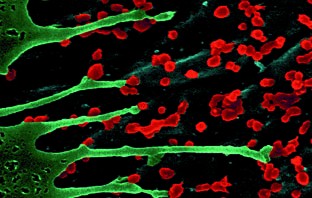
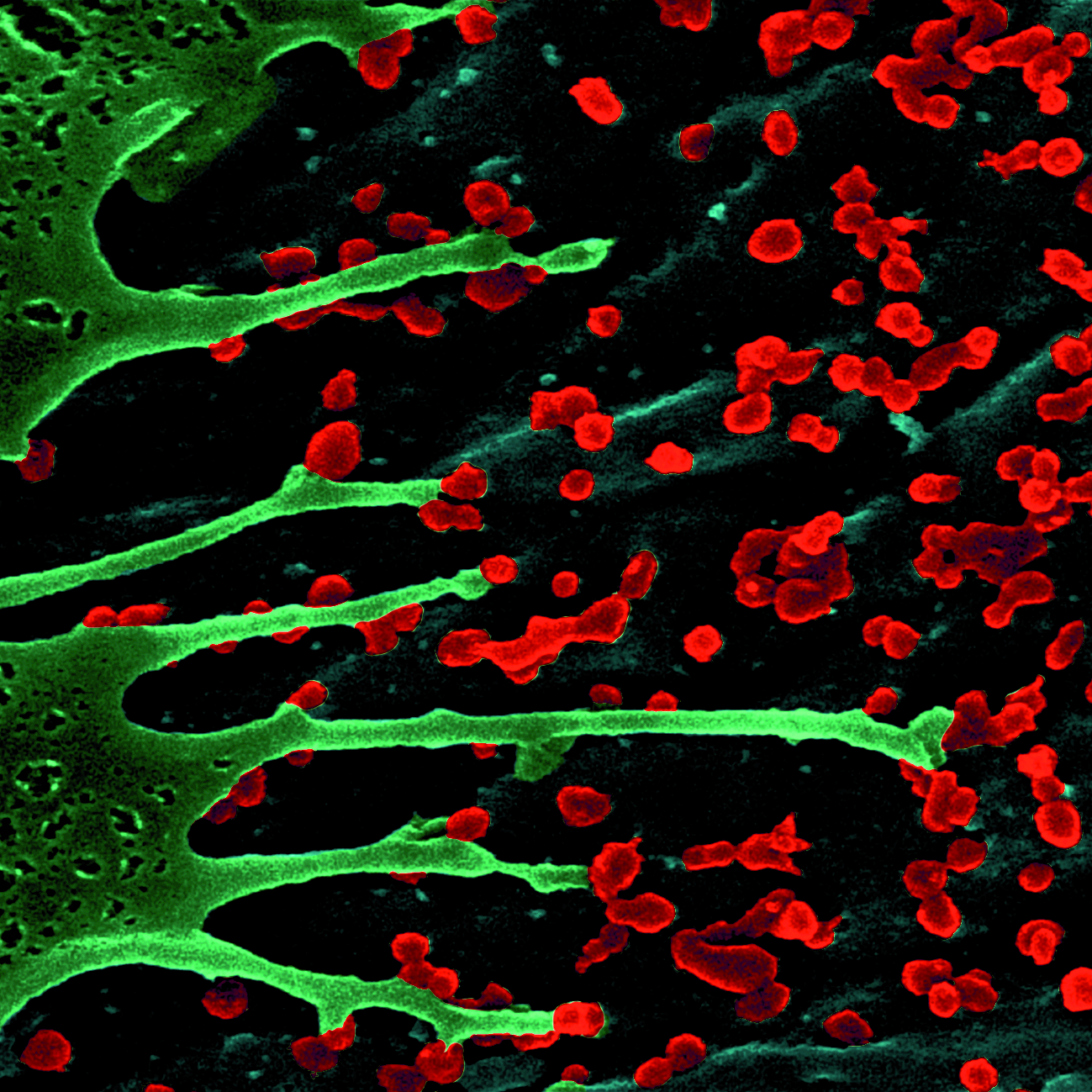
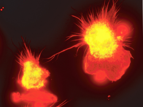
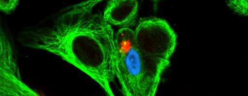
Antimicrobial resistance represents a serious and growing world threat to human and animal health. According to the World Health Organization, “resistance to antibiotics is about to become a public health emergency of yet unknown proportions”. In Europe, resistance to antibiotics is responsible for more than 25,000 deaths annually and the cost of treatment is estimated at 1.5 billion euros annually. New forms of resistance emerge every day, leaving clinicians increasingly devoid of solutions to fight infections. Despite the recognised need to develop new antibiotic weapons, only two new classes of medication have entered the market in the last thirty years.
In November 2011, the European Commission, as part of its Action Plan to combat the increasing threat of antimicrobial resistance, called for “unprecedented collaborate research and development effort to bring new antibiotics to patients” including the launch of the sixth IMI Call for proposals in May 2012 as part of the programme entitled “ New Drugs For Bad Bugs”.
IMI : a unique public-private partnership programme
IMI (Innovative Medicines Initiative) is a unique European public-private partnership between the European Commission and the EFPIA (European Federation of Pharmaceutical Industries and Associations), each contributing one billion euros to finance various projects through issuing tenders.
The aim of the IMI is to propose a coordinated approach to promote the development of safer and more effective treatments for patients by encouraging collaboration between various academic and industrial partners, the public authorities and patient associations and by increasing European competitiveness.
The research leading to these results has received support from the Innovative Medicines Initiative Joint Undertaking (www.imi.europa.eu) under Grant Agreement n°115523, resources of which are composed of financial contribution from the European Union’s Seventh Framework Programme (FP7/2007-2013) and EFPIA companies’ in kind contribution.


")