Menu

Europe is joining forces against neglected parasitic diseases

The international consortium A-PARADDISE (Anti-Parasitic Drug Discovery in Epigenetics), coordinated by Inserm, has just obtained funds of €6 million from the European Commission to conduct large-scale testing of innovative therapies against four neglected parasitic diseases: schistosomiasis, leishmaniasis, Chagas disease and malaria. The researchers have a common objective: to develop new drugs against the parasites that cause these diseases. The project involves 10 European partners, 5 Brazilian partners (who operate in the region where the diseases are endemic) and 2 Australian partners. They will all be meeting on 17 and 18 March at the Institute of Genetics and Molecular and Cellular Biology (Inserm / CNRS / University of Strasbourg Joint Research Unit), to get the project started.

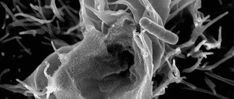
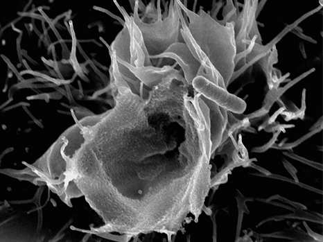
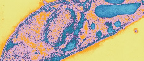
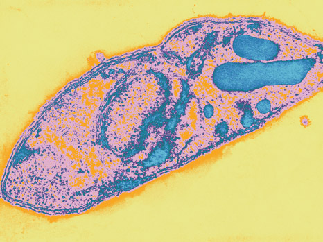
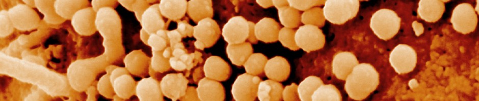
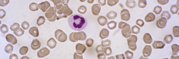
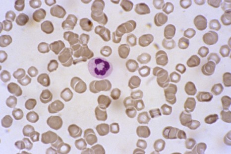
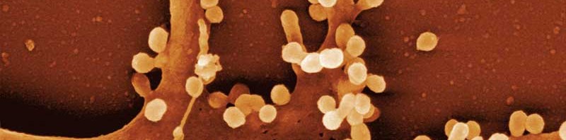
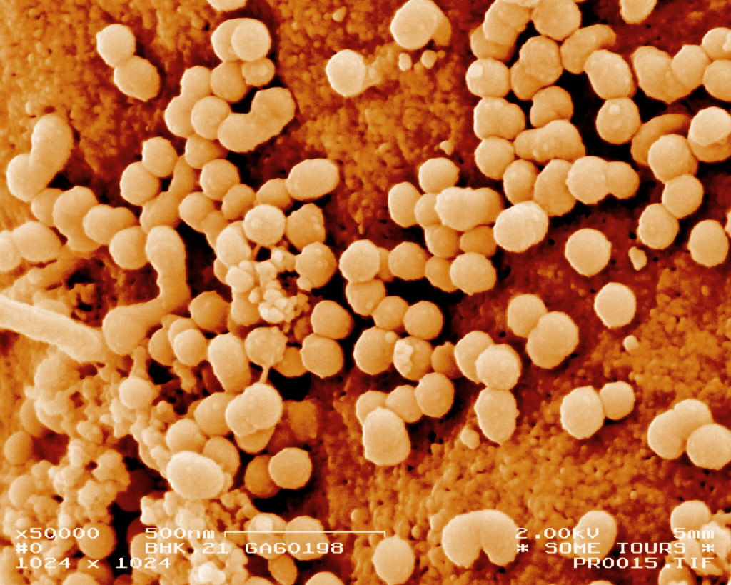
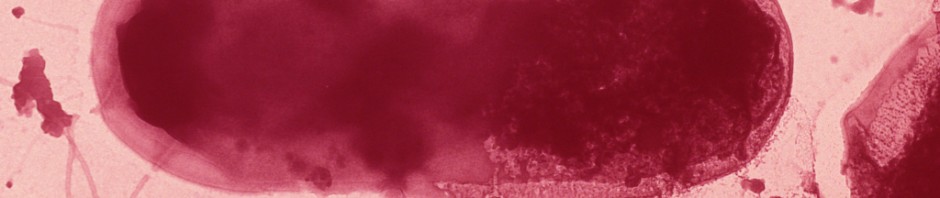
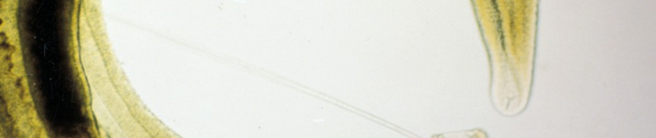
Schistosomiasis, leishmaniasis, Chagas disease and malaria are regarded as neglected diseases because the effort and funds put into developing new treatment and control methods have not been commensurate with their catastrophic human impact. They affect people in developing countries, essentially in Africa, the Middle East, South America and eastern Asia, in tropical and sub-tropical regions. Around one billion people are regularly exposed to these diseases, which cause almost one million deaths every year.

At present, there is no vaccine against these parasites. Furthermore, the efficacy of existing treatments is limited, either by the side effects or by the current or potential development of resistance. Consequently, the A-PARADDISE consortium, which is coordinated by Inserm and headed by Raymond Pierce – Director of Research at the Centre for Infection and Immunity in Lille – is focusing on developing new drugs against these parasitoses.

The A-PARADDISE project will use a methodology that has already been tried and tested during a previous project of a similar scale (SEtTReND), which aimed to develop drugs against schistosomiasis. The researchers investigated histone-modifying enzymes (HME), which determine the structure of the parasite’s chromosomes. It was demonstrated that some HME inhibitors induce cell death, which makes them toxic to this parasite. This research provided the proof of concept that HMEs act on the schistosomiasis parasite, and has led to the development of a bank of candidate compounds which can rapidly be tested against other human parasites.

Thanks to the new project A-PARADDISE, researchers will be able to put the basic principle into practice and build on it by creating a unique platform for testing anti-parasitic drugs targeting HMEs, with a view to incorporating them into a clinical development programme.

The experimental method consists in physically and virtually testing the efficacy and the toxicity of the compounds, in vitro and in vivo.

The ultimate objective of the A-PARADDISE project is to provide several candidate treatments against the four parasites and to pave the way for clinical trials in the near future.

To ensure the success of the project, the participants were all selected for their high level of expertise in their respective fields: high-throughput screening, computer-aided screening, the production of recombinant proteins, next generation sequencing, phenotypic tests, toxicology and pharmacology.

A-PARADDISE: Anti-Parasitic Drug Discovery in Epigenetics

The A-PARADDISE project began on 1 February 2014 and will be backed by the European Union for three years (FP7, grant agreement no. 602080). It is coordinated by Inserm and includes 17 partners based in 5 European countries, Brazil and Australia:

Institut National de la Santé et de la Recherche Médicale, Group Avenir, Paris, France
Centre Européen de Recherche en Biologie et Médecine (CERBM*), France
Martin Luther Universität Halle- Wittenberg (MLU), Germany
Universidade Federal do Rio de Janeiro (UFRJ), Brazil
Universidade de Sao Paulo (USP), Brazil
Albert Ludwigs Universität Freiburg  (ALU-FR), Germany
Fundação Oswaldo Cruz, Centro de Pesquisas René Rachou (Fiocruz), Brazil
Fundação Oswaldo Cruz, Instituto Carlos Chagas (Fiocruz), Brazil
Inserm Transfert SA (IT), France
KANCERA AB (KAN), Sweden
Adlego Biomedical AB (Adlego), Sweden
GriffithUniversity (GU), Australia
University of Queensland (UQ), Australia
Università degli Studi di Roma La Sapienza  (UNIROMA1), Italy
University of East Anglia (UEA), Great Britain
Fundação Arthur Bernardes – Universidade Federal de Viçosa (UFV), Brazil
Institut Pasteur Paris (IPP), France

* The CERBM is the European branch of the Institute of Genetics and Molecular and Cellular Biology (IGBMC, Inserm/CNRS/Université de Strasbourg)

Malaria – “Wake and kill”: a new concept for the elimination of relapse

A team of researchers coordinated by Prof Dominique Mazier (AP-HP, UPMC, Inserm Unit 1135, CNRS ERL 8255) and Dr Georges Snounou, Research Director at CNRS (UPMC, Inserm Unit 1135, CNRS ERL 8255) has succeeded in culturing the dormant hepatic stage of the malaria parasite, previously inaccessible to researchers. The initial results from this technical breakthrough have enabled the development of a new concept for the elimination of malaria relapse due to the activation of these dormant forms. It should enable the establishment of a new strategy for the management of this illness, which would involve combining a drug capable of activating the dormant parasite with one of the many drugs effective against the parasite.
These results have just been published in the journal Nature Medicine.

Present-day management of malaria

After the bite from an infected mosquito, the parasite that causes malaria reaches the liver, where it multiplies. It then propagates in the bloodstream, where its proliferation causes a potentially fatal illness. In some cases, including that of the parasite Plasmodium vivax in humans, a fraction of hepatic parasites may remain “dormant” for a year or more, hence their name, hypnozoites. These subsequently “wake,” or reactivate, over time, and give rise to a bloodstream infection. This feature is probably the source of the belief that malaria persists for life.

The hypnozoite constitutes a two-fold problem in terms of controlling/eliminating malaria—a greater number of cases needing treatment, and increased transmission. Unfortunately, primaquine, and a recently developed analogue, tafenoquine, the only drugs capable of killing hypnozoites, have adverse effects on the body that are sometimes serious. The identification of reliable alternative drugs therefore constitutes a public health emergency. Until now, the search for new anti-hypnozoite drugs has been based on observations made in humans infected with P. vivax, or in monkeys infected with a parasite related to P. vivax, Plasmodium cynomolgi.

Methodology

Through a collaboration with teams from the national IDMIT Center[1] at the French Atomic Energy and Alternative Energies Commission (CEA), and those of the Biomedical Primate Research Centre(BPRC) in the Netherlands, the team led by Prof Dominique Mazier and Dr Georges Snounou first succeeded in maintaining cultures of infected hepatic cells for 40 days, i.e. nearly four times longer than is usually achieved. The team then demonstrated the persistence of dormant stages throughout the duration of culture, with some reactivating over the time, thus mimicking what happens in humans. It also tested new drugs (discovered at the Institut Pasteur in Paris), which inhibit epigenetic factors, on these hypnozoites. These drugs act by targeting histone methyltransferases, and are able to kill the blood stage of the parasite. Paradoxically, one of them activated the hypnozoites. This unexpected result led the team to formulate a new strategy, “Wake and Kill,” which involves combining a drug that activates the dormant parasite with one of the many available treatments known to be effective against the multiplying parasite.

Results provide hope for the management of malaria 

Thanks to this methodology, developed via an international and multi-institute collaboration (Inserm, CNRS, CIMI, CEA, UPMC, AP-HP, Institut Pasteur Paris), it will now be possible to screen drugs in vitro for their anti-hypnozoite effect, thus limiting the need for animals. The challenge is to adapt this technique to screening a large number of compounds. In addition, the possibility of growing hypnozoites in culture will finally allow scientists to study this enigmatic parasite stage, described 100 years after the discovery of the causative agent of malaria by Laveran in 1880.


[1] Infectious Diseases Models For Innovative Therapies

A good outcome for the CHILD-INNOVAC project: successful test in humans of a nasal vaccine against pertussis

The CHILD-INNOVAC European research programme, coordinated by Inserm, has enabled the development of an innovative vaccine that can be administered intranasally, to combat pertussis, which has shown a resurgence in developed countries in recent years. The research consortium, headed by Camille Locht, Director of the Centre for Infection and Immunity of Lille (a joint Unit involving Inserm, CNRS, Institut Pasteur de Lille and University of Lille Nord de France), today published promising results from Phase I clinical trials of the vaccine in human subjects in the online journal PLOS ONE

Researchers from the CHILD-INNOVAC European project, which brought together 10 European partners*, evaluated the efficacy and safety of a new concept in intranasal vaccination against pertussis. They also carried out clinical trials in humans, which provided conclusive results. 

Pertussis is a wrongly “forgotten” disease, according to Camille Locht, a Research Director at Inserm and Director of Scientific Affairs of the Institut Pasteur de Lille. The disease currently affects several tens of millions of individuals, and kills approximately 300,000 children annually worldwide. The associated morbidity and mortality are increasing throughout the world. Its resurgence has become a matter for major concern since 2010 in some developed countries, such as the USA, Australia, the UK, the Netherlands and France. 

The CHILD-INNOVAC project is more specifically focused on combating two major respiratory pathogens: Bordetella pertussis (the bacterium that causes whooping cough) and respiratory syncytial virus (which causes bronchiolitis in infants). These pathogens mainly affect infants aged 0 to 6 months, who are poorly protected by the vaccines presently available. The project also provides proof of concept that this vaccine may be applied to other respiratory infections.

The researchers from the CHILD-INNOVAC project have succeeded in testing in humans, for the first time, a live bacterial vaccine, genetically attenuated and specially designed for intranasal administration to combat major respiratory pathogens. “This original method of administration will make the vaccine accessible to greater numbers of people at a smaller cost,” explains the project coordinator, Camille Locht.

CHILD-INNOVAC: a European success
The main success of this European project is the achievement of a vaccine for which the immunogenicity and safety could be tested in humans in only two and a half years (compared with 5-7 years for most projects of this type). This is a very short time, which Camille Locht explains is due to “the skills and motivation of the consortium, which brought together experts in their respective areas of specialisation from seven European countries. It was possible to relay the data in a flexible and efficient manner at the different stages of the project.” The project received a budget of €5 million, awarded by the European Commission under FP7. 

The Phase I trials in humans allowed the immunogenicity and safety of the vaccine to be measured in comparison with a placebo, under double-blind conditions. They took place in Sweden, which has the most “naive” population with respect to vaccination against pertussis, given that vaccination was abandoned in that country for several years, for reasons of inefficacy. 
The main objective of these trials was to record all possible adverse events, namely cough, sneezing, nasal discharge, effects on general health, etc. These measurements were examined by an independent data monitoring committee.
The second objective was to assess colonisation of the nasal mucosa by the vaccine, and the triggering of an immune response. 
It was possible to test three different doses of the vaccines: a low, intermediate and high dose. 
Results obtained after monitoring the vaccinated subjects for 6 months, and analysing 60,000 data points, showed that the vaccine induced no adverse events compared with the placebo, even at the high dose. The vaccine colonised the nasal mucosa best at the high dose. Moreover, immune responses were triggered in all subjects whom the vaccine had colonised. “It is of special interest that a single nasal administration was able to induce an immune response that was maintained for at least 6 months, i.e. for the duration of the study,” comments Camille Locht. 

The next step will involve administering higher volumes in an effort to increase the level of colonisation of the nasal mucosa by the vaccine. Camille Locht and his collaborators also hope to improve the stability of vaccine over time, with a view to industrial development in the near future. 

Inserm Transfert, in charge of the valorization of the IP portfolio related to the BPZE-based technology, recently entered into an agreement with a biotech partner to further develop the technology.

Further information

CHILD-INNOVAC

The CHILD-INNOVAC project was aimed at developing innovative intranasal vaccines against the two main respiratory pathogens, pertussis and respiratory syncytial virus (RSV). The project has provided prototypes for polyvalent vaccines that can be administered intranasally, based on attenuated B. pertussis. The immunity induced by the BPZE1 vaccine was studied in detail, together with its genetic and biological stability and safety.
CHILD-INNOVAC began in 2008, and was supported by the EU (FP7) for 4 years. It was coordinated by Inserm, along with 27 other European projects. The project involved 10 partners, including 2 private companies and 8 laboratories, based in 7 European countries:

Inserm (coordinator), France: https://www.inserm.fr/
Inserm Transfert, France: https://www.inserm-transfert.fr/
Université Libre de Bruxelles, Belgium: https://www.ulb.be/
Innogenetics, Belgium: https://www.innogenetics.com
National University of Ireland, Maynooth: www.immunology.nuim.ie
Istituto Superiore Di Sanità, Italy: https://www.iss.it/
Swedish Institute for Communicable Disease Control: https://www.smittskyddsinstitutet.se/in-english/
Netherlands Vaccine Institute
National Institute for Public Health and the Environment, the Netherlands: https://www.rivm.nl/
Imperial College of Science, Technology and Medicine, England: http:/www3.imperial.ac.uk/

A prime target for the development of anti-inflammatories

For the first time, scientists from the Institut Pasteur and Inserm have demonstrated the key role played by a particular molecule in intestinal infection. The study was published online in Immunity on December 12, 2013. The molecule, known as ATP, serves as a trigger signal for the inflammatory response targeting pathogenic agents. Using the Shigella flexneri model, the scientists have also shown how this bacterium is able to block the release of ATP in order to escape this defense reaction.

Discovery of this blocking mechanism could be a milestone in therapeutics: the development of new drugs that mimic this process could open up new possibilities for the treatment of chronic inflammatory diseases, such as Crohn’s disease.

shigella

© Inserm/Tran Van Nhieu, Guy

It has been known for some years that the extracellular presence of ATP – a molecule normally found inside cells – is able to alert the body to danger and trigger an inflammatory defense response. Such may be the case when tissue is damaged and cell contents are released. Yet, whether an infectious agent would be capable of triggering the same mechanism remained unclear.

The team led by Philippe Sansonetti, who heads the Molecular Microbial Pathogenesis Unit (Inserm U786 / Institut Pasteur), in association with scientists from the University of Toulouse and the Collège de France have recently shown in vitro and in vivo that infection with enteric pathogenic bacteria – in this study Shigella, Salmonella and enteropathogenic E. coli – induces the cell to actively release ATP into the extracellular environment: in the presence of the bacterium, intestinal epithelial cells open cell surface channels, allowing ATP molecules to escape. By binding to extracellular receptors, ATP triggers a chain of reactions that drive the inflammatory immune response designed to eliminate the threat of infection.

The scientists also proved that the Shigella flexneri bacterium is capable of blocking this release of ATP by injecting an enzyme directly into the infected cell. The enzyme acts by closing the channels. This is the first time a pathogen has been observed to have the ability to suppress this mechanism, thus allowing it to escape defenses put in place by the body. This discovery underlines the importance of ATP as a key regulator of intestinal inflammation.

Although inflammation is a natural defense mechanism that plays an essential role in tissue response to attack, it persists abnormally in some cases. It then becomes chronic, and can lead to inflammatory diseases. These diseases result in a malfunction of the immune system, which attacks the body’s normal components. By showing this potential to halt the inflammatory process, the study highlights this process as a new, prime therapeutic target for the development of anti-inflammatory drugs.

Currently, there are no anti-inflammatory drugs on the market that targets the release of ATP. However, this release mechanism could have an important, as yet undefined part to play in the future treatment not only of certain chronic inflammatory diseases of the intestine, such as Crohn’s disease, but also other pathologies such as cancer, obesity, type 2 diabetes or arteriosclerosis.

Chemotherapy: when our intestinal bacteria provide reinforcement

Research jointly conducted by investigators at Institut Gustave Roussy, Inserm, Institut Pasteur and INRA (French National Agronomic Research Institute) has led to a rather surprising discovery on the manner in which cancer chemotherapy treatments act more effectively with the help of the intestinal flora (also known as the intestinal microbiota). Indeed, the researchers have just shown that the efficacy of one of the molecules most often used in chemotherapy relies to an extent on its capacity to mobilise certain bacteria from the intestinal flora toward the bloodstream and lymph nodes. Once inside the lymph nodes, these bacteria stimulate fresh immune defences which then enhance the body’s ability to fight the malignant tumour.

Results of this work are published in the journal Science on 22 November 2013

bacteria - blue version

©Fotolia

The intestinal microbiota is made up of 100,000 billion bacteria. It is a genuine organ, since the bacterial species that comprise it carry out functions crucial to our health, such as the elimination of substances that are foreign to the body (and potentially toxic), or keeping the pathogens that contaminate us at bay. They also ensure the degradation of ingested food, for better intestinal absorption and optimal metabolism. These millions of bacteria colonise the intestine from birth, and play a key role in the maturation of the immune defences.

However, the bacterial species that make up the intestinal microbiota vary from one individual to another, and the presence or absence of one or another bacterial species seems to influence the occurrence of some diseases, or, conversely, may protect us.

In the cancer area, the French team directed by Prof Laurence Zitvogel, Director of Inserm Unit 1015,Tumour Immunology and Immunotherapy,” at Institut Gustave Roussy, in close collaboration with Institut Pasteur (Dr Ivo Gomperts Boneca, “Biology and Genetics of the Bacterial Cell Wall” Unit) and researchers at INRA (Drs Patricia Lepage and Joël Doré, Micalis Unit, “Food Microbiology in the Service of Health”), has just provided evidence that the intestinal flora stimulates an individual’s immune responses to combat cancer during chemotherapy.

Cyclophosphamide is one of the most widely used drugs in chemotherapy. However, like any treatment, it involves side effects (inflammation of the mucosa etc.), and disrupts the normal balance of the intestinal microbiota. Certain bacteria (of the Gram+ group of bacteria) can pass the intestinal barrier and enter the bloodstream and lymph nodes.

These bacteria, once in the general circulation of the body, may be considered harmful, and the body generates an immune response.

“This chain reaction, a side effect of the treatment, actually turns out to be very useful,” explains Laurence Zitvogel. “Surprisingly, the immune response directed against these bacteria helps the patient to better fight his/her tumour, by stimulating fresh immune defence mechanisms.”

More specifically, immunisation against bacteria leads to the recruitment of effector lymphocytes different to those mobilised by chemotherapy. Their role consists of helping anti-tumour lymphocytes to stem the growth of tumours.

To verify these observations in mice, researchers suppressed all Gram+ bacteria from their intestinal microbiota. Results showed that the efficacy of the chemotherapy was reduced. The researchers also suggest that some antibiotics used during chemotherapy may destroy these Gram+ bacteria, and thus negate their beneficial effect.

“Now that these “beneficial” bacteria that potentiate the anti-tumour immune response have been identified, we should soon succeed in supplying more to the body, especially via pro- or prebiotics and/or a specific diet,” the researcher concludes.

This work has received support from the French National Cancer League, the French National Cancer Institute (lNCa; SIRIC SOCRATES) and from LABEX Onco-Immunology

Identification of a new mechanism in the most commonly used immunotherapy for lymphoma

Using innovative dynamic imaging technique, scientists at the Institut Pasteur, Inserm and the VU Medical Center in Amsterdam have uncovered the mode of action of anti-CD20, an antibody therapy frequently used in the treatment of lymphomas (cancers of the immune system) as well as some auto-immune diseases. In a lymphoma model, the scientists have been able to carry out real time in vivo imaging of the cellular events activated by the treatment and resulting in the destruction of tumor cells. These discoveries should help optimize the efficacy of future therapies involving anti-CD20 antibodies. This work is the subject of an article published online November 1 on the Journal of Clinical Investigation website.

A lymphoma usually develops as a result of abnormal proliferation of one of two types of immune cells:  B lymphocytes (in the vast majority of cases) or T lymphocytes. For the last fifteen years or so, anti-CD20 antibody therapy has frequently been used in the treatment of B-cell lymphomas (in particular those known as non-Hodgkin lymphomas), in combination with conventional chemotherapy. These antibodies are directed against B lymphocytes, bind to cancer cells and mark them for depletion by other immune cells. Anti-CD20 antibody therapy also triggers a decrease in the normal B lymphocyte population, dampening immune responses. For this reason it is also used to treat autoimmune diseases. However, how anti-CD20 antibody therapy works in vivo was not fully understood.

A study led by Philippe Bousso, head of the Dynamics of Immune Responses Unit (Institut Pasteur / Inserm U668), along with researchers at Inserm and the VU Medical Center in Amsterdam, has provided the first conclusive answers. Using dynamic imaging techniques developed at the Institut Pasteur, the scientists have carried out real time in vivo imaging of the destruction of cancerous and normal B lymphocytes during anti-CD20 antibody treatment. The scientists noticed that the phenomenon of B lymphocyte depletion resulting from anti-CD20 antibody therapy primarily takes place in the liver and involves a specific cell type, known as Kupffer cells. The images produced by the scientists clearly show Kupffer cells (in green) capturing cancerous B lymphocytes (in orange) and preventing their circulation before destroying them.

These discoveries provide important insight for optimizing the efficacy of future treatments using anti-CD20 antibodies. Non-Hodgkin lymphomas affect 10,000 people per year in France, and account for 10% of pediatric cancers.

Fine-tuning the approach to malaria and toxoplasmosis research

A study carried out by teams from the Institut Pasteur, the Institut Cochin (Inserm, CNRS, Paris Descartes University), and the Wellcome Trust Centre for Molecular Parasitology at the University of Glasgow, may very well redefine current approaches to malaria and toxoplasmosis research in terms of treatment development. Their research which focuses on the role played by the protein AMA1 (present in both parasites) was published october 9, on the Nature Communications website. For many years AMA1 has been the focus of studies aiming to develop malaria treatments and vaccines. However, the authors of this study express their reservations about strategies that focus strictly on blocking AMA1 and show that malaria and toxoplasmosis parasites without AMA1 are still capable of developing normally. 

photo CP toxoplasmose

Stade mérozoïte de Plasmodium falciparum – Copyright Institut Pasteur

One million people die each year from malaria, making it the most widespread parasitic disease in the world today. Toxoplasmosis, which often presents no symptoms in healthy subjects, is a parasitic disease that mainly affects immunedeficient individuals. Pregnant women are also particularly at risk for this disease and if infected for the first time during their pregnancy could spread infection to the fetus.

Plasmodium and Toxoplasma, genera of apicomplexan parasites, are the agents responsible for these diseases. They both contain the same protein, AMA1, which many studies consider necessary for parasites to enter host cells and propagate infection. Because of this, numerous studies conducted since the discovery of the protein have made it their primary focus in the development of anti-parasitic treatments. However, this trend may soon change, thanks to the collaborative efforts of Robert Ménard (Institut Pasteur, Paris), Isabelle Tardieux (Institut Cochin, Paris), Markus Meissner (University of Glasgow), and their teams. These scientists recently showed that in the total absence of AMA1 Plasmodium berghei and Toxoplasma gondii are still capable of entering infected cells and multiplying. This discovery could have a significant impact on how research is conducted in the development of treatments for malaria and toxoplasmosis.

In 2011, the scientists had already shown that parasites genetically modified with very low levels of AMA1 were still able to infect host cells. In today’s study, parasites were created free of all AMA1 using “reverse genetics” (a technique never before used in this field). The scientists showed that in the absence of AMA1, during the blood and hepatic stages of Plasmodium berghei, as well as during the replication stage of Toxoplasma gondii (the stage where the disease spreads to humans), the parasites were still able to enter host cells. What was affected by the lack of AMA1, however, was the parasites’ ability to bind to host cells (this is the step that precedes cell entry). Because of this, the scientists concluded that AMA1 is not a requirement for cell entry but rather is used to bind parasites to the host cells. These observations have prompted the scientists to put forward new recommendations for optimizing research that focuses on AMA1 for the development of new treatments. In particular, they suggest that therapeutic strategies and vaccine development should focus on other proteins in addition to AMA1.

Circumcision effectively reduces the risk of HIV infection “in real life”

The ANRS-12126 “Bophelo Pele” Project implemented in the township of Orange Farm in South Africa has confirmed the effectiveness of a large-scale program of voluntary medical male circumcision in prevention of heterosexually acquired HIV infection. The follow-up of over 3300 men shows a 57% to 61% reduction in the rate of new HIV infections in circumcised men compared with uncircumcised men. This study, headed by Prof Bertran Auvert and his colleagues, also shows that a circumcision program can be rapidly and effectively implemented in African communities where circumcision is not a social norm. These results, published in PLoS Medicine, argue for accelerated roll-out of voluntary male circumcision programs on the African continent in order to improve prevention of HIV transmission.

Three randomized trials have shown that male circumcision has a protective effect on the risk of HIV infection in men. The first publication dates from 2005 (ANRS-1265 study in South Africa) and its results were subsequently confirmed in Kenya (2007) and Uganda (2007). These studies showed that the risk of circumcised men being infected by HIV was reduced by 50% to 60%. These results led UNAIDS/WHO to recommend in 2007 the circumcision of adult males as a strategy of additional HIV prevention in communities with a high prevalence of HIV and a low prevalence of circumcision.

There remained, however, a need to show that the roll-out of circumcision “in real life” reduces both the incidence (rate of occurrence of new infections) and the prevalence (proportion of people infected in the total population) of HIV infection in men. This had been suggested by preliminary results from the ANRS-12126 “Bophelo Pele” Project of Bertran Auvert and colleagues presented at the International AIDS Society Conference 2011. These results are now confirmed in the PLoS Medicine article, the first scientific publication to show that adult male circumcision “in real life” effectively reduces the risk of HIV infection in men (1).

Between 2007 and 2011, the ANRS-12126 “Bophelo Pele” Project was conducted by Prof Bertran Auvert (UMRS-1018 Inserm, Hôpital Ambroise Paré and Université de Versailles Saint-Quentin) and his colleagues from the National Institute for Communicable Diseases, the Social Sciences Faculty, and Progressus (Johannesburg, South Africa), and from Johns Hopkins University (Baltimore, USA) and the Bichat Hospital (Paris, France). Free, medical circumcision was offered to all male volunteers aged from 15 to 49 years in a population of 110 000 adults in the township of Orange Farm in South Africa. More than 20 000 circumcisions were performed, accompanied by a large information provision and prevention program.

An anonymous questionnaire on sexual practices was administered to 3338 men recruited from the township population who were invited to undergo HIV screening, which included a test to determine, in the event of seropositivity, whether the infection was recent.

The proportion of circumcised men in this sample of 3338 increased from 12% at the start to 53%, and was 58% in the 15- to 29-year-olds. Importantly, sexual behavior, in particular condom use, did not differ between circumcised and uncircumcised men. In contrast, the prevalence and incidence of HIV infection were much reduced in the circumcised men.

The researchers considered that without the voluntary circumcision program, HIV prevalence would have been 19% higher in the study population. This effect is more marked in the 15- to 29-year-olds, in whom the prevalence would have been 28% higher. Also apparent was a decrease in the number of recent infections among the circumcised men. Circumcision was therefore associated with a 57% to 61% reduction in the rate of new infections.

Prof Bertran Auvert considers that “these results are important in two ways. First, they confirm the efficacy of the circumcision practiced on a population scale in reducing HIV transmission appreciably among the men of this population. Second, they show that it is possible to achieve this result in just a few years, including in populations where circumcision is not a common practice.”

The ANRS-12126 Project provides an argument for speeding up the roll-out of voluntary circumcision programs, notably in sub-Saharan Africa, which is home to the vast majority of the 2.2 million people infected by HIV every year worldwide. Prof Jean-François Delfraissy, Director of the ANRS, considers that “given the impact observed in this study on limiting the risk of HIV acquisition in circumcised men, the scale-up of circumcision should more than ever be a public health priority in South and East Africa.”

The ANRS-12126 Project is continuing with the aim of elucidating the effect of circumcision on infection risk reduction in the general population, and in particular among women.

Neutrophils: the Unsung Heroes of Immunotherapy Cancer Treatment

Scientists at the Institut Pasteur and Inserm have identified the group of cells within the immune system that make immunotherapy treatment (therapeutic antibodies) effective. Immunotherapy is frequently used to treat breast cancer. In animal models they showed that neutrophils, the most common white blood cells in the body, are not only necessary but suffice on their own to eliminate tumor cells. The scientists produced these results using both skin and breast cancer models. If confirmed in humans, these discoveries should lead to the optimization of current treatments for several cancers. This work is being published online August 26, 2013 by the medical journal Blood.

With its millions of victims each year, cancer remains one of the leading causes of death worldwide. In women, breast cancer is the most common offender. Nearly one in nine women will develop breast cancer in their lifetime. In France alone, over 50,000 new cases are diagnosed each year.

Between 20 and 30% of breast cancer cases are deemed eligible for immunotherapy treatments which involve the injection of antibodies. Until now, the effectiveness of this type of treatment was attributed to various immune system components such as Natural Killer cells and macrophages. However, work by Pierre Bruhns, head of the Antibodies in Therapy and Pathology Laboratory (Institut Pasteur/Inserm Unit 760), and his team, in collaboration with Professor Clifford A. Lowell of the University of California Department of Laboratory Medicine and two other teams from the Institut Pasteur, has cast serious doubt on this theory.

Etude de la moelle osseuse et de l'hématopoièse

Neutrophile observed by optical microscopy ©Inserm/E.Cramer

The scientists have shown in an animal model that neutrophils alone are effective enough to induce the therapeutic effect of immunotherapy treatments often used to treat breast cancer. Neutrophils are drawn to the tumor after the injection of therapeutic antibodies and are activated after coming into contact with these antibodies. Once activated the neutrophils are capable of destroying the tumor cells. This discovery represents an important step towards the optimization and development of immunotherapy treatments used to treat these cancers.

The scientists made the following observations: the tumor mass did not regress in mice that were neutropenic (having a significantly low level or total lack of neutrophils) or in mice with neutrophils that could no longer be activated through contact with therapeutic antibodies. Then, scientists showed that the anti-tumor effect achieved via immunotherapy could be reproduced in deficient mice by administering them with neutrophils taken from healthy mice. These observations were made using models for both skin and breast cancer leaving us to assume that neutrophils play an important role in immunotherapy treatments used to fight various other cancers.

In their conclusion, the scientists highlighted current immunotherapy treatments used to fight cancer. These treatments are often coupled with other treatments. Because of this, a good strategy might be to favor treatment pairings that increase neutrophil count and activity.

The way the virus continually changes complicates the development of a vaccine against HIV

A team from INSERM Mixed Research Unit 966 Morphogenesis and Antigenicity of HI and Hepatitis Viruses” headed by Martine Braibant and Francis Barin in Tours, has confirmed, with the support of the ANRS, that the AIDS virus has gradually adapted itself to the immune response of the human population during the course of the epidemic. Starting from the time it was isolated in the 1980s to the present day, the virus has become less and less sensitive to neutralising antibodies, complicating the creation of an effective preventive vaccine. Researchers have also shown that the combination of two powerful neutralising antibodies remains capable of neutralising in vitro the most recent HIV variants. Details of the research are published in the journal PLoS Pathogens.


The development of a vaccine that is effective against HIV remains one of the major challenges in the fight against AIDS. Considerable progress has been made in recent years with the identification of human neutralising antibodies capable of blocking in vitro infection caused by the very numerous variants of HIV -1. The scientific community is considering performing clinical trials in humans, using some of these antibodies. One of the issues that remains to be resolved is that of the choice of antibodies and the best combination of antibodies to be used in order to afford the widest possible protection in view of the huge variability of possible forms of HIV -1 currently circulating in the world.

Particules virales du virus de l'immunodéficience humaine (VIH)


© Inserm /  Philippe Roingeard

The research, headed by Martine Braibant and Francis Barin (INSERM-Université de Tours Mixed Unit) with the support of the ANRS, confirms that during the course of the epidemic HIV-1 has gradually adapted itself to the immune response of the human population by becoming less and less sensitive to certain neutralising antibodies. In order to arrive at these results, the researchers were able to analyse viruses taken from samples in patients infected between the late 1980s and the year 2010[1].

“Although it was known that, at an individual level, the virus was able to adapt and circumvent the individual’s own means of defence, our work confirms that the pressure of selection exercised on the virus has had repercussions on a population-wide scale”, explains Martine Braibant.

Despite this depressing prospect, in the same publication, researchers identify a combination of two powerful neutralising monoclonal antibodies developed by Caltech and the Scripps Research Institute (NIH45-46G54W and PGT128) that still remain capable of neutralising in vitro the most recent variants and do so at a concentration compatible with their use in humans.

This study stresses the need to monitor the development of the sensitivity of HIV -1 variants to the various neutralising antibodies. The work of the researchers opens up interesting prospects for teams involved in researching a vaccine to prevent HIV .

Neutralising antibodies: one of the research pathways for developing an anti-HIV vaccine

Current anti-viral vaccination concepts are largely based on the fact that certain so-called neutralising antibodies, by attaching themselves to viral proteins, inhibit the early stages of HIV infection. When blocked in this way, the virus cannot reproduce and is eliminated.

Scientists have currently identified the immune response to be induced via the identification and characterisation of wide-spectrum neutralisation antibodies. They have not yet succeeded in enabling this type of antibody to be produced in the human body through a standard vaccination. One of the research pathways being pursued is the introduction of gene coding for these antibodies in viral expression vectors so as to cause them to be secreted directly into the circulation. This antibody approach has been tested in France as part of the ANRS/VRI vaccine research programme (Vaccine Research Institute)


[1] PRIMO and SEROCO ANRS cohorts, collaboration with Laurence Meyer and Cécile Goujard, Université Paris-Sud – INSERM U1018.


(French) : Découverte d’une nouvelle cible thérapeutique chez E. coli

fermer