Menu

Where and how are fear-related behaviours and anxiety disorders controlled?

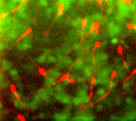
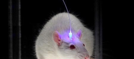
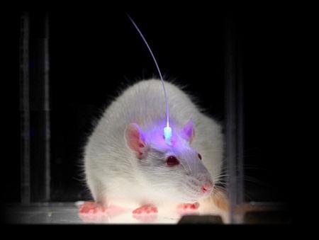
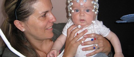
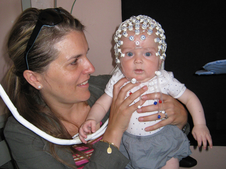
A team of researchers at Inserm led by Cyril Herry (Inserm Unit 862, “Neurocentre Magendie,” Bordeaux) has just shown that interneurons located in the forebrain at the level of the prefrontal cortex are heavily involved in the control of fear responses. Using an approach combining in vivo recordings and optogenetic manipulations in mice, the researchers succeeded in showing that the inhibition of parvalbumin-expressing prefrontal interneurons triggers a chain reaction resulting in fear behaviour. Conversely, activation of these parvalbumin interneurons significantly reduces fear responses in rodents. 

This research is published in the journal Nature

Some traumatic events may lead to the development of severe medical conditions such as anxiety disorders or posttraumatic stress disorder (PTSD).

Anxiety disorders have a prevalence of approximately 18% worldwide.

Despite successful treatments, some patients relapse, and the original symptoms reappear over time (fear of crowds, recurring nightmares, etc.). An understanding of the neuronal structures and mechanisms involved in this spontaneous recovery of traumatic responses is essential.

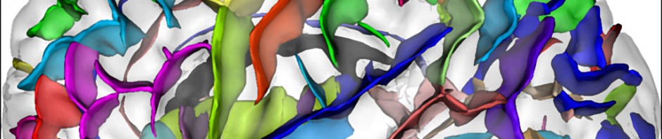
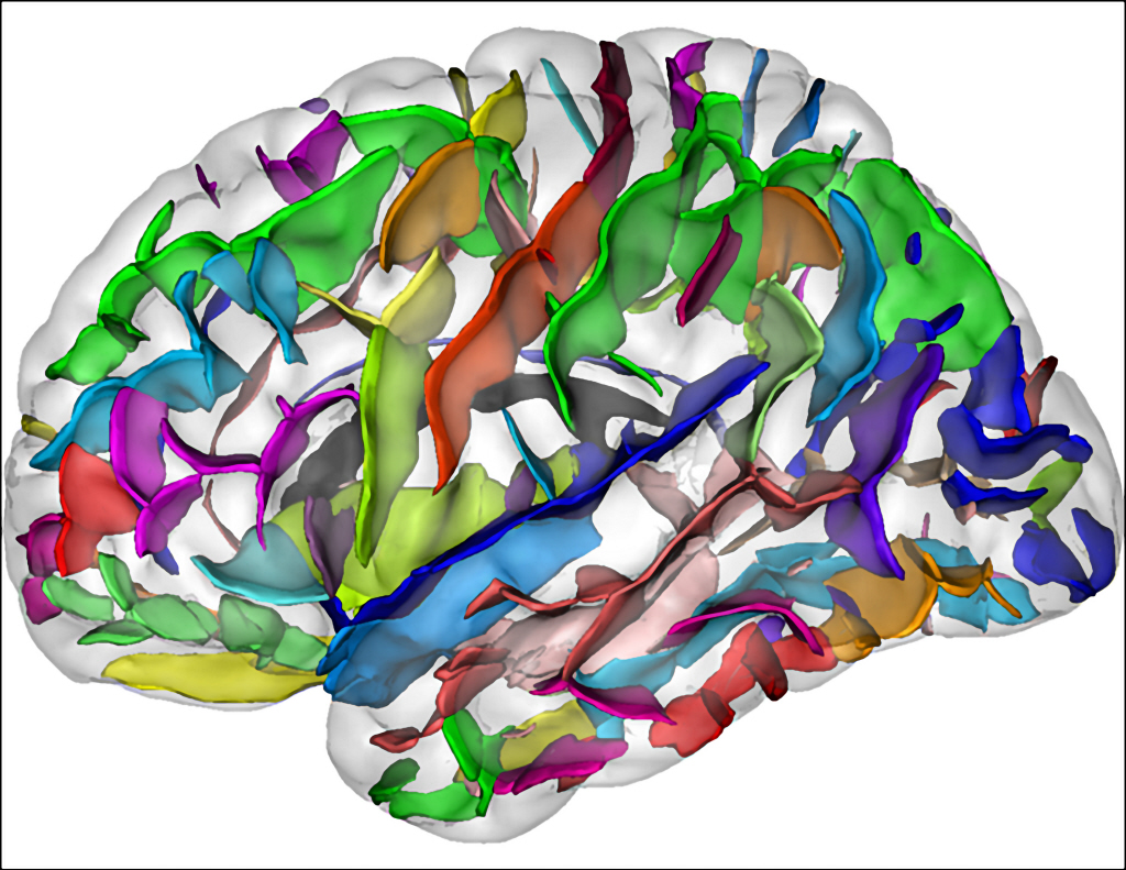
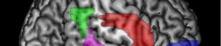
All observations made by researchers indicate that fear behaviours are controlled in the forebrain at the level of the dorsomedial prefrontal cortex. This control of fear behaviour is based on the activation of neurons in the prefrontal cortex that are in contact with specific areas of the amygdala.

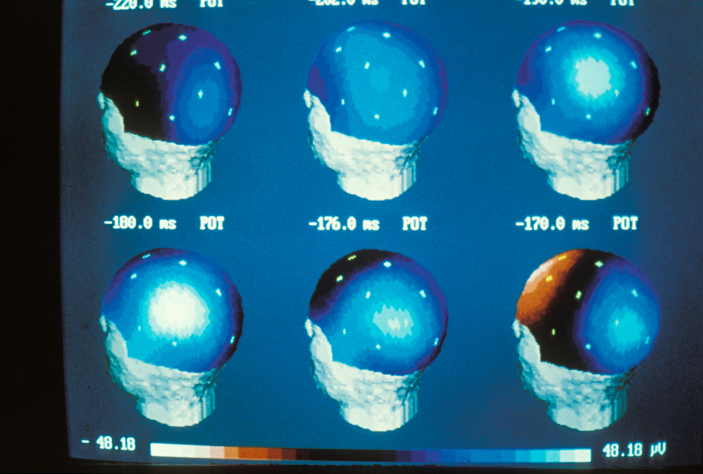
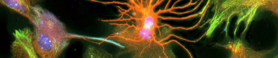
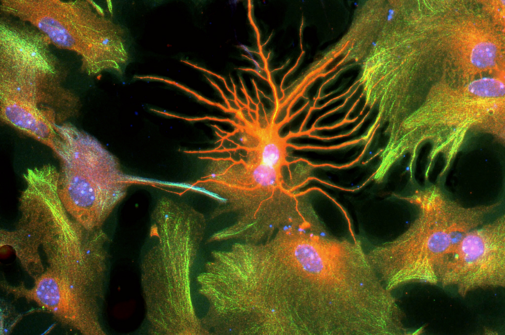
Using an innovative approach combining electrophysiological recording techniques, optogenetic manipulations and behavioural approaches, the researchers were able to demonstrate that fear expression is related to the inhibition of highly specific interneurons—the parvalbumin-expressing prefrontal interneurons.

More specifically, inhibition of their activity disinhibits the activity of the prefrontal projection neurons, and synchronises their action.

Synchronisation of the activity of different neuronal networks in the brain is a fundamental process in the transmission of detailed information and the triggering of appropriate behavioural responses. Although this synchronisation had been demonstrated as crucial to sensory, motor and cognitive processes, it had not yet been examined in relation to the circuits involved in controlling emotional behaviour.

“Our results identify two complementary neuronal mechanisms mediated by these specific interneurons, which accurately coordinate and increase the neuronal activity of prefrontal projection neurons, leading to fear expression,” explains Cyril Herry. 

The identification and better understanding of these neuronal circuits controlling fear behaviour should allow the development of new treatment strategies for conditions such as posttraumatic stress disorder and anxiety disorders. “We could, for example, imagine the development of individual markers for these specific neurons, or the use of transmagnetic stimulation approaches to act directly on excitatory or inhibitory cells and reverse the phenomena.”

How is fear analysed in an animal?

From an experimental standpoint, the classic Pavlovian conditioning procedure involves associating one stimulus, such as a sound, with another, unpleasant stimulus, such as a small electric shock. This first step allows the animal to establish a persistent aversive memory. In other words, the animal comes to remember and learn that the sound is associated with an unpleasant state, and an immobility response is routinely triggered, which is a good indication of fear in an animal.

In the second step, the extinction procedure involves repeated presentation of the sound alone, inducing a temporary inhibition of the conditioned fear responses. This inhibition is only temporary, as the mere passage of time favours the spontaneous recovery of the conditioned fear responses, which, from the clinical standpoint, may be associated with the phenomenon of relapse into traumatic responses seen following the treatment of posttraumatic stress disorder using exposure-based therapies.

Major advance in understanding Alzheimer’s disease: 11 new genetic susceptibility factors discovered

The largest international study ever conducted on Alzheimer’s disease, as part of the IGAP (International Genomics of Alzheimer Project) international consortium, coordinated by the joint research unit comprising Inserm, the Pasteur Institute at Lille-University Lille Nord de France ‘Public health and molecular epidemiology of diseases associated with ageing’ and LabEx DISTALZ, directed by Philippe Amouyel, has identified eleven new regions of the genome involved in the appearance of this neuro-degenerative disease. This work provides an overview of the molecular mechanisms at the root of the disease, revealing a better understanding of the physiopathology of this curse. These results, described in an article published in the journal Nature Genetics dated 27 October 2013, were obtained through a unique global collaborative effort by the best researchers in the field.

Since 2009, 10 genes linked to Alzheimer’s disease have been discovered, providing a better understanding of this dreadful illness. However, a large part of individual susceptibility to develop this disease still remains unknown. Characterising this individual susceptibility carried by our genome required being able to compare patients’ DNA with that from healthy people, to find a few hundred variations among more than 3.5 billion genes making up our genome. Such an approach meant analysing the genomes of thousands of individuals, which cannot be done at a team scale or even that of a single country.

For this reason, in February 2010, managers from the four largest international research consortia on the genetics of Alzheimer’s disease decide to join forces to accelerate the discovery of new genes. In less than 3 years, under the IGAP programme, researchers have succeeded in identifying more genes than over the previous 20 years. They set up their study in two phases. The first consisted of reanalysing all their existing data based on common criteria, a total of more than 17,000 cases of Alzheimer’s disease collected in Europe and North America, comparing them to some 37,000 non-diseased controls. Using advances in human genome sequencing (1000 Genomes project), they were able to compare the distribution of more than 7 million mutations between these cases and controls, so as to select only 11,632 of them from this first phase.

In the second phase, researchers verified these results in independent samples from 11 different countries, totalling 8,572 patients and 11,312 controls. This confirmed the discovery of 11 new genes in addition to those already known and to locate 13 others still being confirmed.

These 11 new genes offer new opportunities for understanding the appearance of Alzheimer’s disease. One of the most significant associations has been found in the HLA-DRB5/DRB1 region of the major histocompatibility complex. This discovery is interesting for more than one reason. Firstly, it confirms the immune system’s involvement in the disease. In addition, this same regions is also found associated with two other neuro-degenerative diseases, multiple sclerosis and Parkinson’s disease. Another link could also be made with the SLC24A4 locus, which codes a protein involved in development of the iris and in the colour variation of hair and skin, and which is associated with the risk of high blood pressure.

Some of these new genes confirm known hypotheses about Alzheimer’s disease, particularly the role of the amyloid pathway (SORL1, CASS4) and the Tau protein (CASS4, FERMT2). The role of the immune response and inflammation (HLA-DRB5/DRB1, INPP5D, MEF2C), already implicated by previous work from Inserm unit 744 (CR1[i], TREM2[ii]), is strengthened, as well as the role of cellular migration (PTK2B), lipid transport and endocytosis (SORL1). New hypotheses have also appeared, associated with hippocampal synaptic function (MEF2C, PTK2B), the cytoskeleton and axonal transport (CELF1, NME8, CASS4), as well as myeloid and microglial cell functions (INPP5D).

For the researchers, this discovery results in three main consequences. Firstly, this observation provides a better understanding of the physiopathology of Alzheimer’s disease, an essential step to discovering new treatments. Furthermore, this genome analysis better identifies the genetic profile of patients with a risk of developing Alzheimer’s disease. Finally, this work demonstrates that, faced with the complexity of such a disease, only the combined strength of global research will allow solutions to be found more quickly for this 21st century curse.

About Alzheimer Disease

These results, which demonstrate the many advances in understanding Alzheimer’s disease, involved teams from LabEx Distalz, and were able to be obtained through the support of the National foundation for Scientific Cooperation on Alzheimer’s disease and similar diseases, as well as the genotyping and analytical capabilities of the CEA National Genotyping Centre, the Study Centre for Human Polymorphism and the Pasteur Institute at Lille.

With the increasing life expectancy of human populations, the number of patients affected by Alzheimer’s disease is tending to rise in France and throughout the world. The leading cause of disorders of the memory and intellectual functions in the elderly, this illness is therefore a major challenge for public health.

Alzheimer’s disease is one of the main causes of dependency in the elderly. It results in a deterioration of neurons in different regions of the brain. It is manifested by growing alteration in memory and cognitive functions, as well as by behavioural problems leading to progressive loss of independence. In France, Alzheimer’s disease affects more than 850,000 people and represents a major social and economic cost.

Alzheimer’s disease is characterised by development of two types of brain lesions: amyloid plaques and neurofibrillary degeneration. Amyloid plaques arise from extracellular accumulation of a peptide, the amyloid β peptide (Aβ), in specific areas of the brain. Neurofibrillary degeneration is intra-neural lesions arising from abnormal aggregation, in the form of filaments, of a protein called the Tau protein.

Identifying genes that contribute to the appearance of Alzheimer’s disease and its development will lead more rapidly to tackling the physiopathological mechanisms underlying this disease, to identifying target proteins and metabolic pathways for new treatments, and to offering methods to identify subjects at greatest risk when effective preventive treatments are available.

The International Genomics of Alzheimer Project, IGAP

In February 2011, researchers from the four leading research consortia into the genetics of Alzheimer’s disease were brought together to speed up the discovery and mapping of genes involved in Alzheimer’s disease. Their research work was conducted in European and North American universities. They combined the knowledge, staff and resources of the European Alzheimer’s Disease Initiative (EADI) in France, directed by Philippe Amouyel, doctor and researcher, Director of the joint research unit comprising Inserm, the Pasteur Institute at Lille-University Lille 2 ‘Public health and molecular epidemiology of diseases associated with ageing’ and LabEx DISTALZ. The Alzheimer’s Disease Genetics Consortium (ADGC) in the United States, directed by Gerard Schellenberg, researcher at the Pennsylvania University Faculty of Medicine. The Genetic and Environmental Risk in Alzheimer’s Disease (GERAD) in the United Kingdom, directed by Julie Williams, researcher at Cardiff University. Cohorts for Heart and Aging Research in Genomic Epidemiology (CHARGE), directed by Sudha Seshadri, doctor at Boston University.


[i] Genome-wide association study identifies variants at CLU and CR1 associated with Alzheimer’s disease.

Lambert JC et al. Nature Genetics 2009. 41: 1094-1099.

[ii] TREM2 variants in Alzheimer’s disease.

Guerreiro R, et al. N Engl J Med. 2013; 368(2): 117-27.

Memory brain systems help resisting temptations

Memory brain systems help resisting temptations – found a recent study led by researchers of the Brain & Spine Institute in Paris. How can some people resist the attraction of immediate pleasures and pursue long-term goals, while others easily succumb and compromise their ultimate expectations? One secret factor might lie in the activity of a deep brain structure: the hippocampus.

Geschäftsmann, Erfolg, Geschäftsidee, Icons


© Fotolia

Economists have been interested for decades in the conflict between smaller-sooner and larger-later rewards. Understanding how humans make inter-temporal choices, such as drinking tonight versus good health later, is crucial for designing insurance policy or anti-alcohol campaigns. This issue has recently been taken to brain scanners, in which volunteers were asked to make choices between monetary payoffs, for instance $10 now versus $11 tomorrow. Using this type of paradigm, scientists found that the dorsolateral part of the prefrontal cortex, a region known to implement behavioral control, was crucial for making patient choices – waiting for higher but delayed payoffs.

However, these paradigms miss an essential feature of the inter-temporal conflicts we have to face in everyday life, says study leader Mathias Pessiglione: immediate rewards can be perceived through our senses, whereas future rewards must be represented in our imagination. To reproduce this situation in the lab, the authors used more natural rewards like food items (for instance, a beer today or a bottle of champagne in a week for now). Volunteers were confronted to choices between immediate rewards presented as pictures, and future rewards presented as texts. In this case specifically, the ability to select future rewards was linked to the amount of hippocampus activity. To complete the demonstration, patients with hippocampus damage due to Alzheimer’s disease were tested in the same choice task. Contrary to patients with prefrontal degeneration, who exhibited excessive impulsivity in all sorts of choices, Alzheimer’s patients were specifically biased towards immediate rewards when future rewards had to be imagined.

This is because the hippocampus is necessary for imagining future situations with a richness of details that make them attractive enough – says Dr Pessiglione. Indeed, this structure has long been considered as essential for storing past episodes, but scientists have recently discovered that it is also involved in simulating future situations. The consequence is that patients with hippocampus damage suffer not only from memory deficits but also from a difficulty in imagining goals that would counter the attraction of immediate rewards and motivate their actions on the long run.

An analgesic molecule discovered in its natural state in Africa

A team of researchers led by Michel De Waard, Inserm Research Director at the Grenoble Institute of Neurosciences  (Inserm, University Joseph Fourier, CNRS), has discovered that an African medicinal plant produces large quantities of molecules with analgesic properties. Even more surprising, analysis show that the molecule is identical to Tramadol, a wholly synthetic medication that is used world-wide as a painkiller. According to the research team, this is the first time ever that a synthetic medication produced by the pharmaceutical industry has been discovered in strong concentrations in a natural source. This unexpected discovery had just been published in the chemical journal’ “Angewandte Chemie.

Nauclea latifolia (also know as the pin cushion tree) is a small shrub that is widely abundant throughout Sub-Saharan Africa. In traditional medicine, in particular in Cameroon, this plant is used to treat different pathologies including epilepsy, fevers, malaria and pain.

In order to identify the presence and the type of potential active substances in this plant, Michel De Waard, Inserm Research Director, organised joint scientific research with the Grenoble Institute of Neurosciences (Inserm unit 836 UJF/CEA/CHU), the Department of Molecular Pharmacological Chemistry (UMR UJF/CNRS 5063, Pr Ahcène Boumendjel) and the University of Buea (Dr. Germain Sotoing Taiwe).

Thanks to this work, the researchers were able to isolate and determine the properties of the component in the plant that was responsible for the presumed analgesic effects, by analysing part of the root bark. And to everyone’s surprise, they found that this component was already commercially available under the name: Tramadol.

The biggest surprise in this study was the fact that this molecule was a known one. “It was identical to Tramadol, a synthetic medication developed in the seventies and often used to treat pain”, explained Michel De Waard, Inserm research director. This medication is used world-wide, because although it is a derivative of morphine, it has less side effects than morphine, in particular addiction problems.

Tramadol1 is in fact a simplified form of morphine that has conserved the elements needed to produce analgesic effects.

structure tramadol vs morphine

crédit : Structure of Tramadol versus structure of morphine

In order to confirm their results, the researchers tested different processes with the aim of proving that the substance discovered was of natural origin. Their analyses were confirmed by three independent laboratories that had received different samples at different times of the year.

“All results converge and confirm the presence of Tramadol in the root bark of Nauclea latifolia. On the other hand, no trace of this molecule was detected in the aerial part of the shrub (leaves, trunk or branches)“, explained the researcher.

Finally, in order to exclude the possibility of accidental contamination of the samples by synthetic Tramadol, the researchers took samples from inside the roots themselves and thus confirmed the presence of the molecule.

From a quantitative point of view, the concentration of Tramadol in the dried bark extracts was measured at 0.4% and 3.9%. These are extremely high levels of active substance.

In addition to the unprecedented aspect of this discovery (the first ever potentially exploitable case where a hitherto synthetically produced medication has been discovered in a natural form and in high quantities), this major result opens up prospects for local populations, giving them access to a source of cheap treatment and validating the concepts of traditional medicines (as decoctions made from barks and roots).

There are over 10 different varieties of this shrub in Africa, so we can envisage repeating the tests in order to determine which varieties contain Tramadol”, concludes Michel De Waard.

This study can also be used to provide a warning as to the risks of drug dependency linked to over-consumption of the roots of this plant. Tramadol is listed as an opiate product, just like morphine from which it is derived.

plant de citronnier


1 No medication is without risks and they all have potentially harmful side effects. It is impossible to predict with any certainty the effects of treatment by any medication. All medication products have both beneficial effects, but also a risk of being harmful. We can reduce this risk as far as possible by ensuring that the prescribed medication is of the correct quality, is safe, efficient, administered to the people who need it, at the right doses and at the right times. Source: WHO

(French) : Journée mondiale de la maladie d’Alzheimer

A gene has been identified that is at cause in several forms of epilepsy with language dysfunction.

A team of researchers from Inserm led by Pierre Szepetowski (INMED: “Institut de Neurobiologie de la Méditerranée” combined inserm/ University of Aix-Marseille unit) has just succeeded in identifying a gene whose mutations are responsible for a wide spectrum of epilepsies and epileptic encephalopathies with language dysfunction in children.
This work has been published in the journal “Nature Genetics”.

Épilepsie

An epilepsy crisis is caused by sudden, short-lived, excessive activity of a group of neurons. It causes paroxysmal clinical symptoms, such as convulsions. Normally, epilepsy does not alter the cognitive capacities. However, in certain forms known as epileptic encephalopathies, the epileptic component can cause or worsen serious cognitive and behavioural problems (mental handicaps, language dysfunctions, autistic regression, etc.). This is the difference between these disorders and “conventional” epilepsy.

The team and the network of researchers led by Pierre Szepetowski tried to get a better understanding of the relationships between epilepsy and the numerous other problems related to the illness: autistic problems, cognitive problems, language dysfunction, speech impairment, dyslexia, voluntary movement disorders, migraines, etc.

Up until now, the cause of three rare forms of epilepsy and epileptic encephalopathies (acquired epileptic aphasia, continuous wave spike in slow sleep syndrome, and Rolandic epilepsy with speech disorders), had been under debate for over fifty years in the medical and scientific world and had remained unknown.

Thanks to a wide-ranging genetic analysis, the researchers, working as part of an extended network of epileptologists and scientists associating different hospitals and research centres , have just demonstrated that 20% of these cases of epilepsy often associated with language dysfunction have a common genetic cause. In all these forms of the disorder, there is mutation of the gene GRIN2A that codes for a glumatate receptor, a crucial neurotransmitter in the brain.

According to Pierre Szepetowski, this new light shed on the problem shows that “these three symptoms can be viewed as different clinical expressions of one and the same pathology at the crossroads between epilepsy, language dysfunction and cognitive and behavioural disorders”.

Identifying the gene GRIN2A as a major gene responsible for these epileptic encephalopathies provides the first crucial indications towards future understanding of the underlying mechanisms.

“These encephalopathies normally start around 4-5 years of age, after a period of normal development. Thereafter, development is variable and highly unpredictable. The identification of a first major cause will help us to better explain to parents how the disorder occurs, in particular for genetic counselling. We can also hope to see early therapeutic strategies set up in the future once we have a better understanding of the mechanisms. These will be crucial to improving the prognosis in cases of associated neuropsychological deficiencies”‘ adds Pierre Szepetowski.

Will in utero prevention of later epilepsy be possible one day?

During pregnancy, defects in the development of the cerebral cortex of the future baby can tigger subsequent epilepsy crises. Pierre Szepetowski’s team has just demonstrated that the absence of a protein known as Srpx2 disturbs the migration of neurons in the brains of developing rats. Even better, they succeeded in counter-acting these defects and their post-natal epileptic consequences in rats by administering Tubacin to the mother. Tubacin is capable of modifying the functioning of tubulins – molecules that are vital to building the architecture of neurons and to neuron migration.
These works, published in early July in the journal “Brain”, look like being a first step in the direction of theoretically preventing in utero the occurrence of certain types of epilepsy.

Caffeine consumption during pregnancy and its effects on the brain during development

Caffeine is the most consumed psychoactive substance in the world, including during pregnancy. Christophe Bernard, Inserm research director, and his team within the “1106 Institut de Neurosciences des Systèmes” unit (Inserm/Aix-Marseille University), have recently described certain harmful effects after caffeine consumption by female mice during pregnancy on the brains of their offspring. This work, despite performed in rodents, suggests that careful studies should be performed to assess the consequences of caffeine consumption by women during pregnancy.

These results are being published in the Science Translational Medicine review of 7th of August 2013.

Many substances have a direct effect on brain function, by modifying the activity of neurons. This applies to antidepressants, anti-anxiety drugs, nicotine, alcohol and recreational drugs such as cannabis, heroin, cocaine, etc. These substances, known as psychoactive substances, bind to proteins present in brain cells and modify their activity. When consumed during pregnancy some of these psychoactive substances can affect the construction of the fetal brain, as the proteins to which they bind play key roles in brain development. The consumption of some of these substances is thus strongly discouraged during pregnancy.

Researchers from the “1106 Institut de Neurosciences des Systèmes” unit (Inserm/Aix-Marseille University) reproduced regular coffee consumption in female mice (equivalent to 3 cups of coffee a day for a human), throughout pregnancy until the weaning of the offspring, by adding caffeine to the drinking water.

“The baby mice showed enhanced susceptibility to epileptogenic conditions and, when reaching adulthood, we detected significant spatial memory problems, i.e. difficulty in identifying their position in their environment” explained Christophe Bernard, Inserm research director.

Migration Neurones

Migration of neurons©Inserm / Christine Métin – Christophe Bernard

The research team managed to identify the mechanism responsible for the deleterious effects of caffeine on the developing brain. During development, some cells are generated in specific cerebral regions, and later migrate to the regions where they will function. This is the case for neurons releasing GABA – a principal chemical mediator in the brain – which later migrate to, among other locations, the hippocampus, a brain region that plays a key role in memory formation.

Caffeine directly influences the migration of these neurons, which contain a particular receptor called A2A. When caffeine binds to these A2A receptors, the migration speed of these neurons is decreased. The cells therefore reach their intended destination later than planned. This delayed migration affects the construction of the brain with effects seen at birth (cellular excitability and susceptibility to seizures) and during adulthood (loss of neurons and memory deficits on certain tests).

©Inserm / Christine Métin – Christophe Bernard

Given their observations in mice, the authors suggest developing longitudinal studies—both short-term and, above all, long-term—to assess the consequences for newborns. Newborns may be exposed to caffeine, either during pregnancy and/or during breastfeeding, or if the child is treated for sleep apnea using a caffeine citrate based treatment.

“This study is the first demonstration of the harmful effects of exposure to caffeine on the developing brain. Although this study raises the question of caffeine consumption by pregnant women, it is necessary to reiterate the difficulty of extrapolating these results obtained in rodents to the human population without taking into consideration the differences in brain development and maturation between the species”

, highlighted the main author of the study.

Optogenetics is proving to be highly promising in the treatment of obsessive-compulsive disorders

Thanks to work carried out by the Massachusetts Institute of Technology in Boston, Eric Burguière, an Inserm researcher working in the MHI research centre and his co-workers have succeeded in reducing the compulsive behaviour of mice using optogenetics, a technique that combines light stimulation with genetic engineering. By applying light stimulation to highly specific neurons in the brain, the researchers managed to re-establish normal behaviour in mice that had beforehand presented pathological repetitive behaviour similar to that observed in human patients suffering from obsessive-compulsive disorders.

These results are published in the journal Science of June 7th 2013. 

Repetitive behaviour is characterised by a certain number of neuro-psychiatric disorders, in particular obsessive-compulsive disorders, that develop to such an extent that they become a real handicap to daily life (for example, washing hands up to 30 times a day; or checking excessively that a door is locked, etc.). Obsessive-compulsive disorders affect 2 to 3% of the population and in France, it is estimated that over one million persons are affected by this disorder.

The usual treatment for obsessive-compulsive disorders is to use pharmacological treatments (anti-depressants, neuroleptics) and/or behavioural psychotherapy. However, in spite of these therapeutic combinations, severe symptoms persist in around one third of patients. So it is necessary to gain better understanding of the cerebral mechanisms that cause these repetitive behaviour patterns in order to provide better treatment.

Previous neuroimaging studies allowed us to identify dysfunctioning in neuron circuits located between the front of the brain (the orbitofrontal cortex) and more deep-seated cerebral structures (the ganglions at the base on the brain), in certain persons suffering from obsessive-compulsive disorders.

In this new study, Eric Burguière and his co-workers (in the laboratory of Pr. Ann Graybiel in the MIT) concentrated their research on this neuron circuit in order to both examine its function in detail and also to develop an approach to treating obsessive-compulsive disorders in a mutant mouse model.

In these mutant mice, the obsessive behaviour was expressed by repeated grooming all day long, to such an extent that it caused cutaneous lesions.

 From a physiological point of view, these animals are failing to express a protein (caused by the absence of a gene Sapap3) that is normally present in the stratial neuron synapses, a structure that is part of the ganglions at the base of the brain and is involved in functions such as learning sequences, developing habits, or decision-making.

Thanks to these mouse models, initial observations allowed the researchers to show that the emergence of compulsive behaviour in mutant mice was caused by a deficiency in behavioural inhibition. The mice are unable to stop the act of grooming, even when it is no longer necessary. The researchers then used recordings of the neuron activity to show that the dysfunction of communication in the brain between the neocortex and the striatum leads to hyperactivity of stratial neurons in mice.

The use of lighT

In order to check this hypothesis, optogenetics was used. This method consists in modifying the previously identified neurons so as to make them express light-sensitive proteins known as opsins. Since these neuron cells are more sensitive to light, it becomes possible to control their activity by exciting them or inhibiting them using a simple light beam.

When the researchers used light stimulation to excite the neurons in the cortex that send messages to the striatum, the compulsive disorders of the mice were greatly attenuated. On the other hand, when there was no stimulation, the compulsive behaviour recurred.

“Our discoveries show that selective stimulation of the circuit can re-establish normal behaviour in mice that originally presented pathological repetitive behaviour, similar to the type of behaviour observed in certain patients suffering from obsessive-compulsive disorders”, stated Eric Burguière.

This study is promising from a methodological point of view, since it shows that the approach using optogenetics may allow us to identify the role played by neuron circuits in the brain that, if found to be dysfunctional, are liable to cause pathological behaviour.

For researchers, this study has an added interest in the light of its clinical prospects. “I have indeed decided to return to France as part of an Inserm[1] team so that I can run a parallel study on the physiological and behavioural effects of deep cerebral stimulation on patients suffering from obsessive-compulsive disorders, and on mice using the optogenetics technique, in order to get a better understanding of how light stimulation works.”

souris optognétique

©K. Deisseroth, Stanford University

Optogenetics: a recent, highly promising technique

Discovered in the mid 2000’s, optogenetics is a technique that combines both optics and genetic engineering. It consists in genetically modifying certain neuron cells to make them sensitive to light. What is the aim of this? The aim is to remotely activate or inhibit a specific sub-population of neurons using a light beam, and, unlike electrical stimulation, to do it without affecting the surrounding cells. What is the purpose? The purpose is to study the causal relationships between the activity of the targeted brain pathways and the behaviour controlled by these.


[1] BEBG team led by Dr. Luc Mallet at the French Brain and Spinal Institute using funds granted by the FondaMental Foundation.

Musical memory deficits start in auditory cortex

Congenital amusia is a disorder characterized by impaired musical skills, which can extend to an inability to recognize very familiar tunes. The neural bases of this deficit are now being deciphered. According to a study conducted by researchers from CNRS and Inserm at the Centre de Recherche en Neurosciences de Lyon (CNRS / Inserm / Université Claude Bernard Lyon 1), amusics exhibit altered processing of musical information in two regions of the brain: the auditory cortex and the frontal cortex, particularly in the right cerebral hemisphere. These alterations seem to be linked to anatomical anomalies in these same cortices. This work, published on XX April 2013 in the journal Brain, adds invaluable information to our understanding of amusia and, more generally, of the “musical brain”, in other words the cerebral networks involved in the processing of music.

Etude des structures cérébrales de sujets sains ou pathologiquesEtude des structures cérébrales de sujets sains ou pathologiques©Auzias/Inserm

Congenital amusia, which affects between 2 and 4% of the population, can manifest itself in various ways: by difficulty in hearing a “wrong note”, by singing “out of tune” and sometimes by an aversion to music. For some of these individuals, music is like a foreign language or a simple noise. Amusia is not due to any auditory or psychological problem and does not seem to be linked to other neurological disorders. Research on the neural bases of this impairment only began a decade ago with the work of the Canadian neuropsychologist Isabelle Peretz.

Two teams from the Centre de Recherche en Neurosciences de Lyon (CNRS / Inserm / Université Claude Bernard Lyon 1) have studied the encoding of musical information and the short-term memorization of notes. According to previous work, amusical individuals experience particular difficulty in hearing the pitch of notes (low or high) and, although they remember sequences of words normally, they have difficulty in memorizing sequences of notes.

In a bid to determine the regions of the brain concerned with these memorization difficulties, the researchers conducted magneto-encephalographs (a technique that allows very weak magnetic fields produced by neural activity to be measured at the surface of the head) on a group of amusics while they were performing a musical task. The task consisted in listening to two tunes separated by a two-second gap. The volunteers were asked to determine whether the tunes were identical or different.

The scientists observed that, when hearing and memorizing notes, amusics exhibited altered sound processing in two regions of the brain: the auditory cortex and the frontal cortex, essentially in the right hemisphere. Compared to non-amusics, their neural activity was delayed and impaired in these specific areas when encoding musical notes. These anomalies occurred 100 milliseconds after the start of a note.

These results agree with an anatomical observation that the researchers have confirmed using MRI: amusical individuals have an excess of grey matter in the inferior frontal cortex, accompanied by a deficit in white matter, one of whose essential constituents is myelin. This surrounds and protects the axons of the neurons, helping nerve signals to propagate rapidly. The researchers also observed anatomical anomalies in the auditory cortex. This data lends weight to the hypothesis according to which amusia could be due to insufficient communication between the auditory cortex and the frontal cortex.

Amusia thus stems from impaired neural processing from the very first steps of sound processing in the auditory nervous system. This work makes it possible to envisage a program to remedy these musical difficulties, by targeting the early steps of the processing of sounds and their memorization.

Are babies endowed with consciousness?

Babies have long been considered as beings with limited skills and behaviors that are principally automatic and of a reflex type, and are not accompanied by a subjective conscious experience. Nevertheless, CNRS scientists in the Laboratoire de Sciences Cognitives et Psycholinguistiques (CNRS/Ecole Normale Supérieure, Paris/EHESS), working in collaboration with scientists from NeuroSpin (Inserm/CEA) have now shown that as from an age of 5 months, infants are endowed with form of consciousness similar to that seen in adults. These findings are published in Science on 19 April 2013.

Un bébé de 5 mois qui a participé à cette étude avec sa mère.

A 5-month old baby who participated in the study with his mother.

© Sofie Gelskov


How can we determine whether babies are conscious of their environment even though they do not yet know how to talk and are incapable of communicating their thoughts? To solve this complex problem, the scientists used an alternative approach which consisted in determining whether the neural markers of consciousness seen in adults might also be present in babies. Indeed, recent research in adults has revealed a two-stage response by the brain to the perception of an external event. During the first 200 to 300 milliseconds, perceptual processing is wholly unconscious and accompanied by neural activity that increases in a linear manner; i.e. according to an amplitude which increases constantly depending on the length of time the objects are presented to them. A later, second stage (after 300 ms) is characterized by a non-linear response corresponding to the threshold of consciousness. Only periods of presentation that are sufficiently long to reach this threshold will give rise to a later response and be accompanied by conscious perception. This late and non-linear response by the brain is considered to be a neural marker of consciousness.

During this study, the presence of this marker of consciousness was tested in 80 infants aged 5, 12 and 15 months. To achieve this, they were asked to look at faces presented to them for varying periods of time (or in other words, for periods shorter or longer than their threshold of perception), while the electrical responses of their brains were recorded by electroencephalography. In all the age groups, the scientists saw the same late and non-linear response as in adults, thus confirming the presence of this “neural signature of consciousness” in the babies. However, although this response is recorded at around 300 ms in adults, it occurred much later in the babies, only being established after at least a second in the youngest infants.
These findings reveal that the cerebral mechanisms underlying perceptive consciousness are already present at a very early stage in infants. But at that time they are relatively slow, before accelerating gradually during development.

Producing new neurones under all circumstances: a challenge that is just a mouse away ….

Improving neurone production in elderly persons presenting with a decline in cognition is a major challenge facing an ageing society and the emergence of neuro-degenerative conditions such as Alzheimer’s disease. INSERM and CEA researchers recently showed that the pharmacological blocking of the TGFβ molecule improves the production of new neurones in the mouse model. These results incentivise the development of targeted therapies enabling improved neurone production to alleviate cognitive decline in the elderly and reduce the cerebral lesions caused by radiotherapy.

The research is published in the journal EMBO Molecular Medicine

Neurone en orange entouré d'astrocytes en vert orangé, les noyaux sont bleus.

©L Simonneau/Inserm

New neurones are formed regularly in the adult brain in order to guarantee that all our cognitive capacities are maintained. This neurogenesis may be adversely affected in various situations and especially:

– in the course of ageing,
– after radiotherapy treatment of a brain tumour. (The irradiation of certain areas of the brain is, in fact, a central adjunctive therapy for brain tumours in adults and children).

According to certain studies, the reduction in our “stock” of neurones contributes to an irreversible decline in cognition. In the mouse, for example, researchers reported that exposing the brain to radiation in the order of 15 Gy[1] is accompanied by disruption to the olfactive memory and a reduction in neurogenesis. The same happens in ageing in which a reduction in neurogenesis is associated with a loss of certain cognitive faculties. In patients receiving radiotherapy due to the removal of a brain tumour, the same phenomena can be observed.

Researchers are studying how to preserve the “neurone stock”. To do this, they have tried to discover which factors are responsible for the decline in neurogenesis.

Contrary to what might have been believed, their initial observations show that neither heavy doses of radiation nor ageing are responsible for the complete disappearance of the neural stem cells capable of producing neurones (and thus the origin of neurogenesis). Those that survive remain localised in a certain small area of the brain (the sub-ventricular zone (SVZ)). They nevertheless appear not to be capable of working correctly.

Additional experiments have made it possible to establish that in both situations, irradiation and ageing, high levels of the cytokine[2] TGFβ cause the stem cells to become dormant, increasing their susceptibility to apoptosis (PCD) and reducing the number of new neurones.

“Our study concluded that although neurogenesis reduced in ageing and after a high dose of radiation, many stem cells survive for several months, retaining their ‘stem’ characteristics”, explains Marc-Andre Mouthon, one of the main authors of the research, that was conducted in conjunction with José Piñeda and François Boussin.

The second part of the project demonstrated that pharmacological blocking of TGFβ restores the production of new neurones in irradiated or ageing mice.

For the researchers, these results will encourage the development of targeted therapies to block TGFβ in order to reduce the impact of brain lesions caused by radiotherapy and improving the production of neurones in the elderly presenting with a cognitive decline.


[1] Molecule synthetised by immune system cells that is essential for communication between cells.


[2] The radiotherapy dosage received by living tissue is measured in grays (Gy): 1Gy corresponds to a transfer of 1 joule per 1 kilogram of matter.

fermer