Menu

A chip that is 100% biocompatible to measure brain activity

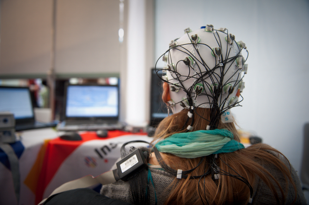
Interpreting the signals emitted by the brain and translating them into commands usable by humans is a goal that researchers have been pursuing in the development of what is known as brain-machine interfaces. With respect to health, these interfaces could be used by people suffering from paralysis. Up to now, researchers have encountered technological difficulties because the sensors used to record brain activity do not yet do so with enough accuracy.

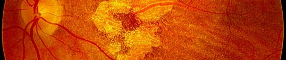
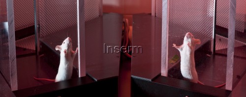
With the support of the Bioelectronics Department of the St Etienne Ecole des Mines, a research team headed by Christophe Bernard of INSERM Unit 1106 “Institut de neurosciences des systèmes” have designed a system for sensing brain activity that is 100% biocompatible and made of organic matter. The medium, which is only a few microns thick, is as thin and flexible as cellophane, and yet very tough. The model was tested on an animal suffering from epilepsy. The quality of the brain signal recorded was 10 times better than the traditional brain activity recording systems. The research was published in the Nature Communications journal.
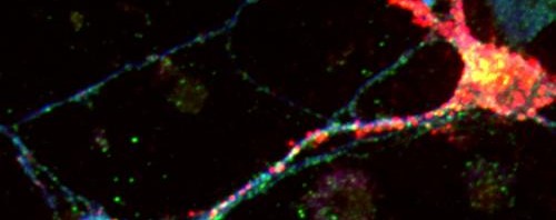
Des nouvelles techniques au service de la santé, OpenVibe. Une interface cerveau-ordinateur ou ICO (en anglais Brain-Computer Interface ou BCI) permet à son utilisateur d'envoyer des commandes à un ordinateur ou à une machine uniquement par la pensée. © P Hirsch/Inserm

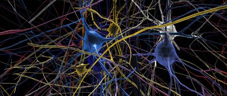
Man-machine interfaces have been playing a central role for several years in the diagnosis and treatment of certain conditions, in the movements of artificial limbs (exoskeletons) and even in the design of artificial sensory organs. In the case of brain-machine interfaces, the problem consists in detecting the signals emitted by the brain and translating them into commands that are usable by humans. These signals are used for diagnostic purposes (such as, for example, to determine whether a person is epileptic and which regions of the brain are responsible for the attacks), to link an artificial eye to the regions of the brain that process visual information or to control the movement of exoskeletons in people who are paralysed by recording the neurons in the regions of the brain that control the motor function of limbs.

For the capture of the maximum number of signals emitted by the brain, there is a need for direct contact with the central nervous system. This is very hard to achieve when using non-invasive measurement systems (i.e. electrodes placed on the head). Another disadvantage is that most of sensors used today are not biocompatible, thus triggering a defence reaction in the tissues, resulting in loss of signal after a certain amount of time. Finally, and this is the most important aspect, the signals detected are pre-amplified at a distance from the source, and this results in the presence of considerable interference on the recordings, thus preventing them being used to their best advantage.

One solution: organic transistors

The Bioelectronics Department of the St Etienne Ecole des Mines in Gardanne, the Institut de Neuroscience des Systèmes (INSERM Unit 1106) and the Microvitae SME at Gardanne have contributed a technological solution to these problems.

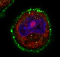
The researchers designed a system of brain activity sensors made of an organic material (based on carbon composites) that is 100% biocompatible. The medium is only a few microns thick, as thin and flexible as cellophane, and very tough.

But the revolutionary technology lies in enabling the recording site to work as an organic transistor that amplifies the signal locally. The system was tested in an animal model suffering from epilepsy. The quality of the signal was amplified tenfold in comparison with the traditional systems an amount that the researchers claim to be considerable.

A technological solution such as this makes it possible to record numerous neurons and interface with brain structures in the long term. The immediate clinical applications could include assistance with diagnosing epilepsy and functional mapping in the context of neurosurgery for brain tumours. Naturally, these transistors could also be used for non-invasive recordings in direct contact with the head.

In addition to disease control, the technology will enable major advances to be made in basic research, and especially in the context of the Human Brain Project which has received one billion euros of finance from the European Union. Recording systems based on organic transistors are precursors to the Man-machine interfaces of tomorrow.

Seven genetic risk factors associated with AMD

An international group of researchers has discovered seven new regions in the human genome associated with an increased risk of developing age-related macular degeneration (AMD), one of the main causes of blindness. Thierry Léveillard, INSERM’s director of research at the Institut de la Vision (INSERM / UPMC / CNRS), is coordinator of the European AMD Gene Consortium group, an international network of researchers representing 18 research groups. The results were placed online on 03 March 2013 in the journal Nature Genetics.


DMLA atrophique

© Inserm

AMD affects the macula, the region of the retina responsible for central vision. It is thanks to the macula that a human being can perform certain tasks requiring good visual acuity, such as reading, driving and recognising facial features. As AMD progresses, the performance of such tasks becomes harder and is eventually impossible. Although certain forms of AMD are treatable if the condition is detected sufficiently early, there is no cure.

Scientists have shown that age, diet and smoking affect the risk of a person developing AMD. Genetics also plays an important role. AMD is often hereditary and is most common in certain population groups.

In 2005, researchers showed that certain variations of the coding gene for factor H of the complement – a component of the innate immune system – are associated with a serious risk of developing AMD.

In this new study, the AMD Gene Consortium collected data from 18 research groups in order to increase the power of previous analyses. An analysis by the consortium consisted of data taken from more than 17,000 AMD sufferers, and these were compared with data from more than 60,000 individuals not suffering from AMD. The current analysis identified seven new genetic regions associated with the condition. As in the case of the 12 regions previously discovered, these seven regions are dispersed throughout the genome, indicating genes and functions that affect AMD.

“The challenge represented by the genetic complexity of AMD could be overcome by an association between all the centres working on this condition that causes blindness throughout the world; this is a demonstration of how union is strength”

, explains Thierry Léveillard, INSERM director of research at the Institut de la Vision (INSERM/UPMC/CNRS), co-ordinator of the EU-JHU sub-consortium, whose members consist of various European and U.S. centres that have played an important role in this study.

Since 2005, a total of 19 regions have been identified as being associated with AMD. They involve a variety of biological functions, including regulation of the innate immune system, maintaining cell structure, the growth and permeability of blood vessels, the metabolising of lipids and atherosclerosis.

As with other common conditions such as type II diabetes, the risk of an individual developing AMD is probably determined by not one but several genes. A more complete analysis of the DNA of areas surrounding the 19 regions identified by the AMD Gene Consortium could indicate several rare genetic variations that might have a decisive effect on the risk of AMD. The discovery of such genes could considerably improve scientists’ understanding of the pathogenesis of AMD and contribute significantly to the search for more effective treatments.

According to José-Alain Sahel, Director of the Institut de la Vision (INSERM / UPMC / CNRS): “

Without the methodical and coordinated clinical identification work performed at all the centres, the identification of such markers would be random. These clinical relationships will soon be very important in the application of predictive and personalised medicine.” 

“Simplified” brain lets the iCub robot learn language

The iCub humanoid robot on which the team directed by Peter Ford Dominey, CNRS Director of Research at Inserm Unit 846 known as the “Institut pour les cellules souches et cerveau de Lyon” [Lyon Institute for Stem Cell and Brain Research] (Inserm, CNRS, Université Claude Bernard Lyon 1) has been working for many years will now be able to understand what is being said to it and even anticipate the end of a sentence. This technological prowess was made possible by the development of a “simplified artificial brain” that reproduces certain types of so-called “recurrent” connections observed in the human brain. The artificial brain system enables the robot to learn, and subsequently understand, new sentences containing a new grammatical structure. It can link two sentences together and even predict how a sentence will end before it is uttered. This research has been published in the Plos One journal. 

robot ICub apprentissage

© P Latron/inserm

INSERM and CNRS researchers and the Université Lyon 1 have succeeded in developing an “artificial neuronal network” constructed on the basis of a fundamental principle of the workings of the human brain, namely its ability to learn a new language. The model was developed after years of research in the INSERM 846 Unit of the Institut de recherche sur les cellules souches et cerveau, through studying the structure of the human brain and understanding the mechanisms used for learning.

One of the most remarkable aspects of language-processing is the speed at which it is performed. For example, the human brain processes the first words of a sentence in real time and anticipates what follows, thus improving the speed with which humans process information. Still in real time, the brain continually revises its predictions through interaction between new information and a previously created context. The region inside the brain linking the frontal cortex and the striatum plays a crucial role in this process.

Based on this research, Peter Ford Dominey and his team have developed an “artificial brain” that uses a “neuronal construction” similar to that used by the human brain.

Thanks to so-called recurrent construction (with connections that create locally recurring loops) this artificial brain system can understand new sentences having a new grammatical structure. It is capable of linking two sentences and can even predict the end of a sentence before it is provided.

To put this advance into a real-life situation, the INSERM researchers incorporated this new brain into the iCub humanoid robot.

In a video demonstration, a researcher asks the iCub robot to point to a guitar (shown in the form of blue object) then asking it to move a violin to the left (shown by a red object). Before performing the task, the robot repeats the sentence and explains that it has fully understood what it has been asked to do.

For researchers, the contribution that this makes to research into certain diseases is of major importance. This system can be used to understand better the way in which the brain processes language. “We know that when an unexpected word occurs in a sentence, the brain reacts in a particular way. These reactions could hitherto be recorded by sensors placed on the scalp”, explains Peter Ford Dominey. The model developed by Dr Xavier Hinaut and Dr Peter Ford Dominey makes it possible to identify the source of these responses in the brain. If this model, based on the organisation of the cerebral cortex, is accurate, it could contribute to possible linguistic malfunctions in Parkinson’s disease.

This research has another important implication, that of contributing to the ability of robots to learn a language one day. “At present, engineers are simply unable to program all of the knowledge required in a robot. We now know that the way in which robots acquire their knowledge of the world could be partially achieved through a learning process – in the same way as children”, explains Peter Ford Dominey.

©P Latron/Inserm

Treatrush (TreatRetUsher): combating Usher Syndrome blindness – European collaboration in the service of a rare disease

On the occasion of International Rare Disease Day: 28 February 2013

How has research into Usher Syndrome – a particularly debilitating hereditary disease that damages the two main senses, hearing and sight – overcome a major hurdle in understanding the mechanisms causing pigmentary retinopathy? How has it managed to improve clinical diagnosis and develop molecular diagnosis? How has it prepared gene therapy tests for the retinopathy aspect of the syndrome? Scientists who have joined forces in the European TREATRUSH (TreatRetUsher) project have been reporting progress. Twelve partners in seven countries came together to form this network, financed by the European Commission and coordinated by the Pierre and Marie Curie University (UPMC). In France, it brings together researchers from the Collège de France, INSERM, the Institut Pasteur, the CNRS and the UPMC, working at the Institut de la Vision and the Institut Pasteur, as well as clinicians from the XV-XX Hospital and the Armand-Trousseau Hospital.

Usher Syndrome is a hereditary disease attacking hearing and sight. It affects about one person in 10,000. It is the main cause of deafness associated with a loss of vision in young people. The three clinical types of the syndrome, USH1, USH2 and USH3, are distinguished by the severity of hearing loss, early onset of retinitis pigmentosa and the presence or otherwise of a vestibular (inner ear) attack that translates into difficulties with balance. In each of these aspects of the disease the USH1 form is the most incapacitating.

Going beyond the major advances previously achieved in identifying the genes responsible for the syndrome and understanding the pathogenesis of the damage done to hearing by the syndrome, the Treatrush project has set itself the aim of improving diagnosis, understanding the cellular and molecular mechanisms behind retinopathy and preparing gene therapy trials.

  • Responding to the imperative of early diagnosis. 

A reduction in vision only manifests years after the loss of hearing, so the absence of early diagnosis of the syndrome can cause parents to make the wrong choice, and cause their children to learn sign language as young as possible, rejecting early cochlear implantation. Consequently, the European Consortium’s ENT specialists and ophthalmologists have jointly drawn up clinical investigation protocols that should result in a diagnosis being available for children under one year old. At the same time, a robust and swift molecular diagnosis of all forms of the syndrome has been developed. An initial study[1] enabled the detection of mutations in 54 patients. It was supplemented by the introduction of a new method[2] that now makes it possible to analyse a cohort of more than 350 patients recruited throughout Europe (France, Germany, Italy, Slovenia, Spain).

  • Clarifying the physio-pathological mechanisms that cause retinopathy.

While mutant mice in which there are defective Usher-1 genes are profoundly deaf, they do not present with retinopathy. Consequently, the defective mechanisms causing retinopathy in those suffering from the syndrome were totally unknown. Three years ago, teams headed by Professor Christine Petit, co-ordinator of the Treatrush Project, and Professor José-Alain Sahel, attempted to understand the physio-pathological mechanisms causing retinitis pigmentosa in patients suffering from Usher Syndrome, by using other animal models[3]. They were successful and thus laid the foundation for discovering a new type of mechanism that causes pigmentary retinopathy, one that also involves the structures of photoreceptors, an aspect hitherto neglected, the role of which is still unknown. This data is essential for the creation of any protocol designed to improve treatment.

Project teams in Italy, France and the United States are currently working to develop gene therapy via viral vectors associated with the adenovirus (AAV) capable of effectively transferring tiny therapeutic genes into retinal photoreceptors with the aim of preventing their degeneration. These vectors have already proven their worth in other forms of retinitis. They are currently being tested on animals to identify some of the Usher genes.

The advances achieved in curing this rare illness open the way to an understanding and treatment of commoner causes of deterioration in vision and hearing. As Christine Petit and José-Alain Sahel explain, “frequent disorders are of a more complex origin and are thus harder to study than rare monogenic diseases. Yet many of the contributing factors should belong to the same elementary mechanisms as those which, when defective, lead to a particular, rarer disease. As proof, take the example of the USH1 Syndrome. The molecules encoded by these genes lie at the heart of the auditory transduction machinery, machinery that converts a sound signal into an electric signal in the auditory sensory cells. It is self-evident that in cases of frequent damage to hearing, regardless of origin, this machinery is also the target of deficiency. A constellation of rare diseases should thus contribute to creating the model for the pathogenic processes occurring in a common disease”. 

The Usher Syndrome, an attack of the two main senses – hearing and sight

This syndrome is a hereditary sensory disability and the main cause of combined loss of hearing and blindness. It is responsible for 3% to 6% of infant deafness and about 50% of cases of severe deafness associated with adult blindness. Deafness is generally congenital, while the clinical  manifestations of retinitis pigmentosa occur slightly later in life. This syndrome has been divided into three sub-types, USH1, USH2 and USH3; the USH1 form is the most serious. In children suffering from the USH1 form, damage to sight is frequently not discovered until around the age of eight or ten. This late diagnosis makes it very hard to provide the appropriate medical treatment.

Important scientific progress was made by geneticists and hearing physiologists about fifteen years ago, when they identified the ten genes responsible and the discovery of deficient cellular and molecular mechanisms explained the loss of hearing caused by the syndrome. Conversely, the pathogenesis of retinitis pigmentosa in the syndrome remained unknown. A major advance occurred recently in understanding the damage caused to the retina, as part of the European Treatrush Project, through research conducted by teams headed by Professors Christine Petit and José-Alain Sahel. The patient exploration protocols were improved and standardised to allow for early diagnosis. A new molecular diagnostic tool was developed and more than 400 patients from various European countries were tested. In 1995, Professor Christine Petit’s laboratory identified the first gene causing the USH1 Syndrome. This is the gene that encodes VIIa myosin and is responsible for most cases of USH1. It is precisely in this form of the syndrome that a gene therapy retinopathy trial will soon be conducted by Professor José-Alain Sahel.

TREATRUSH – Combatting blindness caused by the Usher Syndrome

The aim of TREATRUSH is to treat and combat blindness caused by the Usher Syndrome. https://www.treatrush.eu

The project began in February 2010 and lasted for four years, the total budget of about 6 million euros being funded by the European Union (PC7). The project involves twelve partners based in seven countries:

Université Pierre and Marie Curie (UPMC), Paris, France: https://www.upmc.fr/

Institut National de la Santé et de la Recherche Médicale (Inserm), Paris, France : www.inserm.fr/

Eberhard Karl University of Tübingen  (EKUT), Tübingen, Germany: https://www.uni-tuebingen.de/en

Medical Research Council (MRC), Oxford, UK: https://www.mrc.ac.uk

Fondazione Telethon (FTELE.IGM), Naples, Italy: https://www.telethon.it/

Amsterdam Molecular Therapeutics (AMT), Amsterdam, Netherlands: https://www.amtbiopharma.com/

Novartis Forschungsstiftung, Zweigniederlassung Friedrich Miescher-Institut for Biomedical Research, Basle, Switzerland: https://www.fmi.ch/

Faun Foundation (FAUN), Nuremberg, Germany

The Trustees of the University of Pennsylvania (UPENN), Philadelphia, USA : https://www.upenn.edu/

Institut de la Vision-Fondation Voir et Entendre, Paris, France: https://www.institut-vision.org    https://www.fondave.org/

Johannes Gutenberg University of Mainz, Mainz, Germany: https://www.uni-mainz.de/eng/

Massachusetts Eye and Ear Infirmary, Bos


[1] Bonnet C, Grati M, Marlin S, Levilliers J, Hardelin JP, Parodi M, Niasme-Grare M, Zelenika D, Délépine M, Feldmann D, Jonard L, El-Amraoui A, Weil D, Delobel B, Vincent C, Dollfus H, Eliot MM, David A, Calais C, Vigneron J, Montaut-Verient B, Bonneau D, Dubin J, Thauvin C, Duvillard A, Francannet C, Mom T, Lacombe D, Duriez F, Drouin-Garraud V, Thuillier-Obstoy MF, Sigaudy S, Frances AM, Collignon P, Challe G, Couderc R, Lathrop M, Sahel JA, Weissenbach J, Petit C, Denoyelle F. (2011) Complete exon sequencing of all known Usher syndrome genes greatly improves molecular diagnosis. Orphanet J Rare Dis. 6:21.

[2] Fakin A, Jarc-Vidmar M, Glavač D, Bonnet C, Petit C, Hawlina M. (2012) Fundus autofluorescence and optical coherence tomography in relation to visual function in Usher syndrome type 1 and 2. Vision Res. 75: 60-70.

[3] Sahly I, Dufour E, Schietroma C, Michel V, Bahloul A, Perfettini I, Pepermans E, Estivalet A, Carette D, Aghaie A, Ebermann I, Lelli A, Iribarne M, Hardelin JP, Weil D, Sahel JA, El-Amraoui A, Petit C. (2012) Localization of Usher 1 proteins to the photoreceptor calyceal processes, which are absent from mice. J Cell Biol. 15;199(2):381-99.

Where does the energy come from that is needed as a carrier in neuron extensions?

The movement of molecules in the neuron extensions known as axons is a process that is vital for the survival of cells and the smooth operation of the nervous system. It is performed by vesicles that travel fast thanks to the energy-hungry molecular engines. At the “Signalling, neurobiology and cancer” Laboratory (Institut Curie/CNRS/Inserm) at the Institut Curie, the team headed by Frédéric Saudou[1], INSERM Director of Research, has shown that the vesicles have their own energy production system needed for travelling and do not depend on the mitochondria that are the main source of cell energy. This mechanism works by means of glycolysis, the first stage in the conversion of glucose and for the huntingtin protein, the protein that mutates in Huntington’s Disease, a neurodegenerative condition. The results were published on 31 January 2013 in the Cell journal.

Unlike carcinomas in which the cell proliferates, neurodegenerative conditions such as Alzheimer’s Disease, Parkinson’s Disease and Huntington’s Disease are due to the accelerated death of neurones. At the “Signalling, neurobiology and cancer” Laboratory (Institut Curie/CNRS/Inserm), part of the Institut Curie, the research team headed by Frédéric Saudou is studying the function of the huntingtin protein which mutates in Huntington’s Disease. “When it is altered, by a process that is still not fully understood, huntingtin causes the accelerated death of neurons in the striatum, the region of the brain in which Huntington’s disease first manifests” explains Frederic Saudou.

His team has shown the essential role played by huntingtin in the swift travel of vesicles through the neuron extensions or axons. axones. Huntingtin stimulates the progress of these vesicles by interacting with molecular engines, enabling them to travel to specific regions of the brain such as the striatum, the brain structure that is attacked in victims of Huntington’s Disease.

ATP, the engine essential for transporting the vesicles

So where does the cell energy come from that is vital for ensuring the transport of the vesicles in the axons over long distances, that may in some cases be as long as one metre? The adenosine triphosphate (ATP) molecule is an energy source shared by all animal and plant species. In humans, it is mostly produced by specialist organelles in the cells, known as the mitochondria. “In this project we have shown that a process other than the mitochondria is involved in the supply of energy to the molecular engines[1] responsible for movement along the axons” explains Frédéric Saudou. In fact, the inhibition of the mitochondrial function has no effect on this swift movement. On the other hand, the genetic inactivation of an enzyme that is essential for glycolysis, the first stage in the conversion of glucose into energy significantly reduces movement.

A mechanism that is dependent on the huntingtin protein

The enzymes responsible for glycolysis are situated directly on the vesicle and produce the energy needed for the movement of the axons. We then tried to discover the mechanism responsible for fixing it on the vesicle membrane. Our researches have established that attachment to the vesicle is performed by the huntingtin protein. We do not yet know, however, whether this function is disrupted in Huntington’s disease” stresses Frédéric Saudou. Researchers do not exclude the existence, however, of other mechanisms that link the glycolysis enzymes to the vesicle membrane.

Image taken via microscopy showing the position of the vesicule of a glycolysis enzyme, the protein GAPDH (in red), with huntingtin (in green) shown in a neuron from a rat’s cortex. The vesicles have been coloured blue.

©Diana Zala/Institut Curie

More about Huntington’s Disease

Huntington’s Disease is a rare neurological disorder that affects one person in 10,000 and only manifests in adulthood. The most typical symptoms are mental disturbance (anxiety, irritability, depression), a progressive deterioration in intellectual capacity culminating in dementia, associated with abnormal involuntary jerking movements of the limbs, head and neck.

The genetic anomaly that causes Huntington’s disease is an abnormal increase in the repetition of the three nucleic acids (C, A and G – known as the CAG triplet) in the coding gene for the huntingtin protein. This results in an abnormal expansion of a repetition of an amino acid (polyglutamine or polyQ repetition) in the huntigtin protein. The mechanisms leading to the manifestation of the disease are still little understood and at present there is no treatment to prevent the emergence of symptoms inpatients. A better understanding of the cell processes that occur within the neurons should make it possible to identify new treatment strategies for this neuro-degenerative disease. Understanding these mechanisms could also help in the treatment of other conditions such as cancer.


[1] The molecular engines responsible for the movement of the molecule or cell structures throughout the cell skeleton are the proteins kinesin and dynein.


[1] Frédéric Saudou is head of the “Cell Signalling and Neurobiology” team in the Cell Signalling, Neurobiology and Cancer Unit at the Institut Curie/CNRS UMR 3306/Inserm U1005

Le Human Brain Project gagne la compétition du plus grand fonds scientifique européen

OpenViBE2 : A major French project involving Brain-Computer Interfaces applied to video games

OpenViBE2 (2009-2013) is a collaborative research project, supported by finance from the ANR, that is based on the potential of the technologies known as “brain-computer interfaces”(ico) in the field of video games.This is a project that has brought together the scientific expertise needed through a multi-disciplinary consortium consisting of nine partners – the university laboratories who pioneered the field (INRIA, INSERM, cea, GIPSA-Lab), well-known video-game manufacturers (ubisoft, blacksheep studio, kylotonn games) and specialists in usage and transfer (lutin, clarte).After three years of work and achieving numerous scientific advances associated with the development of innovative industry prototypes, OpenViBE2 has made it possible to have greater control over the future of such technologies on the French market as well as internationally.

© Inserm / Hirsch, Philippe

  • Acting on thought thanks to brain-computer interfaces 

[break]A brain-computer interface enables users to send commands to a computer purely by using their mental activity. BCIs use electroencephalographic (EEG) devices based on electrodes that are placed on the surface of the head, and that record electrical signals corresponding to exchanges of electric current between the neurones. The electrical activity produced by the brain is then analysed in real time and translated into a command sent to the computer or any other automated system thus making it possible to move a cursor to the right or left, simply by mentally imaging the movements a hand would make.

Initiated in 2005 by INRIA and INSERM, the first OpenViBE (2005-2009) project, which was also supported by the ANR, was completed in 2009 with the creation of open-source software that made it possible to easily design, develop and test brain-computer interfaces (https://openvibe.inria.fr). Today, this is recognised software that is used throughout the world. The  OpenViBE2 software is a sequel to this initial project and is aimed at exploring these technologies more thoroughly by exploring them in a particularly complex applicational context –  that of video games. This is a relatively new field of application for BCIs. But the advent of low-cost EEG  helmets has opened the way to such applications.

The purpose of OpenViBE2 is thus to improve the current abilities of BCIs and test their use and their potential in the field of video games.

  • Video games: a growing market

[break]As a development of the “gesture-controlled” command (joystick, mouse, gamepad), the “mental” command is a new and very promising route in the field of video-gaming. Since the early 2000s, scientists have been tacking the scientific issue of how to incorporate BCI technologies to enable them to interact with virtual environments.

In this context, the aim of the OpenViBE2 project is to identify and use the gamer’s mental state and brain responses to interact with the game and/or adapt the actual content of the video game accordingly. A very original approach suggested in the project was to consider BCIs not as a substitution technique for the classic interfaces (joystick, mouse, gamepad) but rather to consider using the BCIs as a new way of playing that complements the traditional techniques. In this way, the gamer can continue to use a joystick while at the same time or at certain key moments in the game, he/she can also mobilise his/her brain activity for playing.

  • Advances in the Open ViBE2 project: promising innovations 

[break]Whether in respect of diseases and neurological syndromes, the world of entertainment or daily life, OpenViBE2 opens the way to innovative technologies and major interests for human beings. During the course of the OpenViBE2 project, major scientific advances have been obtained in these three fields:

In Neuroscience: identification of new mental activities linked to the attention processes.

In the first leg of the OpenViBE project, the  INSERM team known as “Mental Dynamics and Cognition (Dycog)” at the Lyon Neuroscience Research Centre (INSERM/CNRS) participated in the development of software capable of “writing through thought”, thus facilitating communication by people suffering from a motor handicap. OpenViBE2 offers new advances in the field of neurosciences that makes it possible to use interfaces brain-computer for therapeutic purposes in order to improve certain neurological disorders such as attention deficit disorders. Thanks to the device, researchers were able to analyse:

–   attention paid to the outside world by measuring in real time and selectively the level of engagement of the cerebral network responsible for seeking specific information in a visual scene.
–   attention paid by the user to an internal representation, c’est-à-dire i.e. the level of engagement of the cerebral network responsible for maintaining mental representation.
–    The level of distraction of a person by determining the real time in which a person is distracted and for how long.

The principle of virtual reality, used mainly in the serious game ADHD developed thanks to OpenViBE2, enabled researchers to obtain results with respect to attention deficit disorder. The virtual environment is similar to the real environment and the user has to concentrate in order to perform a task that he/she knows from the real world. This virtual training employs the process known as NeuroFeedback in which the user is required to self-regulate his/her mental activity.

The serious game, which is associated with augmented reality, has a positive retraining effect that persists beyond the training session and into the real environment”, stresses Jean Philippe Lachaux, Director of Research at INSERM.


Progress is still necessary with respect to the device so that it can be accessible
to all.

In processing signals: the CEA and GIPSA-Lab researchers have found new techniques to better filter and interpret electrical signals from the brain.

These make it possible in particular to eliminate noise interference (especially when it is linked to muscle activities such as facial muscles, blinking or jaw-clenching), and extracting the relevant brain activity as accurately and in as focused a manner as possible. These techniques are very useful in a gaming context, in which gamers may be very mobile.

In virtual reality and man-machine interaction: OpenViBE2 has enable the INRIA researchers to invent new concepts to interact with video games in the most original and effective way and suggest new world firsts:

–    The brain-computer “multi-player” interface: the INRIA researchers designed the very first  collaborative or competitive gaming application  in which the brain activities of two gamers were analysed simultaneously. Both players could play on the same side or in opposition to each other, in a simplified video football game.
–   The natural incorporation of BCI into the virtual world: researchers studied how to best incorporate the stimulation necessary for certain brain-computer interfaces based on  mental responses, by incorporating visual stimuli into the virtual world. For example, some BCIs “visual flashes” that are recognisable in the user’s brain. These can be incorporated into a video game, for example in the form of butterflies that beat their wings at different frequencies.
–   Automatic adaptation of the virtual world to the gamer’s mental state: Finally, researchers have offered radically new approaches in which elements of the virtual environment have been automatically modified depending on the player’s mental state. In a virtual maze game, guides are thus activated automatically if the user presents with too great a mental burden. This work has benefited internationally through the award of several science prizes (Best Paper Award, Eurohaptics 2012, BCI Award 2012 nominee).

  • The gaming room of the future

[break]The OpenViBE2 project has made it possible to develop numerous proofs of the academic concept and commercial video game prototypes all of them activated by the brain.

Video game manufacturers have worked directly with the INRIA university laboratories, as well as with INSERM, CEA and the GIPSA-Lab, to develop video games based on brain activity. This collaboration has made it possible to better define scientific research initiatives throughout the project and bring all the research together into solutions that are best suited to the technological constraints of the field of application.

During the course of the project, the partners introduced a huge campaign of experiments involving “brain-computer interfaces and video games” run by CHART  at the Cité des Sciences in the course of which nearly 400 testers tested the prototypes. The results of these experiments made it possible to highlight the attractions of these technologies to a wider public, and provide the consortium of partners with a large amount of user feedback. A standardisation project was also run by UBISOFT to define the “ideal” helmet best suited to the video-games market.

Here are a few examples of proofs of the concept developed by the university labs:

– MindShooter (INRIA): In this game, the gamer controls a spaceship shown at the bottom of the screen and he/she has to destroy the enemy ships at the top of the screen. Three commands are available: move right, move left, and fire. To activate them, the gamer must concentrate on the part of the ship that corresponds to the action he/she wishes to trigger.

Brain Invaders (GIPSA-LAb): This game is inspired by the Japanese “Space Invaders” game. Once again, the aim is to destroy the spaceships on the screen, thanks to a mental response that is produced as soon as a rare but expected event occurs.

Brain Arena (INRIA): This simplified “multi-player” football game enables two users to play together or one against the other using their mental powers.

Three commercial prototypes of video games based on mental activity have also been developed by the consortium.

Cocoto Brain (Kylotonn Games): A “Casual game” based on mental activity, Entertainment for the Nintendo Wii console. The gamer needs to protect a fairy by preventing all her enemies from getting near her. The gamer has to concentrate on targets positioned over the enemies in order to neutralise them.

– BCI Training Center (Black Sheep Studio), a game of the “mental training” type using the EEG developed by Black Sheep Studio. The game enables the gamer to practice brain-training gaming activities (visual searching, finding a word in a grid) associated with a real-time adaptation of the game, depending on the player’s mental state measured by the EEG device (relaxation and concentration).

A serious game for the treatment of attention deficit disorder (CLARTE): a prototype is designed for children destined for children suffering from attention deficit hyperactivity disorder (ADHD) has been developed by CLARTE. The virtual environment of this platform is a classroom in which the child suffering from ADHD is invited, for example, to watch a video on a screen in the class, of which the perceived quality is directly linked to his/her mental activity. Thus, if he/she wants to continue to watch the video under optimal conditions, he/she must continue to maintain his/her activity under the conditions indicated by the therapist. Several mini-games are also offered to improve his/her attentional ability using the EEG.

©Lachaux/Inserm

In conclusion, the OpenViBE2 project made it possible to acquire scientific expertise and technological know-how that is unique in the world, concerning the use of this very promising technology in a developing market. Important scientific advances have been obtained in many different fields, such as neuroscience, electrical signal processing from the brain, man-machine interfaces and virtual reality. The prototypes made it possible to illustrate numerous scientific results for the project in actual commercial settings.

The results of the project were capitalised in the form of know-how, methods and software such as the free OpenViBE software that ought to make it possible to have better future control over the opening up of these BCI technologies to the public at large. Discussions are currently taking place concerning the marketing of prototypes in the near future with help of the French start-up MENSIA TECHNOLOGIES.

More than 50 scientific papers have been presented at conferences or published in journals of reference.

Please find photographs to illustrate the project in the Inserm photothèque.

Comment des souris subissant des agressions répétées développent une aversion sociale

La protéine à l’origine de la maladie de Huntington impliquée dans les tumeurs mammaires

Detrimental effect of obesity on lesions associated with Alzheimer’s disease

Researchers from Inserm and the Université Lille/Université Lille Nord de France have recently used a neurodegeneration model of Alzheimer’s disease to provide experimental evidence of the relationship between obesity and disorders linked to the tau protein. This research was conducted on mice and is published in the Diabetes review: it corroborates the theory that metabolic anomalies contribute massively to the development of dementia.

In France, more than 860,000 people suffer from Alzheimer’s disease and related disorders, making them the largest cause of age-related loss of intellectual function. Cognitive impairments observed in Alzheimer’s disease result from the accumulation of abnormal tau proteins in nerve cells undergoing degeneration (see the picture below). We know that obesity, a major risk factor in the development of insulin resistance and type 2 diabetes, increases the risk of dementia during the aging process. However, the effects of obesity on ‘Taupathies’ (i.e. tau protein-related disorders), including Alzheimer’s disease, were not clearly understood. In particular, researchers assumed that insulin resistance played a major role in terms of the effects of obesity.

The “Alzheimer & Tauopathies” team from mixed research unit 837 (Inserm/Université Lille 2/Université Lille Nord de France) directed by Dr. Luc Buée, in collaboration with mixed research unit 1011 “Nuclear receptors, cardiovascular diseases and diabetes”, have just demonstrated, in mice, that obese subjects develop aggravated disorders. To achieve this result, young transgenic mice, who develop tau-related neurodegeneration progressively with age, were put on a high-fat diet for five months, leading to progressive obesity.

“At the end of this diet, the obese mice had developed an aggravated disorder both from the point of view of memory and modifications to the Tau protein”

explains David Blum, in charge of research at Inserm.

This study uses a neurodenegeneration model of Alzheimer’s disease to provide experimental evidence of the relationship between obesity and disorders linked to the tau protein. Furthermore, it indicates that insulin resistance is not the aggravating factor, as was suggested in previous studies.

“Our research supports the theory that environmental factors contribute massively to the development of this neurodegenerative disorder” underlines the researcher. “Our work is now focussing on identifying the factors responsible for this aggravation” he adds.

The degeneration of neurons in Alzheimer’s disease

© Wikipedia – Zwarck  / licence Creative Commons  CC-BY-SA-2.5

In the case of healthy neurons (top), the Tau protein is normal.
In the case of diseased neurons (bottom), abnormal tangles of Tau proteins (phosphorylated) form, causing degeneration.

This research was supported by LabEx DISTALZ (development of Innovative Strategies for a Transdisciplinary Approach to Alzheimer’s Disease) within the framework of future investments.

Why good resolutions about taking up a physical activity can be hard to keep

Physical inactivity is a major public health problem that has both social and neurobiological causes. According to the results of an Ipsos survey published on Monday 31 December, the French have put “taking up a sport” at the top of their list of good resolutions for 2013. However, Francis Chaouloff, research director at Inserm’s NeuroCentre Magendie (Inserm Joint Research Unit 862, Université Bordeaux Ségalen), Sarah Dubreucq, a PhD student and François Georges, a CNRS research leader at the Interdisciplinary Institute for Neuroscience (CNRS/Université Bordeaux Ségalen) have just discovered the key role played by a protein, the CB1 cannabinoid receptor, during physical exercise. In their mouse studies, the researchers demonstrated that the location of this receptor in a part of the brain associated with motivation and reward systems controls the time for which an individual will carry out voluntary physical exercise. These results were published in the journal Biological Psychiatry

©fotolia

The collective appraisal conducted by Inserm in 2008 highlighted the many preventive health benefits of regular physical activity. Such activity is limited, however, by our lifestyle in today’s industrial society. While varying degrees of physical inactivity may be partly explained by social causes, they are also rooted in biology.

“The inability to experience pleasure during physical activity, which is often quoted as one explanation why people partially or completely drop out of physical exercise programmes, is a clear sign that the biology of the nervous system is involved”, explains Francis Chaouloff.

But how exactly? The neurobiological mechanisms underlying physical inactivity had yet to be identified.

Francis Chaouloff (Giovanni Marsicano’s team at the NeuroCentre Magendie; Inserm joint research unit, Université Bordeaux Ségalen) and his team have now begun to decipher these mechanisms. Their work clearly identifies the endogenous cannabinoid (or endocannabinoid) system as playing a decisive role, in particular one of its brain receptors. This is by no means the first time that data has pointed to interactions between the endocannabinoid system, which is the target of delta9-tetrahydrocannabinol (the active ingredient of cannabis), and physical exercise. It was discovered ten years ago that physical exercise activated the endocannabinoid system in trained sportsmen, but its exact role remained a mystery for many years. Three years ago, the same research team in Bordeaux observed that when given the opportunity to use a running wheel, mutant mice lacking the CB1 cannabinoid receptor, which is the principal receptor of the endocannabinoid system in the brain, ran for a shorter time and over shorter distances than healthy mice. The research published in Biological Psychiatry this month seeks to understand how, where and why the lack of CB1 receptor reduces voluntary exercise performance (by 20 to 30%) in mice allowed access to a running wheel three hours per day.

The researchers used various lines of mutant mice for the CB1 receptor, together with pharmacological tools. They began by demonstrating that the CB1 receptor controlling running performance is located at the GABAergic nerve endings. They went on to show that the receptor is located in the ventral tegmental area of the brain (see diagram below), which is an area involved in motivational processes relating to reward, whether the reward is natural (food, sex) or associated with the consumption of psychoactive substances.

Longitudinal section of the mouse brain (top) and diagram of interactions between the endocannabinoid, GABAergic and dopaminergic systems during voluntary physical exercise (bottom)

©Inserm/F. Chaouloff

Left: stimulating the CB1 receptors excites the dopaminergic neurons in the ventral tegmental area involved in motivation.
Right: the absence of CB1 receptors lowers performance by 20 to 30% owing to reduced motivation.

VTA: Ventral tegmental area/NAcc: nucleus accumbens/PFC: prefrontal cortex/DA: dopamine 

Based on the results of this study and earlier work, the Bordeaux team suggests the following neurobiological explanation: at the beginning and for the duration of physical exercise, the CB1 receptor is constantly simulated by the endocannabinoids, lipid molecules that naturally activate this receptor in response to pleasant stimuli (rewards) and unpleasant stimuli (stress). Endocannabinoid stimulation of the CB1 receptor during physical exercise inhibits the release of GABA, an inhibitory neurotransmitter that controls the activity of the dopamine neurons associated with the motivation and reward processes. This stimulation of the CB1 receptor “inhibits inhibition”, in other words, it activates the dopaminergic neurons in the ventral tegmental area. The CB1 receptor must therefore be stimulated before the exercise can go on for longer and the body must receive the necessary motivation.

Conversely, without these CB1 receptors, the “GABAergic brake” continues to act on the dopaminergic neurons in the ventral tegmental area, leading to the reduced performance levels observed above.

It is already known that CB1 receptors play a regulatory role in the motivation to consume rewards, whether natural or not. What is original about this research is that it shows that physical exercise can be added to the array of natural rewards regulated by the endocannabinoid system. “If confirmed, this motivational hypothesis would imply that the role played by the CB1 receptor has more to do with ‘staying power’ in the exercise than with actual physical performance levels” explain the researchers.

This work reveals that the endocannabinoid system plays a major role in physical exercise performance through its impact on motivational processes. It thus opens up new avenues of research into the mediators of pleasure – and even addiction – associated with regular physical exercise. “After endorphins, we now need to consider endocannabinoids as another potential mediator of the positive effects that physical exercise has on our mood,” the researchers conclude.

fermer