Menu

Confirmation of the neurobiological origin of attention – deficit disorder

A study, carried out on mice, has just confirmed the neurobiological origin of attention – deficit disorder (ADD), a syndrome whose causes are poorly understood. Researchers from CNRS, the University of Strasbourg and INSERM1 have identified a cerebral structure, the superior colliculus, where hyperstimulation causes behavior modifications similar to those of some patients who suffer from ADD. Their work also shows noradrenaline accumulation in the affected area, shedding light on this chemical mediator having a role in attention disorders. These results are published in the journal Brain Structure and Function.

Attention – deficit disorder affects between 4 – 8% of children. It manifests mainly through disturbed attention and verbal and motor impulsiveness, sometimes accompanied by hyperactivity. About 60% of these children still show symptoms in adulthood. No cure exists at this time. The only effective treatment is to administer psychostimulants, but these have substantial side effects, such as dependence. Persistent controversy surrounding the neurobiological origin of this disorder has hindered the development of new treatments.

The study in Strasbourg investigated the behavior of transgenic mice having developmental defects in the superior colliculus. This structure, located in the midbrain, is a sensory hub involved in controlling attention and visual and spatial orientation. The mice studied were characterized by duplicated neuron projections between the superior colliculus and the retina. This anomaly causes visual hyperstimulation and excess noradrenaline in the superior colliculus. The effects of the neurotransmitter noradrenaline, which vary from species to species, are still poorly understood.

However, we do know that this noradrenaline imbalance is associated with signific ant behavioral changes in mice carrying the genetic mutation. By studying them, researchers have observed a loss of inhibition: for example mice hesitate less to penetrate a hostile environment. They have difficulties in understanding relevant information and demonstrate a form of impulsiveness. These symptoms remind us of adult patients suffering from one of the forms of ADD.

Currently, the fundamental work on ADD uses mainly animal models obtained by mutations that disturb dopamine production and transmission pathways. In mice with a malformed superior colliculus, these pathways are intact. The changes occur elsewhere in the neural networks of the midbrain. By broadening the classic boundary used to research its causes, using these new models would allow a more global approach to ADD to be developed. Characterizing the effects of noradrenaline on the superior colliculus more precisely could open the way to innovative therapeutic strategies.

Colliculus supérieur

In this image, marking shows the axons in retinal neurons (in red) that innervate the superior colliculus (in blue) in a “normal” mouse
© Michael Reber / Inserm Institut des neurosciences cellulaires et intégratives

(1) From the Laboratoire de Neurosciences Cognitives et Adaptatives (CNRS/Université de Strasbourg) and the Institut des Neurosciences Cellulaires et Intégratives (CNRS)

Caffeine and Alzheimer’s disease : a link with tau protein

Researchers at Inserm and University of Lille 2/University of Lille Nord de France directed by David Blum, Inserm Research Fellow, have provided experimental evidence of the beneficial effects of caffeine in an animal model of Alzheimer’s disease. This work, carried out on mice and published in Neurobiology of Aging, supports the idea that caffeine has a protective effect in some brain pathologies.

Post-it note with smiley face sticked on cup

©fotolia

Affecting more than 800,000 people in France, Alzheimer’s disease and related diseases are the leading cause of age-related loss of intellectual function. The cognitive impairment seen in Alzheimer’s disease mainly results from the accumulation of abnormal tau proteins in degenerating nerve cells. Regular caffeine consumption is known to reduce age-related cognitive decline, and the risk of developing dementia. However, the effects of caffeine on pathologies associated with tau protein, known as tauopathies, one of which is Alzheimer’s disease, had not been clearly elucidated.

Dr. David Blum, from the Alzheimer & Tauopathies laboratory at Joint Research Unit 837 (Inserm/University of Lille 2/University of Lille Nord de France), directed by Dr. Luc Buée, has just shown in mice that regular caffeine consumption prevents memory deficits and some of the modifications to tau protein. To arrive at this result, young transgenic mice that progressively develop age-related neurodegeneration associated with tau protein were given caffeine orally for 10 months.

“Mice treated with caffeine developed a less severe pathology from the point of view of memory, tau protein modifications, and neuroinflammation” explains David Blum, a research fellow at Inserm.

This study provides experimental evidence of a link between caffeine consumption and pathologies associated with tau protein in a neurodegeneration model of Alzheimer’s disease. This study also indicates that caffeine may act on different types of brain dysfunction involved in Alzheimer’s disease in order to exert its beneficial effects.

“This work indicates a significant role for environmental factors in the development of Alzheimer’s disease, emphasises the researcher. In the light of these results, we now hope to identify the molecular target responsible for these beneficial effects of caffeine on the one hand, and initiate a clinical trial to test the effects of caffeine on patients with Alzheimer’s disease on the other hand,” he adds.

This work received support from LabEx DISTALZ (Development of Innovative Strategies for a Transdisciplinary Approach to Alzheimer’s Disease) under the Investissements d’Avenir (Investment for the Future) programme, and from the France Alzheimer and LECMA/AFI associations.

Friedreich’s ataxia – an effective gene therapy in an animal model

The team led by Hélène Puccio, director of research for Inserm at the Institute of Genetics and Molecular and Cellular Biology (IGBMC) (Inserm / CNRS / University of Strasbourg) in close collaboration with Patrick Aubourg’s team (Inserm and Professor of Neuropaediatrics at Bicêtre Hospital) has demonstrated, in the mice, the efficacy of gene therapy for treating the heart disease associated with Friedreich’s ataxia, a rare hereditary neuro-degenerative disorder. The transfer, via a viral vector, of a normal copy of the gene deficient in patients, allowed to fully and very rapidly cure the heart disease in mice. These findings are published in Nature Medicine on 6 April, 2014.

Friedreich’s ataxia is a severe, rare hereditary disorder which combines progressive neuro-degeneration, impaired heart function and an increased risk of diabetes. The condition affects one in every 50,000 birth. There is currently no effective treatment for this disease. In most cases, Friedreich’s ataxia starts in adolescence with impaired balance and coordination (ataxia) of voluntary arm and leg movements, confining the majority of patients to a wheelchair after 10-20 years’ progression. However, complications affecting the heart are the major cause of death in 60% of patients, most often before the 35 years of age.

The disease is caused by a common mutation in the FXN gene which leads to a dramatic decrease in the production of the protein named ‘frataxin’. The reduced frataxin level disturbs the activity of mitochondria. These organelles are essential to cells and play a fundamental role in energy production. The nerve tissue (cerebellum, spinal cord etc.) and heart tissue are particularly vulnerable to this shortage of energy, which can lead to fatal heart failure.

The teams led by Hélène Puccio, director of research at Inserm and Patrick Aubourg have developed a therapeutic approach based on the use of an adeno-associated virus (AAV)[1], which is known to efficiently target and express a therapeutic gene in heart cells. The virus was modified to make it harmless but nevertheless capable of introducing a normal copy of the FXN gene in the heart cells, thus leading to the expression of frataxin.

Hélène Puccio’s team tested the efficacy of this treatment in a mouse model that faithfully reproduces the heart symptoms of patients suffering from Friedreich’s ataxia. The results show that a single intravenous injection of AAVrh10 expressing frataxin is not only capable of preventing the development of heart disease in animals before the appearance of symptoms, but also, more impressively, of fully and rapidly curing the hearts of animals at an advanced stage of heart disease. After three weeks of treatment, the heart become fully functional again; mitochondrial function and the appearance of heart tissue being very similar to those of healthy mice. “This is the first time that gene therapy has prompted full, lasting remission of heart disease so quickly in an animal model.” explains Hélène Puccio.
Figure 3 post review

The illustrations show a measurement of the activity of a mitochondrial protein (in blue) essential for cellular energy production, which is impaired when frataxin is absent (no staining in the untreated heart). Using gene therapy expressing frataxin, the activity of this essential protein can be corrected across the heart’s entire surface. © Inserm / H. Puccio

As the central nervous system is also a target of AAV vectors, Hélène Puccio and Patrick Aubourg’s teams are investigating whether a similar approach using gene therapy could be as effective for the spinal cord and cerebellum as it is for the heart.
Based on these promising results, work has begun on the necessary developments to propose to patients suffering from Friedreich’s ataxia and presenting a progressive cardiomyopathy a treatment by gene therapy. To this end, three of the paper’s authors have set up AAVLife, a French company specialising in gene therapy for rare diseases, to translate to clinic these important laboratory findings. A patent application has been submitted by Inserm Transfert for this gene therapy approach.

This study was backed by FARA[2], AFAF[3] and AFM[4].


[1] AAV: specifically the serotype AAVrh10.

[2] Friedreich’s Ataxia Research Alliance, an American association specialising in the treatment of Friedreich’s ataxia.

[3] French Friedreich’s ataxia association

[4] French muscular dystrophy association

Neonates are able to make connections between space, time and number

From birth, babies already have a representation of space, time and number. This has been proven by Dr Maria Dolores de Hevia, Dr Véronique Izard, Aurélie Coubart, Professor Elizabeth Spelke and Professor Arlette Streri from the Psychology and Perception Laboratory (Paris Descartes University/CNRS/Inserm) in a study published in PNAS.

The origin of our understanding of space, time and number is a subject examined by various disciplines such as philosophy, experimental psychology, developmental psychology and cognitive science. Space, time and number are connected both in the real world and the human mind, but how do we come to understand these connections? Do we learn to connect these concepts based on sensory experience by observing their correlations in the world around us or are our minds capable of understanding them innately from birth?

4 month old baby


© Fotolia

To answer this question, a test protocol was set up in the maternity unit of Bichat Hospital which recorded the visual attention of 96 neonates with an average age of 2 days (aged between 7 and 96 hours). The experiment put them in a situation where they had to use two of their senses: sight and hearing. In the first phase, a sequence of sounds evoking a numerical quantity (6 or 18 syllables) and/or a time period (1.4 or 4.2 seconds) was played to the babies over a period of one minute while they watched a moving line on a screen. In the second phase, the researchers presented new visual and auditory events, which were different to the first phase. These events were changed either congruently (1) in the same direction (e.g. longer line, larger number of sounds) or incongruently in opposite directions (e.g. longer line, smaller number of sounds).

The results show that neonates respond when these values change congruently. They are therefore able to make the connection between a numerical quantity and/or a time period and a length in space.

This test protocol proved that just hours after birth, humans are already sensitive to the common structure of time, space and number, which corroborates philosophical theories such as those espoused by Kant.

We still need to establish whether this applies to other quantitative dimensions such as light and sound and to determine the cerebral bases of these predispositions.

(1) With different elements matching and corresponding

How does light affect our brain’s performance?

Who would have thought that our brains are better equipped to process cognitive tasks if we are exposed to light several hours beforehand? Researchers in Inserm Unit 846 “Stem Cells and Brain” and the Cyclotron Research Centre at the University of Liege (Belgium) have recently proven that this ‘delayed effect’ is due to a sort of light memory (or photic memory). The results of this work are published in the PNAS journal.


It has long been known that light exerts powerful effects on the brain and on our well-being. Light is not only required for vision but is also essential for a wide range of “non-visual” functions including  synchronization of our biological clock to the 24h day-night cycle. Light also conveys a powerful stimulating signal for human alertness and cognition and has been routinely employed to improve performance, counter the negative impact of sleepiness or “winter blues”.

The mechanisms underlying these positive effects of light still remain largely unknown.

However, within the last decade or so, researchers have discovered a new type of light sensitive cell (photoreceptor) in the eye called melanopsin. This novel photoreceptor has been shown to be an essential component for relaying light information to a set of so-called non-visual centers in the brain. In the absence of this photoreceptor, animal research showed that non-visual functions are disrupted, the biological clock becomes deregulated and ‘free-runs” independent from the 24 day-night cycle and the stimulatory influence of light is impaired.

The melanopsin photopigment is unusual in many aspects and differs from our rods and cones since it shows invertebrate-like properties and is maximally sensitive to blue light. In humans, it has not been possible to apply genetic tools to selectively isolate the precise role of this new photoreception system and consequently the role of melanopsin in human cognition and alertness has not been established.

However, researchers from the Cyclotron Research Centre of the University of Liège (Belgium) and of the Department of Chronobiology of the INSERM Stem Cell and Brain Research Institute (Bron, France) have just provided evidence demonstrating the involvement of melanopsin in the impact of light on cognitive brain function.

By exploiting the specific invertebrate-like responses of melanopsin combined with state-of-the-art functional Magnetic Resonance Imaging (fMRI) recording strategies, they were able to show that impact of light on brain areas recruited to perform an ongoing cognitive task depended on the specific color of previous light exposures.

Prior exposure 1-hr earlier to an orange light enhanced the subsequent impact of the test light, while prior exposure to blue light had the reverse outcome.

image IRM

FIGURE: 16 young and health participants performed an auditory cognitive task while exposed to a test light. Brain areas in orange responded more to the test light if participants had been exposed to orange light 70 minutes earlier. 1. Thalamus; 2. Dorsolateral prefrontal cortex; 3. Ventrolateral prefrontal cortex.© Inserm/ Howard Cooper

These areas are important in the regulation of alertness and complex cognitive processes.

The phenomenon of prior light effects on a subsequent response to light is typical of melanopsin as well as certain photopigments of invertebrate and plant, and has been referred to as a “photic memory”.

“Humans may therefore have an invertebrate or plant-like machinery within the eyes that participates to regulate cognition. It may also explain what human chronobiologists have described as “previous light history effects”, a form of long term adaptation to previous lighting conditions,” explains Howard Cooper.

These findings emphasize the importance of light for human cognitive brain functions and constitute compelling evidence in favor of a cognitive role for melanopsin. More generally, the continuous change  of light throughout the day also changes us. Ultimately, these findings argue for the use and design of lighting systems to optimize cognitive performance.

Why does the brain remember dreams?

Some people recall a dream every morning, whereas others rarely recall one. A team led by Perrine Ruby, an Inserm Research Fellow at the Lyon Neuroscience Research Center (Inserm/CNRS/Université Claude Bernard Lyon 1), has studied the brain activity of these two types of dreamers in order to understand the differences between them. In a study published in the journal Neuropsychopharmacology, the researchers show that the temporo-parietal junction, an information-processing hub in the brain, is more active in high dream recallers. Increased activity in this brain region might facilitate attention orienting toward external stimuli and promote intrasleep wakefulness, thereby facilitating the encoding of dreams in memory.

The discovery in video ( subtitles in english ) :

The reason for dreaming is still a mystery for the researchers who study the difference between “high dream recallers,” who recall dreams regularly, and “low dream recallers,” who recall dreams rarely. In January 2013 (work published in the journal Cerebral Cortex), the team led by Perrine Ruby, Inserm researcher at the Lyon Neuroscience Research Center, made the following two observations: “high dream recallers” have twice as many time of wakefulness during sleep as “low dream recallers” and their brains are more reactive to auditory stimuli during sleep and wakefulness. This increased brain reactivity may promote awakenings during the night, and may thus facilitate memorisation of dreams during brief periods of wakefulness.

In this new study, the research team sought to identify which areas of the brain differentiate high and low dream recallers. They used Positron Emission Tomography (PET) to measure the spontaneous brain activity of 41 volunteers during wakefulness and sleep. The volunteers were classified into 2 groups: 21 “high dream recallers” who recalled dreams 5.2 mornings  per week in average, and 20 “low dream recallers,” who reported 2 dreams per month in average. High dream recallers, both while awake and while asleep, showed stronger spontaneous brain activity in the medial prefrontal cortex (mPFC) and in the temporo-parietal junction (TPJ), an area of the brain involved in attention orienting toward external stimuli.

Jonction-temporo-parietal


Temporo-parietal jonction (TPJ) © Perrine Ruby / Inserm

“This may explain why high dream recallers are more reactive to environmental stimuli, awaken more during sleep, and thus better encode dreams in memory than low dream recallers. Indeed the sleeping brain is not capable of memorising new information; it needs to awaken to be able to do that,”

explains Perrine Ruby, Inserm Research Fellow.

The South African neuropsychologist Mark Solms had observed in earlier studies that lesions in these two brain areas led to a cessation of dream recall.  The originality of the French team’s results is to show brain activity differences between high and low dream recallers during sleep and also during wakefulness.

“Our results suggest that high and low dream recallers differ in dream  memorization, but do not exclude that they  also differ in dream production. Indeed, it is possible that high dream recallers produce a larger amount of dreaming than low dream recallers” concludes the research team.

Mechanism elucidated: how smell perception influences food intake

A research team led by Giovanni Marsicano, a Inserm Research Director at Unit 862 (NeuroCentre Magendie, Bordeaux), has succeeded in elucidating how the endocannabinoid system controls food intake through its effects on the perception of smells. These results are due to appear in the journal Nature Neuroscience on 9 February 2014.

Nature-Neuro Nose_illustration

© Charlie Padgett

In animals, as in humans, hunger mechanisms are known to stimulate food intake. Hunger triggers a set of mechanisms that encourage feeding, for example by increasing sensory perceptions such as the sense of smell. The researchers have now succeeded in revealing what links hunger and increased smell perception in the brain, and the resulting urge to eat.

The researchers have discovered how this mechanism is initiated in the endocannabinoid system in mice. This system interconnects receptors located in the brain and involved in different sensations such as euphoria, anxiety, or even pain, that are also sensitive to cannabinoid substances, such as cannabis.

The researchers discovered that the CB1 cannabinoid receptors control a circuit that connects the olfactory bulb (the region in the nervous system that initially handles olfactory information, located above the nose) to the olfactory cortex (higher structures of the brain). When the sensation of hunger is felt, it triggers the activity of the cannabinoid receptors, which in turn activate the olfactory circuit, which then becomes more responsive.

It is therefore this biological mechanism that brings about the increased sensitivity to smell during hunger, explaining one of the reasons for food intake and attraction to food.

The researchers expect that the circuit involved in the olfactory system is altered in obese or anorexic patients, and that sensitivity to smell may be more or less strong compared to normal. Elucidation of the biological mechanism will allow better management of these types of pathologies.

This work was funded by ERC (European Research Council).

Autism: birth hormone may control the expression of the syndrome in animals

The scientific community agrees that autism has its origins in early life—foetal and/or postnatal. The team led by Yehezkel Ben-Ari, Inserm Emeritus Research Director at the Mediterranean Institute of Neurobiology (INMED), has made a breakthrough in the understanding of the disorder. In an article published in Science, the researchers demonstrate that chloride levels are elevated in the neurons of mice used in an animal model of autism, and remain at abnormal levels from birth. These results corroborate the success obtained with the diuretic treatment tested on autistic children by the researchers and clinicians in 2012, and suggest that administration of diuretics to mice before birth corrects the deficits in the offspring. They also show that oxytocin, the birth hormone, brings about a decrease in chloride level during birth, which controls the expression of the autistic syndrome. 

This work is due to appear in the 6 February 2014 issue of Science.

Neurons contain high levels of chloride throughout the entire embryonic phase. As a result, GABA, the main chemical messenger of the brain, excites the neurons during this phase instead of inhibiting them, in order to facilitate construction of the brain. Subsequently, a natural reduction in chloride levels allows GABA to exercise its inhibitory role and regulate the activity of the adolescent/adult brain. In many brain disorders (childhood epilepsy, cranial trauma, etc.), studies have shown abnormally high chloride levels. Having made various observations, Dr Lemonnier’s team (Brest), and Yehezkel Ben-Ari’s team at Inserm carried out a clinical trial in 2012, based on the hypothesis of high chloride levels in the neurons of patients with autism. The researchers showed that administration of a diuretic to children with autism (which reduces neuronal chloride levels) has beneficial effects[1]. The results of the trial supported this hypothesis, but because high neuronal chloride levels could not be demonstrated in children with autism, it was not possible to prove the mechanism proposed or justify the treatment.

In the present study, the researchers therefore used two animal models of autism, a genetic model, Fragile X syndrome, which is the genetic mutation most frequently associated with autism, and another, generated by injecting the spleen of pregnant mice with sodium valproate, a product known to generate abnormalities in children, including autistic spectrum disorder.

A high level of chloride in the brain

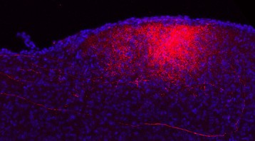
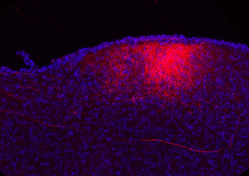
For the first time, the researchers recorded the activity of neurons at the embryonic stage and immediately after birth in order to observe modifications in chloride levels. These observations showed that neuronal chloride levels are abnormally high in both young and adult animals used in the autism model. GABA strongly excites neurons, and the researchers recorded aberrant electrical activities in the brain, which persist in adult animals.

The fall in chloride level, a particularly impressive phenomenon seen at birth in control animals, is absent in both of these animal models, and the neurons have the same chloride level before and after birth. These high levels are due to reduced activity of a chloride transporter, thus preventing transport of chloride out of the neuron. As a result, a major feature of neurons during birth is abolished in animal models of autism.

“Chloride levels during delivery are determinants of the occurrence of autism spectrum disorder,”

 explains Yehezkel Ben-Ari, an Emeritus Research Director at Inserm.

Beneficial effects of the diuretic on brain activity

The researchers administered a diuretic treatment to the mother (in both animal models) for 24 h shortly before delivery to see if this would restore brain inhibition in the offspring. They showed that the drop in chloride level was re-established in the neurons several weeks after a single treatment during birth. According to the research team, antenatal treatment restored brain activity to approximately normal levels, and corrected the “autistic” behaviour in the animals once they became adults.

“These results thus validate the working hypothesis that led us to the treatment we developed in 2012,” states the principal author of the study.

Oxytocin, the birth hormone, naturally reduces chloride levels

The role of oxytocin in reducing neuronal chloride was also studied. The researchers had previously shown in 2006[2] that this hormone, which triggers labour, also has many beneficial actions on the brains of newborns, including protective effects in the event of complications during delivery, and even analgesic properties. Oxytocin acts like the diuretic, reducing the intracellular chloride levels.

In the present study, the team tested the long-term effects of blocking the actions of the hormone before birth. A drug that blocks the signals generated by oxytocin was injected into pregnant mice. The researchers evaluated the effects of this blockage on the offspring, and showed that it reproduced the entire autism-like syndrome in them, both with respect to the electrical and behavioural aspects (identical to the two animal models of autism). As a result, the hormone’s natural actions, just like those of the diuretic, are crucial to this delicate phase, and may control the pathogenesis of autism via the cellular chloride levels.

“These data validate our treatment strategy, and suggest that oxytocin, by acting on the chloride levels during delivery modulates/controls the expression of autism spectrum disorder,”

 states Yehezkel Ben-Ari.

Taken together, these observations suggest that earliest possible treatment is essential for maximum possible prevention of the disorder.

This work raises the importance of carrying out early epidemiological studies in order to better understand the pathogenesis of the disorder, especially through analysing data on deliveries where a drop in chloride has occurred. Indeed, complicated deliveries with episodes of prolonged lack of oxygen, for example, or complications during pregnancy, such as viral infections, are often suggested as risk factors.

Finally, given the role of oxytocin in triggering labour, “although it is true that epidemiological data suggesting that scheduled caesarean deliveries may have increased the incidence of autism are controversial, it nonetheless remains that these studies should be followed up and extended in order to confirm or refute this relationship, which is still possible,” insists Yehezkel Ben-Ari, who concludes, “To treat this type of disorder, it is necessary to understand how the brain develops and how genetic mutations and environmental insults modulate brain activity in utero.


[1] See press release – Inserm press room – Un essai clinique prometteur pour diminuer la sévérité des troubles autistiques (A promising clinical trial for reducing the severity of autistic disorders) (Translational Psychiatry, 2012)

[2] Maternal Oxytocin Triggers a Transient Inhibitory Switch in GABA Signaling in the Fetal Brain During Delivery. Tyzio et al, Science 2006

Parkinson’s disease: an immense step forward thanks to gene therapy

A French and English team (AP-HP, Inserm, UPEC, CEA/Mircen, Oxford Biomedica, Cambridge University) has conducted a clinical phase 1/2 gene therapy study among patients suffering from an evolved form of Parkinson’s disease. Fifteen patients were able to benefit from this new treatment, which involves injecting a vector expressing the genes of three enzymes that are essential for the biosynthesis of dopamine, which is lacking in Parkinson’s disease. Thanks to this therapy certain cells in the brain begin to produce and secrete dopamine again. In all the patients, the motor symptoms of the disease were improved for up to 12 months after administration of the treatment. After a period of four years, this study is at this stage demonstrating innocuousness and tolerance of the lentiviral vector used for the first time in human beings. This study was coordinated by Prof. Stéphane Palfi, head of neurosurgery at Henri-Mondor Hospital (AP-HP) within the framework of the neurolocomotor research cluster directed by Prof. Césaro.
It is the subject of a publication in The Lancet

Palfi opération © APHP

Parkinson’s: a common neurodegenerative disease

With about 120,000 patients in France, Parkinson’s disease is the most common neurodegenerative disorder after Alzheimer’s disease. It essentially manifests itself through motor symptoms that steadily grow and become more severe such as trembling, rigidity of the limbs and diminished movement of the body. This pathology is due to the degeneration of neurons that produce dopamine, a neurotransmitter that participates in motor control .

Currently, the treatment of people affected by this disease consists of taking medication that mimics the action of the dopamine missing in the brains of these patients. While this treatment makes it possible to improve motor activity considerably during the first stages of the disease, severe undesirable effects appear at the end of this time such as fluctuations in the effect of the treatment and abnormal involuntary movements, called dyskinaesia.

Developing a new treatment that permits the physiological restoral of missing dopamine

For several years, experts on Parkinson’s disease, researchers and doctors, have held the hypothesis that the intermittent intake of medication during the day alters the functioning of the brain by stimulating neurons in an excessively irregular manner. This phenomenon would constitute the origin of the complications connected with dopaminergic treatment.

The currently most pressing issues in the treatment of Parkinson’s disease thus concern the development of a technology that would make it possible to induce:

  • sustained dopaminergic stimulation;
  • local dopaminergic stimulation in order to induce beneficial motor effects while avoiding the complications that follow stimulation in other regions of the brain not affected by Parkinson’s disease .

This is why researchers today are turning to gene therapy, which consists of causing a therapeutic gene to be expressed directly by brain cells.

Gene therapy consists of introducing therapeutic genes in vivo so that they express directly in the targeted cells.

It rests on the use of viral vectors such as lentiviruses, adenoviruses and AAVs (adeno-associated viruses), which have the ability to introduce their genetic material into the nucleus of host cells.

Some requirements must be absolutely satisfied for a wild virus to be able to be transformed into a vector with the ability to ensure the transfer of genes of therapeutic interest in complete security. These viral envelopes are stripped of their properties for multiplication and rendered non-pathogenic.

Increasing the synthesis of dopamine through gene therapy

In the majority of cases, Parkinson’s disease does not have a genetic origin. However, the biochemical modifications responsible for the symptoms can be corrected by using a gene therapy strategy of the ‘replacement or restoral of function’ type in order to increase the synthesis of dopamine (by expressing genes involved in the biosynthesis of dopamine) and restore the function of dopaminergic cells partially.

It is this approach that was adopted in the phase I/II biomedical study coordinated by Prof. Stéphane Palfi (Henri-Mondor Hospital, AP-HP), the results of which have just been published.

Fifteen patients were operated on by Prof. Palfi, coordinating investigator, in two centres of excellence in neurosurgery – Henri Mondor Hospital (AP-HP) in France and Addenbrookes Hospital in Cambridge, UK.

For the first time in human beings, the team used a lentiviral vector which expresses the genes of three enzymes – AADC (decarboxylase of aromatic amino acids), TH (tyrosine hydroxylase) and CH1 (GTP-cyclohydrolase 1) – essential in the biosynthesis of dopamine. The product was administered in the area of the brain called the striatum during a heavy surgical operation.

Once in the right place, the genes contained in the lentivirus can express themselves and reprogramme cells, which begin to produce and secrete dopamine in the extracellular environment.

Three increasing dosage levels (1×, 2× and 5×) were tested.

‘This biomedical gene therapy study shows innocuousness over the long-term transfer of genes by the lentiviral vector when it is injected directly into the brain of patients suffering from Parkinson’s disease’, explains Prof. Stéphane Palfi. ‘The clinical analysis suggests that the vector used enables a reduction in motor symptoms depending on the vector dose administered, with the strongest dose being the most effective .

The objective of future clinical developments of the vector will be to confirm an improved viral construction that would make it possible to induce an increased release of dopamine (phase 2a). This phase will be followed by a study of the therapeutic effect of ProSavin® by comparing a group of patients receiving the treatment and another group not receiving the treatment (phase 2b). This study, which is pioneering the use in gene therapy of a lentivirus injected in situ, will definitely open up new therapeutic perspectives for diseases of the nervous system.’

Architecture of phase I/II clinical trial

The local and sustained production of dopamine in vivo was restored in 15 patients suffering from an evolved form of this disease. The long-term monitoring of these patients (4 years) evidenced undeniable innocuousness, tolerance and signs of the therapeutic effectiveness of the viral vector depending on the administered dose, with the strongest dose of the vector inducing the most substantial therapeutic effects.

Key figures

15 patients treated

1 lentiviral vector used for the first time in humans

3 dosage levels tested

Research initiated in 2009

This clinical trial follows on from a preclinical study published in 2009, which showed for the first time the effectiveness and innocuousness of the medication in an animal model. Carried out within the framework of the MIRCen translational platform of the CEA, it has opened the door to the clinical study of ProSavin®.

Molecule discovered that protects the brain from cannabis intoxication

Two INSERM research teams led by Pier Vincenzo Piazza and Giovanni Marsicano (INSERM Unit 862 “Neurocentre Magendie” in Bordeaux) recently discovered that pregnenolone, a molecule produced by the brain, acts as a natural defence mechanism against the harmful effects of cannabis in animals. Pregnenolone prevents THC, the main active principle in cannabis, from fully activating its brain receptor, the CB1 receptor, that when overstimulated by THC causes the intoxicating effects of cannabis. By identifying this mechanism, the INSERM teams are already developing new approaches for the treatment of cannabis addiction.

These results are to be published in Science on 3 January.

Over 20 million people around the world are addicted to cannabis, including a little more than a half million people in France. In the last few years, cannabis addiction has become one of the main reasons for seeking treatment in addiction clinics. Cannabis consumption is particularly high (30%) in individuals between 16 to 24 years old, a population that is especially susceptible to the harmful effects of the drug.

While cannabis consumers are seeking a state of relaxation, well-being and altered perception, there are many dangers associated to a regular consumption of cannabis. Two major behavioural problems are associated with regular cannabis use in humans: cognitive deficits and a general loss of motivation. Thus, in addition to being extremely dependent on the drug, regular users of cannabis show signs of memory loss and a lack of motivation that make quite hard their social insertion.
The main active ingredient in cannabis, THC, acts on the brain through CB1 cannabinoid receptors located in the neurons. THC binds to these receptors diverting them from their physiological roles, such as regulating food intake, metabolism, cognitive processes and pleasure. When THC overstimulates CB1 receptors, it triggers a reduction in memory abilities, motivation and gradually leads to dependence.

didactic fgure vallee et al Vallée et al animation

Developing an efficient treatment for cannabis addiction is becoming a priority of research in the fiend of drug addiction.

In this context, the INSERM teams led by Pier Vincenzo Piazza and Giovanni Marsicano have investigated the potential role of pregnenolone a brain produced steroid hormone. Up to now, pregnenolone was considered the inactive precursor used to synthesize all the other steroid hormones (progesterone, estrogens, testosterone, etc.).

The INSERM researchers have now discovered that pregnenolone has quite an important functional role: it provide a natural defence mechanism that can protect the brain from the harmful effects of cannabis.


Essentially, when high doses of THC (well above those inhaled by regular users) activate the CB1 cannabinoid receptor they also trigger the synthesis of pregnenolone. Pregnenole then binds to a specific site on the same CB1 receptors (see figure) and reducing the effects of THC.

The administration of pregnenolone at doses that increase the brain’s level of this hormone even more, antagonize the behavioral effects of cannabis.

At the neurobiological level, pregnenolone greatly reduces the release of dopamine triggered by THC. This is an important effect, since the addictive effects of drugs involve an excessive release of dopamine.
This negative feedback mediated by pregnenolone (THC is what triggers the production of pregnenolone, which then inhibits the effects of THC) reveal a previously unknown endogenous mechanism that protects the brain from an over-activation of CB1 receptor.

A protective mechanism that opens the doors to a new therapeutic approach.

The role of pregnenolone was discovered when, rats were given equivalent doses of cocaine, morphine, nicotine, alcohol and cannabis and the levels of several brain steroids (pregnenolone, testosterone, allopregnenolone, DHEA etc..) were measured. It was then found that only one drug, THC, increased brain steroids and more specifically selectively one steroid, pregnenolone, that went up3000% for a period of two hours.

Dose pregnenolone

This increase in pregnenolone is a built-in mechanism that moderates the effects of THC. Thus, the effects of THC increase when pregnenolone synthesis is blocked. Conversely, when pregnenolone is administered to rats or mice at doses (2-6 mg/kg) that induce even greater concentrations of the hormone in the brain, the negative behavioural effects of THC are blocked. For example, the animals that were given pregnenolone recover their normal memory abilities, are less sedated and less incline to self-administer cannabinoids.

Experiments conducted in cell cultures that express the human CB1 receptor confirm that pregnenolone can also counteract the molecular action of THC in humans.
Pier Vincenzo Piazza explains that pregnenolone itself cannot be used as a treatment “Pregnenolone cannot be used as a treatment because it is badly absorbed when administerd orally and once in the blood stream it is rapidly transformed in other steroids”.

However, the researcher says that there is strong hope of seeing a new addiction therapy emerge from this discovery. “We have now developed derivatives of pregnenolone that are well absorbed and stable. They then present the characteristics of compounds that can be used as new class of therapeutic drugs. We should be able to begin clinical trials soon and verify whether we have indeed discovered the first pharmacological treatment for cannabis dependence.”
This work was made possible with support from: MILDT, Conseil Régional d’Aquitaine, ERC and INSERM.

Clinical trial launched to treat Sanfilippo B syndrome using gene therapy

A phase I/II gene therapy clinical trial for children suffering from Sanfilippo B syndrome, a rare genetic disease, enrolled a first patient in October of this year. The trial is being carried out and coordinated by the Institut Pasteur (the trial’s sponsor), Inserm, AFM-Téléthon and Vaincre les Maladies Lysosomales (VML). It is being conducted at Bicêtre Hospital (AP-HP) in Paris. If the treatment is successful it will pave the way towards the development of other gene therapy treatments using the same process.

Asthme enfant

© Inserm / Latron

Sanfilippo syndrome is a rare genetic disease (also referred to as an orphan disease) that affects approximately 1 in 100,000 children. It is caused by a gene mutation that affects lysosomes – organelles that play essential roles in cell functions – including digestion and protein recycling mechanisms. The first symptoms of the disease – hyperactivity, speech disorders – arise at roughly 2 years of age and lead to neurodegeneration, progressive hearing loss, gradual loss of autonomy and premature death, in most cases before the age of 20. There is currently no cure or treatments available to address either the symptoms or the progression of the disease.

This clinical trial is the result of 10 years of collaborative research* carried out by Professor Jean-Michel Heard and his team at the Institut Pasteur (Biotherapies for Neurodegenerative Diseases Unit, Institut Pasteur/Inserm U1115) in partnership with AFM-Téléthon and Vaincre les Maladies Lysosomales (VML). It is based on the development of a viral vector capable of delivering one of the four potentially mutated genes in Sanfilippo patients (corresponding to four essential lysosomal enzymes) to the patient’s brain cells. This trial focuses on the B form of the disease. Cells incorporate the missing gene, provided by the viral vector, into their DNA thus enabling them to produce the missing enzyme.

The treatment consists of several intracerebral vector deposits in several areas of the brain. It was administered to the first patient in October 2013 by Professors Marc Tardieu, from the pediatric neurology department at Bicêtre Hospital (AP-HP), and Michel Zerah, from the pediatric neurosurgery center at Necker Hospital (AP-HP). Scientists and medical professionals consider that the patient’s very young age – two and a half years old –increases the chances of the therapy’s success. Three other children will be enrolled into the trial over the coming months thanks to the cooperation and support of Vaincre les Maladies Lysosomales (VML).

The original construction of the viral vector, produced by the company uniQure, uses innovative technology which enables batches to be manufactured with a high level of purity. Because of this, the process is already compatible for large-scale use. uniQure was chosen as a partner  because it is the first company to receive market approval in Europe for a gene therapy treatment, Glybera®.

Due to the slow progression of Sanfilippo syndrome, benefits of the treatment on the natural progression of the disease will not be appreciated before several years. This trial, if successful, could also open the door to future applications of the viral vector in gene therapy treatments, particularly in the treatment of neurodegenerative diseases.

fermer