Sorry, this press release is only available in French.
Category: Neurosciences, cognitives sciences, neurology and psychiatry
Obesity: Does our second brain work too well?

Scientists from Inserm have just demonstrated how a high fat and sugar diet prevents the natural destruction of neurons from the enteric nervous system in mice. It seems that, by slowing down natural ageing of the “second brain”, this particular diet contributes to the development of obesity. This is the surprising conclusion of a joint French/German research project coordinated by Michel Neunlist, Director of Research at Inserm, and Raphaël Moriez from Inserm unit 913 in Nantes, in their paper “Neuropathies of the enteric nervous system and digestive pathologies: involvement of enteric glial cells”. The result is that these neutrons over-proliferate, overwork and make for accelerated gastric emptying. This effect could contribute to the development of obesity by reducing the satiety signals and so increasing the food intake. These works are published in The Journal of Physiology.
© Inserm
In addition to our brain that controls all our physiological functions, we also possess a second brain that regulates the digestive functions. This other brain, known as the enteric nervous system (ENS), runs the length of the digestive tube. It is made up of over 100 million neurons, which makes the digestive tube the second most important neurological organ in our body. The ENS plays a central part in controlling numerous functions, ranging from regulating digestive motility (gastric emptying, colic transit), through intestinal barrier functions that protect from external pathogenic agents, to the absorption of nutrients.
Researchers have been finding out about the key role of the ENS over recent years. It plays a major part in numerous pathologies, not only digestive (functional digestive disorders, chronic intestinal inflammatory disorders), but also extra-intestinal, such as Parkinson’s disease. Surprisingly, despite that fact that obesity is an increasing problem that is posing a stiff challenge to Public Health, very little is known about the involvement of the ENS in this pathology. All the more surprising because the ENS also plays a part in controlling the key functions that help absorb nutrients and regulate the intake of food.
In order to find out more details on this subject, the researchers (1) studied the impact of a high fat and sugar diet on the ENS and its effect on gastric emptying and intestinal transit.
It appears that, by preventing maturation of the second brain, a high fat and sugar diet contributes to the development of obesity.
These works unexpectedly showed up that when this diet is administered to young mice, it inhibited the loss of neurons that is normally observed in the reference population over time.
“We think that by inhibiting the natural development of the enteric nervous system over time, a high fat and sugar diet prevents the digestive tube from adapting to an adult diet by maintaining the young phenotype corresponding to a phase of life where the food intake is at its maximum”, says Raphaël Moriez
On a functional level, the neuroprotection induced by the hypercalorific diet eventually modifies the gastric functions. So in animals that are given a high fat and sugar diet, gastric emptying takes place too fast compared to the reference population, and could be directly related to the development of obesity by decreasing the satiety signals and increasing the food intake. This same phenomenon of accelerated gastric emptying has been observed in obsess patients.
From a physiological point of view, this “neuroprotective” effect is associated to an increase in the gastric production of a neuroprotective factor, GDNF, itself induced by leptin, a hormone that is now well know for its role in regulating the feeling of satiety in human beings.
These works have highlighted the ability of nutrients to modulate the operation of the second brain and the part played by this brain in developing obesity, in particular in the young. We believe that we will eventually be able to prevent neurodegenerative disorders or even central nervous system disorders using nutritional approaches.
(1) From Inserm unit U913 of the University of Nantes working with German researchers (University of Munich) and from the Inserm UMR U773
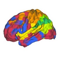
The onset of cognitive decline begins at 45

Abundant evidence has clearly established an inverse association between age and cognitive performance, but the age at which cognitive decline begins is much debated. Until now, the general consensus was that the onset of decline did not begin until 60. A study published in the British Medical Journal, conducted by an Inserm research team, directed by Archana Singh-Manoux (Centre for Research in Epidemiology and Population Health), shows that our memory and capacity for reasoning and understanding start to decline at the age of 45. This research is part of the Whitehall II cohort study and focused on more that 7,000 people over a ten-year period.
Increased life expectancy implies fundamental changes in the composition of populations, with a significant rise in the number of elderly people. These changes are likely to have a massive influence on the life of individuals and on society in general. Abundant evidence has clearly established an inverse association between age and cognitive performance, but the age at which cognitive decline begins is much debated. Recent studies concluded that there was little evidence of cognitive decline before the age of 60.
However, clinical studies demonstrate a correlation between the presence of amyloid plaques in the brain and the severity of cognitive decline. It would seem that these amyloid plaques are found in the brains of young adults.
Few assessments of the effect of age on cognitive decline use data that spans over several years. This was the specific objective of the study led by researchers from Inserm and the University College London.
As part of the Whitehall II cohort study, medical data was extracted for 5,198 men and 2,192 women, aged between 45 and 70 at the beginning of the study, monitored over a 10-year period. The cognitive functions of the participants were evaluated three times over this time. Individual tests were used to assess memory, vocabulary, reasoning and verbal fluency.
The results show that cognitive performance (apart from the vocabulary tests) declines with age and more rapidly so as the individual’s age increases. The decline is significant in each age group.
For example, during the period studied, reasoning scores decreased by 3.6 % for men aged between 45 and 49, and 9.6 % for those aged between 65 and 70. The corresponding figures for women stood at 3.6% and 7.4% respectively.
The authors underline that evidence pointing to cognitive decline before the age of 60 has significant consequences.
“Determining the age at which cognitive decline begins is important since behavioural or pharmacological interventions designed to change cognitive aging trajectories are likely to be more effective if they are applied from the onset of decline.” underlines Archana Singh-Manoux.
“As life expectancy continues to increase, understanding the correlation between cognitive decline and age is one of the challenges of the 21st Century” she adds.
Our endocrine system has a good memory!

On hearing the word ‘memory’, the brain normally springs to mind… or perhaps our immune system, which memorizes data to react more effectively should bacteria infect us a second time. But who would ever have imagined that our endocrine glands also ‘remember’ certain data? A group of Inserm and CNRS researchers, supervised by Patrice Mollard at the Institute of Functional Genomics (1) (Montpellier), have recently demonstrated that, in mice, hypophyseal endocrine cells used to control lactation are organised into a network (like those in the brain) during the first feeding phase. This network is then conserved, or “memorized”, for even greater functional efficiency during the feeding of a second litter. It is the first time that an experience-dependent ‘memory’ has been revealed in the endocrine system. The research is the subject of an article published in the Nature Communications review dated 3 January 2012.
The plasticity of biological systems makes it possible for organisms to dynamically modify their physiology to adapt to existing environmental conditions. In terms of cells, this process is usually associated with the immune system; with regard to tissue, this process was characterized in the brain many years ago and is at the heart of intense neurobiological research.
Besides these two systems used to memorize data over the long-term, there was no evidence suggesting other cells could operate in a similar manner.
The pituitary gland represents an ideal model to check this hypothesis since it has distinct populations of endocrine cells, organised into networks, which secrete different hormones to control a multitude of physiological functions.
Patrice Mollard’s team in Montpellier worked in conjunction with Paul Le Tissier’s team in London (NIMR-MRC) (2) to determine whether the endocrine cell networks had any memorization capacities. The cells that secrete prolactin (lactation hormone) were used as a model. Prolactin secretion commands a range of responses that are crucial for the feeding of baby mice, including milk production.
The production of prolactin (and thus maternal milk) in mice is stimulated by lifting an inhibiting (dopaminergic) signal in the brain and by the suckling phenomenon.
Using bi-photon calcium imaging, the researchers were able to distinguish interaction between the prolactin-producing cells, before, during and after a feeding period.
Before suckling, these cells were weakly interconnected.
During suckling, the cells responded to lactation by increasing coordinated intercellular communication, functional connectivity and tissue production.
The originality of this discovery lies in the fact that three months after weaning, the network is still in place, as if it has been memorized. “In the future”, explains Patrice Mollard, the same stimulus (suckling) will trigger a more coordinated and effective response. The network will secrete more prolactin and will again cause an increase in tissue production”.

However, the network is not created if the suckling stimulus power is reduced. Mice often have large litters (an average of eight babies per litter), if only three babies are positioned for suckling, the stimulus is too low to create the network.
It is the first time that researchers have highlighted an experience-dependent memory in a system of this kind. “It opens open a vast range of possibilities. We think that this discovery could also be applied to other endocrine systems, such as pancreatic beta cells and endocrine cells in the gastrointestinal tract”, conclude the authors.
Notes
(1) (CNRS / Inserm / Universités de Montpellier 1 & 2)
(2) MRC National Institute for Medical Research