Menu

Magnetic stimulation to improve visual perception

Using transcranial magnetic stimulation (TMS), an international team led by researchers from the Centre de Recherche de l’Institut du Cerveau et de la Moelle Epinière (CNRS / Inserm / UPMC) has succeeded in enhancing the visual abilities of a group of healthy subjects. Following stimulation of an area of the brain’s right hemisphere involved in perceptual awareness and in orienting spatial attention, the subjects appeared more likely to perceive a target appearing on a screen. This work, published in the journal PLoS ONE, could lead to the development of novel rehabilitation techniques for certain visual disorders. In addition, it could help improve the performance of individuals whose tasks require very high precision.

TMS is a non-invasive technique that consists in sending a magnetic pulse into a given area of the brain. This results in an activation of the cortical neurons located within the range of the magnetic field, which modifies their activity in a painless and temporary manner. For several years, scientists have been looking at the possibility of using this technique to enhance certain brain functions in healthy subjects.

In this respect, the team led by Antoni Valero-Cabré has carried out research involving the stimulation of a region of the right cerebral hemisphere known as the frontal eye field. Strictly speaking, this is not a primary visual area but it participates in the planning of ocular movements and the orientation of each individual’s attention in the visual space. In a first experiment, a group of healthy subjects tried to distinguish a very low contrast target appearing on a screen for just 30 ms. In some of the tests, the subjects received a magnetic pulse lasting between 80 and 140 ms on this frontal region before the target appeared. The researchers found that the success rate was higher when using TMS. The visual sensitivity of healthy subjects was temporarily increased by around 12%. In a second experiment, the subjects were shown a fleeting visual cue indicating the spot where the target could appear. In this configuration, the enhancement of visual sensitivity, which remained of the same order, was only apparent when the cue indicated the correct location of the target.

Although cerebral functions such as conscious vision are highly optimized in healthy adults, these results show that there is a significant margin for improvement, which can be “enhanced” by TMS. This technique could be tested for the rehabilitation of patients suffering from cortical damage, due for example to a cardiovascular accident, and for that of patients with retinal disorders. The second experiment suggests that rehabilitation based on both TMS and visual cues could be more selective than the use of stimulation alone. The researchers want to further explore this possibility using repetitive TMS, which, in this case, could make it possible to obtain long-lasting modification of cerebral activity.

Furthermore, according to the researchers, TMS could be used in the near future to increase the attentional abilities of individuals performing tasks that require good visual skills.

These experiments benefited from funding from the European ERANET NEURON BEYONDVIS initiative, partly financed by ANR.

(French) La lecture : un vrai travail d’équipe dans le cerveau

Sorry, this press release is only available in French.

Aging of the brain: genetic modifications now identified

The hippocampus is a part of the brain that shrinks as we age, causing memory disorders. An acceleration of this phenomenon is one of the signs of Alzheimer’s disease. Joint international work involving French (1) research teams has shown that genetic mutations are linked to the reduction of the hippocampal volume. These results were obtained from epidemiological studies that analyzed the genomes and MRI scan images of 9232 participants aged from 56 to 84. In France, roughly 2000 MRI scans were performed as part of the study known as the “3-city-study” (2). The results of this work will be published on April 15th 2012 in the review Nature Genetics.

Shrinkage of the hippocampus occurs with age and is caused by the cumulative effect of various factors. Hippocampal atrophy is a recognized biological marker of Alzheimer’s disease, so it is vital that researchers determine the cause of this process.

An international study under the French leadership of Christophe Tzourio looked for genetic variabilities linked to the shrinkage of the hippocampus. To do this, the genomes and MRI scan data of over 9000 persons aged between 56 and 84 were analyzed with a view to detecting a potential link between certain mutations and the reduction in the hippocampal volume. The participants’ data (both subjects presenting dementia and healthy subjects) were extracted from eight cohort studies in Europe and in North America.

The researchers first identified 46 differences in the DNA sequences of the participants, thought to be related to a reduction in the volume of the hippocampus. Eighteen mutations located in different areas of chromosome 12 were found to be strongly linked to shrinkage of the hippocampus. The other links included a mutation on chromosome 2. Then a final mutation on chromosome 9 was also found to be involved in the hippocampal shrinkage in a third and younger sample. These results indicate that “as yet unidentified factors” trigger mutations in precise areas of the genome, causing the reduction of the hippocampal volume.

The hippocampus (shown in red) is a structure located deep down in the brain, that plays an essential role in the memory processes. Its volume decreases with age and this shrinkage occurs more rapidly in cases of Alzheimer’s disease. For this study, computerized analysis of the MRI scan images and calculation of the hippocampal volume had to be developed individually for each participant

© UMR5296 CNRS CEA Université de Bordeaux

The hippocampus (shown in red) is a structure located deep down in the brain, that plays an essential role in the memory processes. Its volume decreases with age and this shrinkage occurs more rapidly in cases of Alzheimer’s disease. For this study, computerized analysis of the MRI scan images and calculation of the hippocampal volume had to be developed individually for each participant.

Once the mutations had been identified, the researchers tried to find out what exactly they modified. They discovered that they changed the structure of genes that were important for numerous functions involved, among others, in cell death (HRK), embryonic development (WIF1), diabetes (DPP), or neuronal migration (ASTN2).

“This study marks a major turning point, since it confirms the fact that genetic factors are linked to a brain structure, the hippocampus, involved in dementia and more generally in the aging of the brain,” explains Christophe Tzourio. This new approach, that studies a targeted area of the brain rather than a disease, will help us to more precisely decipher the mechanisms of Alzheimer’s disease.

The next stages will aim at obtaining a better understanding of how these genetic mutations are actually involved in the overall functioning of Alzheimer’s disease. Although clinical applications are not for the imminent future, these discoveries are a step forward to a better understanding of this disease and of cerebral aging in general.

“This discovery confirms the importance of using sophisticated means such as MRI scanning and genome analysis for the cohort studies. And this is only possible if we have close collaboration between the different disciplines involved,” concludes Christophe Tzourio.

Footnotes

(1) In neuroepidemiology (Inserm U708 – University of Bordeaux, C Tzourio), brain scanning by the neurofunctional imaging group (CNRS/CEA/University of Bordeaux Segalen, B Mazoyer), and in genetics (UMR 744 Inserm University of Lille, P Amouyel).

(2) The 3C study also called the “Three-city-study” denoting the cities of Bordeaux, Dijon and Montpellier, is a large cohort study involving over 9000 persons aged 65, begun in 1999. https://www.three-city-study.com/

A new gene thought to be at cause in early-onset forms of Alzheimer’s disease

A new gene that causes early-onset of Alzheimer’s disease has been discovered by the research team of Dominique Campion at the Insert unit 1079 ”Genetics of cancer and neuropsychiatric diseases” in Rouen. The research scientists showed that in the families of 5 of 14 patients suffering from the disease, mutations were detected on the gene SORL1. This gene regulates the production of a peptide involved in Alzheimer’s disease. The results of this study have been published in the review Molecular Psychiatry issued April 3rd

Precise genetic mutations have been seen to play a part in early-onset forms of Alzheimer’s disease. However, there is a sub-population of patients in whom there is no mutation of these genes. So how can these patients, in whom there are no pre-established mutations, be suffering from early-onset Alzheimer’s?

To reply to this question, the research team working under the leadership of Dominique Campion and Didier Hannequin (Inserm unit 1079 and Centre national de référence malades Alzheimer jeunes, University hospital Rouen), studied the genes from 130 families suffering from early-onset forms of Alzheimer’s disease. These families were identified by 23 French hospital teams within the framework of the “Alzheimer Plan”. Of these families, 116 presented mutations on the already known genes. But in the 14 remaining families, there was no mutation at all observed on these genes.

A study of the genome of the 14 families using new whole DNA sequencing techniques showed evidence of mutations on a new SORL1 gene. The SORL1 gene is a coding gene for a protein involved in the production of the beta-amyloid peptide. This protein is known to affect the functioning of the brain cells (see insert).

Two of the identified mutations are responsible for an under-expression of SORL1, resulting in an increase in the production of the beta-amyloid peptide. “The mutations observed on SORL1 seem to contribute to the development of early-onset Alzheimer’s disease. However, we still need to identify more clearly the way in which these mutations are transmitted on the SORL1 gene within families” states Dominique Campion.

Alzheimer’s disease is one of the main causes of dependency among the elderly. It results from neuron degradation in different areas of the brain. Its symptoms include increased alterations to memory, cognitive functions and behaviour disorders that lead to a progressive loss of independence.

Alzheimer’s disease is characterized by the development of two types of lesion in the brain: amyloid plaques and neurofibrillary degenerescence. Amyloid plaques are caused by extracellular accumulation of a peptide, beta-amyloid peptide (Aβ) in specific areas of the brain. Neurofibrillary degenerescences are intraneuronal lesions caused by abnormal filamentary aggregation of a protein known as a Tau protein.

(French) Une stratégie de prévention pour éviter la mort subite chez les patients atteints de la maladie de Steinert

Sorry, this press release is only available in French.

How sleep reduces our awareness of the outside world

While we sleep, our self-awareness and our awareness of the outside world are reduced. Why? Habib Benali, Director of Research at the Inserm functional imaging laboratory (Unit 678 Inserm/UPMC) and his team have been investigating this phenomenon. The research scientists have shown that during nonrapid eye movement sleep, the brain’s activity is re-organized into networks that communicate less with each other than during wakefulness. The results of this study on brain activity have been published in the PNAS review.

Deep slow sleep is a sleep phase in which our self-awareness and our consciousness of the outside world are considerably reduced. Yet the neurons in our brain are still highly active during this phase. So consciousness is not just associated to brain activity, but rather to its ability to process information. This is the conclusion of the study carried out by the research scientists from the functional imaging laboratory (Inserm/Université Pierre et Marie Curie) of Pitié Salpêtrière, and the Cyclotron research center of Liège.

© H. Benali / Inserm

Graphical representation showing the flow of information inside the brain during

wakefulness (on the left) and during nonrapid eye movement sleep

By measuring the quantity of information exchanged between different areas of the brain, the research scientists were able to show that the flow of information in the brain during sleep was different from that observed during wakefulness.

During sleep, the activity of the brain seems to be organized into a multitude of smaller networks. Inside each network, data exchange is just as active, even more active than during wakefulness, but communication between these networks decreases“, explains Habib Benali, Director of Research at Inserm.

The figure shows that the brain (B) contains different networks (colored circles) that group together several brain areas (small peripheral black dots). The longer the black lines connecting these structures (colored circles and black dots), the more able they are to process information independently. During sleep (situation on the right), the distance between these structures increases, meaning that the information in these small clusters of brain areas is processed on a more local scale.

The research team believes that this is due to a hierarchical reorganization of the flow of information during NREM sleep, since this has been observed repeatedly at several levels in the brain. “We suggest that the modification of the information flows between areas of the brain reduces the brain’s ability to generate a unified representation of the person and the outside World” states Habib Benali.

A step closer to morphine without side effects?

Morphine has been used for centuries to relieve severe pain. However, its pain-relief properties are coupled with considerable side effects. Morphine imitates the action of molecules produced naturally by the brain (endorphins). So why does it have such harmful side effects? This question has recently been elucidated by Sébastien Granier, researcher at the Institute of Functional Genomics (Inserm/CNRS/Universités de Montpellier 1 and 2) and his team, in conjunction with collaborators in the US. The 3D structure of brain receptors is likely to differ, depending on the type of molecule bound upon it (morphine or endorphin): the body’s response will be completely and accordingly modified. Thanks to this discovery, the researchers have now set their sights on retaining the benefits of morphine while eliminating any undesired side effects.

The results were published in the Nature review on 21 March 12.

Opium is a natural poppy (papaver somniferum) extract and is one of the oldest drugs known to man, used for its psychotropic, sedative and analgesic properties. These effects are caused by its major component: morphine. Today morphine has widespread clinical pain-relief applications.

Morphine’s action is relayed by μ-opioid receptors expressed at the surface of cells in the central nervous system. These receptors are part of a superfamily of proteins, G protein-coupled receptors (GPCR), which are the target of around 30% of drugs currently on the market.

At a molecular level, morphine binds with μ-opioid receptors to imitate the action of molecules produced naturally in the brain: endorphins. However, its clinical use is limited by two effects. Firstly, the development of a tolerance phenomenon means that, in the case of repeated injections, the morphine dose must be increased to obtain the same therapeutic effect. Secondly, morphine consumption can lead to drug dependency (heroin, the acetylated form of morphine, is the most obvious example). Furthermore, morphine consumption has serious side effects: respiratory depression, constipation, physical and psychic dependency. These harmful effects can be explained by the fact that morphine triggers a cellular response that differs from that triggered by endorphins. Since morphine and endorphins bind with the same receptor, the two molecules stabilize the μ-opioid receptors in separate spatial conformations that cause the differences in biological responses.

It is therefore essential to understand the structural bases of morphine action, and opioids in general, if we are to develop molecules that retain the beneficial effects of morphine whilst eliminating the side effects,” explains Sébastien Granier, Inserm researcher and project leader.

The research produced by Sébastien Granier (and Brian Kobilka’s team in Stanford) resolved the 3D structure of μ-opioid receptors when associated with a molecule that has a similar chemical structure to morphine.


© Kobilka Lab
3D representation of μ-opioid receptors when associated with morphine antagonistic.

© Kobilka Lab

3D representation of a μ-opioid receptor dimer.

The images of the 3D structure above show that the specific area in the receptor where the opioid molecule binds is mostly open to the external medium, thus explaining the rapid action of these components.

The structure also reveals a very important characteristic of this receptor function: the formation of a receptor dimer (1). “This is the very first time that we have succeed in producing images of the 3D structure of a complex of this kind for this receptor family” he adds. This structure with a μ-opioid receptor dimer opens up avenues of research to study this phenomenon and improve understanding of these functional implications.

The 3D structure resolution of the μ-opioid receptor holds the key to treating pain and addictions, and could, in the long-run, lead to the production of new analgesic medicine with no side effects.

Footnote

(1) Molecule produced by ‘associating’ two identical molecules

How does cannabis affect working memory?

A deterioration of working memory is observed in people who consume drugs containing cannabinoid compounds found in cannabis leaves and buds. A team led by Giovanni Marsicano, Supervisor of Inserm Research Unit 862 (Magendie Neurocentre, University of Bordeaux), in collaboration with a team led by Xia Zhang (University of Ottawa, Canada), has recently identified the mechanism by which these substances affect working memory. These researchers have demonstrated for the first time that the adverse effect of cannabinoids on working memory is exerted via receptors located in the glial cells (brain cells present in large numbers and scarcely studied). This effect is associated with a decrease in neural connections in the hippocampus, the region of the brain that coordinates working memory processes.

These results were published in Cell on 2 March 2012.

Working memory is used perform common cognitive operations (thinking, reading, writing, calculating, etc.) on information stored temporarily (for periods ranging from a few seconds to a few minutes). This allows for the integration audio, visual and spatial information. One of the major effects of intoxication with cannabinoids is the alteration of working memory, as observed in both humans and animals. Cannabis disturbs this function, thus preventing the consumer from performing common daily tasks. Cannabinoid receptors are expressed in the glial cells of the hippocampus, a cerebral structure essential for memory modulation. The cellular mechanisms responsible for the adverse effects of cannabis on this memorization process were previously unknown.

Giovanni Marsicano and his collaborators at the Magendie Neurocentre (Inserm Research Unit 862, University of Bordeaux 2) have successfully identified a mechanism by which cannabis causes adverse effects on working memory. The researchers have demonstrated that cannabinoids, when connected to their receptors, can decrease the strength of neural connections in the hippocampus.

Cannabinoids are a group of approximately 60 compounds present in cannabis leaves and buds. They act on the brain via cannabinoid receptors. In this study, the researchers have focused on cannabinoid receptor CB1, present in large quantities in nerve terminals within the brain (see diagram below). Cannabinoid receptor CB1 is present both in neural membranes (shown in yellow) and in the membranes of astroglial cells (shown in pink) located in the hippocampus (en orange) and used to provide support for neurons.

The connection of cannabinoids (shown in green) to CB1 receptors (shown in pink) activates the transmission of glutamate signals (shown in light blue) to the glutamate receptors (shown in dark blue) of nerve terminals used to transfer information between neurons. This mechanism modulates the strength of connections between hippocampal neurons (signal depression), thereby disturbing working memory.

In order to identify the action mechanisms of cannabinoids, the researchers have evaluated spatial working memory in the presence of THC, the best known cannabinoid (shown in green). Two groups of mice were studied in which CB1 receptors had been suppressed (in astroglial and neural cells, respectively).

When CB1 receptors are suppressed only in neural cells, the presence of THC causes spatial working memory deficits. On the contrary, when CB1 receptors are suppressed only in astroglial cells, spatial working memory performance is preserved. Therefore, the CB1 receptors located in astroglial cells are responsible for the adverse effects of THC on spatial working memory.

“These in vitro and in vivo results surprisingly demonstrate the importance of the activation of the CB1 receptors of astroglial cells, and not those of neural cells, in the mediation of the effects of cannabinoids on working memory”, explains Giovanni Marsicano.

Over the past years, a large number of studies have demonstrated the interest of cannabis for the treatment of various diseases. “The description of cannabinoid-specific action mechanisms in the hippocampus should enable optimization of the therapeutic potential of cannabinoids, which is currently limited by significant adverse effects associated with their consumption”.

Post-traumatic stress disorder (PTSD) defined by an inversion of brain activity induced by stress hormones

We remember, right down to the tiniest detail, a car accident that happened over two years ago, when we feared for our lives; and yet, who among us can still remember a very good meal enjoyed just last year, even if it was really very, very good? Today, there is ever-increasing understanding of the biological bases behind this “adaptive” capacity to remember a stressful. However, until now, we knew very little about the state of post-traumatic stress disorder (PTSD). This pathological state is triggered in some individuals after exposure to a highly stressful event. In this state, patients are gripped by fear even when faced with elements that have no objective danger. Is PTSD specific to the human race, influenced by its history and culture? Or is it a state that is found in various species, resulting in common biological changes? These are the questions that Pier-Vincenzo Piazza, Director of the Neurocentre Magendie in Bordeaux (Inserm/ Université Victor Segalen), and his collaborators, have attempted to answer. Details of their results are provided in the Science review (advance publication on-line on Science Express website on 23 February).

It is easier to memorize a stressful event than an agreeable one. Almost all species capable of behaviour are able to recall negative events, proof that this is likely to be a capacity selected during evolution: it ensures survival in a hostile environment.

However, for some individuals, exposure to very stressful events may trigger a pathological state: post-traumatic stress disorder (PTSD) being the most emblematic. In the United States, estimates suggest that this syndrome afflicts 6.8% of the general population and that 30% of veterans from the Vietnam war and 12% of the Gulf war suffer from it (source National Centre for PTSD).

In this state of stress, the memory of the patient is impaired: it is no longer capable of adapting its reaction of fear to the “right” context and to the “right” predictive elements. Sufferers experience fear in situations where there are no threats. These fears become all-consuming, eventually preventing a normal life. “If you are attacked by a lion in the savannah as a flock of birds flew overhead, it would be normal to feel fear if you were to make a return trip to the savannah” explains Pier-Vincenzo Piazza. “However, you should not be gripped by fear if, when strolling around another natural open space, for example a golf green, you hear a bird cry or catch sight of birds on the horizon,” thethe Inserm research director goes on. If this is the case, you may have developed post-traumatic stress disorder as a consequence to the lion attack.

The teams, led by Pier-Vincenzo Piazza and Aline Desmedt, have demonstrated that such memory impairment associated with PTSD is not specific to humans and is also found in mice. To this end, the researchers conditioned mice to a) anticipate a threat (an electric shock of varying intensity) by using the same specific context (an indicating environment) and b) to distinguish this specific context from stimuli, which although present during the conditioning process, did not predict the threat (a sound).

In normal conditions, the mice showed a reaction of fear when exposed to the specific context (the indicator environment) of the threat, but did not react to the sound (not predictive of threat).

Following this conditioning session, the researchers then administered increasing concentrations of glucocorticoid hormones, the main biological response to stress in mammals. When administration of the glucocorticoids followed an intense threat, as is the case with individuals suffering from post-traumatic stress, the mice were no longer able to restrict their fear reaction to the “right” context and to the correct indicators pronouncing a possible threat. The animals began to show fear: they froze in response to indicators, which although present during the stressful situation, were not predictive of the threat in any way. These results show that PTSD is probably due to a simultaneous overproduction of glucocorticoids in some subjects as the traumatic event occurs.

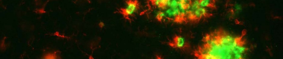
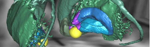
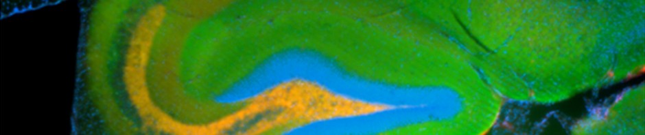
Memory impairment induced by glucocorticoids is accompanied by activity reorganisation in the brain, and, more specifically, the hippocampus-amygdale circuit, which is essential for coding memories associated with fear. In normal conditions, when an individual associates a threat with a context, strong activity is observed in the hippocampus, the structure in the brain required for all knowledge acquisition associating a specific context, area, etc. with an event. However, activity in the amygdale is low. The amygdale is an area of the brain that is also involved in emotional memory, but it memorizes specific indicators, such as sounds, which predict the threat.

When the mice were subject to an increase in glucocorticoids and memory impairment characterising PTSD was observed, activity in the hippocampus was reduced, whereas activity in the amygdale increased. In states of post traumatic stress, the researchers noted an inversion of normal activity in the brain. Normal activity in the amygdale may explain the fact that the subject begins to “over-respond” to presumed indicators, present during the traumatising event, but which are not, themselves, predictive of any danger. Low activity in the hippocampus may explain why subjects no longer recognize the right context: they are therefore incapable of containing their fear to appropriate situations.

PTSD is not only an overbearing memory of the traumatizing situation, but also a memory impairment that prevents patients from containing their reaction of fear to the context that predicts the threat”, explain the researchers. In the case of post-traumatic stress disorder, a vivid memory of the traumatising event is associated with amnesia in terms of the surrounding context of the event. Some contextual elements present during the traumatising event are wrongly considered to be predictive of the event.

To conclude, the authors explain that PTSD-related memory problems seem to be caused by a biological response to abnormal stress suffered by some patients: for these patients, excessive glucocorticoid production at the same time as exposure to intense stress triggers an inversion of normal activity in brain structures that code fear-related memories.

“We have demonstrated that PTSD can occur in species other than humans and that there are shared biological origins. The mice model of this pathology now paves the way for better understanding of the molecular bases of this pathology, which could lead to the development of a treatment,” conclude the Inserm researchers.

Is there a general motivation centre in the depths of the brain?

A team coordinated by Mathias Pessiglione, Inserm researcher at the “Centre de recherche en neurosciences de la Pitié Salpêtrière” (Inserm/UPMC-Université Pierre and Marie Curie/CNRS) have identified the part of the brain driving motivation during actions that combine physical and mental effort: the ventral striatum. The results of their study were published in PLoS Biology on 21 February 2012.

The results of an activity (physical or mental) partly depend on the efforts devoted to it, which may be incentive-motivated. For example, a sportsperson is likely to train with “increased intensity” if the result will bring social prestige or financial gain. The same can be said for students who study for their exams with the objective of succeeding in their professional career. What happens when physical and mental efforts are required to reach an objective?

Mathias Pessiglione and his team from Inserm unit 975 “Centre de recherche en neurosciences de la Pitié-Salpêtrière” examined whether mental and physical efforts are driven by a motivation ‘centre’ or whether they are conducted by different parts of the brain. The researchers studied the neural mechanisms resulting from activities that combine both action and cognition.

To this end, a series of 360 tests, combining mental and physical effort, were performed whilst being monitored by a scanner. The 20 voluntary participants were placed in the supine position, with their heads in a functional MRI scanner. They then had to complete a series of tasks through which they could accumulate winnings. However, in each series the winnings were limited to the first incorrect response. The tasks combined cognitive and motor actions. The participants had to find the highest number from among different-sized numbers and then select it by squeezing a handle located by their left or right hand (depending on the number’s location). At the end of the test, a winnings summary was displayed to motivate the participant.

© M. Pessiglione, Inserm

3D representation of motivational system (striatum ventral) activated during physical or mental effort.

Using images obtained from the MRI scans taken during the test, Mathias Pessiglione and his team identified a general motivational system in the depths of the brain, i.e. a structure capable of activating any effort type, both mental (concentrating on the task in hand) or physical (lifting a load). The researchers observed that the ventral striatum was activated in proportion to the amount of money involved: the higher the degree of motivation, the higher the activation level. Furthermore, the ventral striatum is connected to the median part of the striatum (the caudate nucleus) when the task to be performed is cognitively difficult (when the physical size and the numerical value of the numbers did not correspond). This ventral region solicits the lateral part of the striatum (the putamen) when the difficulty is motor-related (when the handle had to be squeezed very tightly).

The researchers suggest that the expectation of a reward is encoded in the ventral striatum, which can then drive either the motor or cognitive part of the striatum, depending on the task, in order to boost performance. “The ventral striatum may commute connections in accordance with the request, i.e. enhance the neuronal activity in the caudate nucleus for a cognitive operation and in the putamen for a physical action” explains Mathias Pessiglione.

When your left hand mimics what your right hand does: it’s in the genes

Research scientists from Inserm, CNRS, UPMC and AP-HP working for the Centre de Recherche de l’Institut du Cerveau et de la Moelle (CRICM) of la Pitié-Salpêtrière, have just discovered mutations that could be the cause of congenital mirror movement disorders. Persons suffering from this disorder are unable to perform different movements with different hands. Genome sequencing in several members of a French family showed the culprit to be the RAD51 gene. Further work carried out on mice suggests that this gene plays a part in motor network cross-over. Cross-over is a key factor in the transmission of brain signals, because it allows the right side of the brain to control the left side of the body and vice versa. This research has been published in The American Journal of Human Genetics.

Congenital mirror movement disorders by Emmanuel Flamand-Roze

Congenital mirror movement is a rare disease transmitted from one generation to another by dominant inheritance. The affected persons lose the ability to carry out different movements with separate hands: when one hand moves in a certain way, the other hand is “forced” to copy the same movement, even if the person does not wish to do so. So people suffering from this disease are totally incapable of bimanual motor activities, such as piano playing for example. This phenomenon has been observed in children, but generally cleared up spontaneously before the age of 10, no doubt due to maturing of the motoneuron networks. However, in people who are affected by the disorder, the illness starts in early childhood and remains unchanged throughout life.

In 2010, research scientists from Quebec analyzed the genome from the members of a large Canadian family and discovered a gene responsible for the disease. Mutations had been detected in the DCC (Deleted in Colorectal Carcinoma) gene. Following this discovery, the team of researchers and doctors coordinated by Emmanuel Flamand-Roze began to search for mutations in this gene in several members of a French family who were also suffering from congenital mirror movements disease, but without success. “The DCC gene was intact”, explained Emmanuel Flamand-Roze. “We thought we were nearly there and instead we had to start searching for mutation in a different gene”, he adds.

Using an approach that combines conventional genetic analysis and “whole exome” analysis (a new-generation genetic analysis technique that involves entirely sequencing the important part of the genome), scientists demonstrated that the RAD51 gene was responsible for congenital mirror movement disease in a large French family and went on to corroborate this result using the same techniques on a German family suffering from the same disorder.

“The RAD51 gene was already known to the scientific community as a potential catalyst for certain types of cancer and in problems of resistance to chemotherapy”, explains Emmanuel Flamand-Roze. “So we wondered whether it had yet another function that could explain the motor symptoms of CMM disease”.

In humans, the motor system is a cross-control system, which means that the left side of the brain controls the motor functions of the right side of the body and vice versa, with the cross-over taking place at the brainstem. While studying the expression of the RAD51 protein during development of the motor system in mice, the research scientists discovered that this gene could be implanted into the cross-over of the motor network that links the brain to the spinal fluid at the brainstem.

This discovery opens up a whole new field of investigation into the development of the motor system and to achieving better understanding of the cerebral mechanisms that control bimanual motricity. It could also shed light on other motricity disorders related to fine movement organization, such as dystonia or certain genetic neurodevelopmental diseases.

fermer