Menu

Huntington’s Disease: Brain Abnormalities Detectable in Embryos

Section of the human brain (cortex). The progenitor cells in magenta are less engaged in neuronal differentiation than those in green © Monia Barnat/Grenoble Institut des Neurosciences/Inserm, Université Grenoble Alpes

Huntington’s disease is a genetic neurological condition that usually manifests in adulthood. Teams of researchers and clinicians from Inserm, Université Grenoble Alpes, Sorbonne Université, CNRS and AP-HP at the Grenoble Institute of Neuroscience and the Brain Institute, have discovered abnormalities in the brains of human embryos that carry the mutation responsible for Huntington’s disease. This research, to be published in Science, looks at the mechanisms of the silent progression of the disease and when and how to treat patients in the future.

Huntington’s is a rare and hereditary genetic disease of the central nervous system, which usually manifests between 30 and 50 years of age in the form of gradually-worsening psychiatric, cognitive and motor disorders. It is caused by a mutation of the gene coding for the huntingtin protein and is transmitted in an “autosomal dominant” manner: inheriting just one copy of the defective gene is enough to develop the condition. Around 18,000 people in France are concerned, of whom 6,000 are already symptomatic and around 12,000 possess the gene carrying the mutation but are asymptomatic.

The teams of Sandrine Humbert, Inserm research director at the Grenoble Institute of Neuroscience (Inserm/Université Grenoble Alpes), and Alexandra Durr, university professor-hospital practitioner at Sorbonne Université, Pitié Salpêtrière hospital – AP-HP, and the Brain Institute (Inserm/Sorbonne Université/CNRS/AP-HP), are interested in the early stages of Huntington’s disease and the long period that precedes the onset of symptoms. In new research published in Science, they looked at when brain abnormalities could develop.

The teams studied the brains of 13-week-old human embryos donated by parents following medical termination of pregnancy. They observed several differences between the embryos that carry the mutation of the gene coding for huntingtin and those that do not.

In the carriers, the pathological huntingtin protein is abnormally localized in the progenitor cells that generate the neurons of the cortex. This mislocalization is linked, among other things, to problems of junction protein localization in these cells and to alterations in the size, density and orientation of the

cilium, an organelle essential to the functioning of these cells. These abnormalities disrupt the “division-differentiation” balance of the progenitor cells, which are derived from a reservoir of dividing cells, some of which differentiate into neurons while the others continue to divide in order to provide new progenitor cells. In the embryos that carry the mutation, these progenitor cells start differentiating more quickly at the expense of the reservoir of dividing cells.

The researchers repeated the experiment with a mouse model of Huntington’s disease at an equivalent stage of embryonic development and found the same abnormalities. This meant they could validate this animal model in order to further explore the early mechanisms of the disease at other stages of embryonic development or after birth.

“This is the first time that abnormalities of brain development have been identified in this disease. Abnormalities which are also relatively large and extensive, although we are not yet able to determine their direct consequences,” clarify Humbert and Durr, who led this research.

But why do the carriers of the mutation show no symptoms until later down the line? “At this stage, our hypothesis is that very early on the brain implements compensatory mechanisms that allow normal functioning. It could be the same in people who carry mutations linked to other types of degeneration, such as Alzheimer’s disease or amyotrophic lateral sclerosis,” specify the researchers.

They will now continue to describe brain development in mouse models of Huntington’s disease, try to understand how these early defects contribute to the adult disease, and how their compensation could be regulated during the silent symptomless period. “This discovery also has important implications for how and when disease-modifying therapies should from now on be considered,” they conclude.

Coupe de cerveau humain (cortex)

Human brain section (cortex). On the left, the nuclei are marked in blue; on the right, the progenitor cells in magenta are less engaged in neuronal differentiation than those in green.

©Monia Barnat/Grenoble Institut des Neurosciences/Inserm, Université Grenoble Alpes

Star-Shaped Brain Cells Shed Light on the Link Between Cannabis Use and Sociability

In a culture of neurons, feeder cells and astrocytes (star-shaped cell in the center in red) cohabit. Marking of tubulin in green, actin in red, and nuclei in blue. Researchers have focused on these cells to understand the impact of cannabis use on sociability.©Inserm/Saoudi, Yasmina/Ballet, Sandrine

Cannabis use can lead to behavioral changes, including reduced social interactions in some individuals. To better understand the phenomenon, Inserm researcher Giovanni Marsicano and his team from NeuroCenter Magendie (Inserm/Université de Bordeaux), in collaboration with Juan Bolaños’ team from the University of Salamanca, have identified for the first time in mice the cerebral mechanisms underlying the relationship between cannabis and reduced sociability. Their findings have been published in Nature.

Regular exposure to cannabis may have a harmful impact on sociability. For some consumers, studies show that it may lead to withdrawal and reduced social interactions. However, the brain network and the mechanisms involved in this relationship were unclear until now.

In order to learn more about the subject, a group led by Inserm researcher Giovanni Marsicano at NeuroCenter Magendie (Inserm/Université de Bordeaux)[1] has joined forces with a Spanish team from the University of Salamanca led by Juan Bolaños[2].

More broadly, their work is aimed at improving our knowledge of how cannabinoid receptors (the brain receptors that interact with chemical compounds in cannabis) work.

In their study published in the journal Nature, the researchers show that after exposure to cannabis, behavioral changes related to sociability occur as a result of the activation of specific cannabinoid receptors, located in star-shaped cells of the central nervous system called astrocytes.

Cannabinoid receptors and mitochondria

These findings are the result of almost a decade of hard work. In 2012, Marsicano and his team had made a surprising discovery: cannabinoid receptors are not only present on the cell membrane, as previously believed. Some of these receptors are also located on the membrane of the mitochondria, the intracellular organelles whose role is to provide the cells with the energy they need.

This new study comes after the team has identified cannabinoid receptors located on the membrane of the mitochondria within astrocytes. Among other functions, these cells play a very important role in energy metabolism of the brain. They capture glucose from the blood and metabolize it into lactate, which acts as “food” for neurons. “Given the importance of astrocytes and energy use for brain function, we wanted to understand the role of these specific cannabinoid receptors and the consequences for the brain and behavior when exposed to cannabis,” explains Marsicano.

Researchers then exposed mice to the cannabinoid THC, the main psychoactive compound in cannabis. They observed that persistent activation of mitochondrial cannabinoid receptors located in astrocytes resulted in a cascade of molecular processes leading to dysfunction of glucose metabolism in astrocytes.

As a result, the ability of astrocytes to transform glucose into “food” for neurons was reduced. In the absence of the necessary energy intake, the functioning of neurons was compromised in the animals, with a harmful impact on behavior. In particular, social interactions were decreased for up to 24 hours after exposure to THC.

“Our study is the first to show that the decline in sociability sometimes associated with cannabis use is the result of altered glucose metabolism in the brain. It also opens up new avenues of research to find therapeutic solutions to alleviate some of the behavioral problems resulting from exposure to cannabis. In addition, it reveals the direct impact of astrocyte energy metabolism on behavior,” says Marsicano.

At a time when the debate over therapeutic cannabis is returning to the forefront, the researchers also believe that this type of work is needed to better understand how the body’s various cannabinoid receptors interact with the drug, and whether any of them are particularly associated with harmful effects. Such research would make it possible to ensure the optimal management of patients who might need this type of therapy.

[1] With Arnau Busquets-Garcia (now in Barcelona, Spain) and Etienne Hebert-Chatelain (now in Moncton, Canada)

[2] With Daniel Jimenez-Blasco

Publication of a multicentre observational study, in the journal JAMA Neurology, to identify risk factors for the severity of COVID-19 in patients with multiple sclerosis in France

Multiple sclerosis (MS), red cells are dying (cell death). Astrocytes exposed to glycotoxic factor. ©Inserm/RIEGER F.

A study, aimed at identifying the risk factors for the severity of COVID-19 in patients with multiple sclerosis in France, was carried out by teams from the neurology department of the Pitié-Salpêtrière hospital AP -HP, from the Brain Institute (Inserm / CNRS / Sorbonne University) at the Pitié-Salpêtrière hospital AP-HP, from Sorbonne University and the neurology service of the Strasbourg CHU. This work was carried out with the participation of all the centers and neurologists involved in the management of multiple sclerosis, thanks to the national networks FCRIN4MS, the French Observatory of MS (OFSEP) and the Société Francophone of the SEP (SFSEP).

The results of this study, which were published on June 26, 2020 in the journal JAMA Neurology, led to the publication of the first registry on multiple sclerosis and COVID-19. This register made it possible to provide concrete information in real time to guide the management of patients during the epidemic.

Multiple sclerosis (MS) is an inflammatory disease of the central nervous system, which mainly affects young adults, and which can be responsible for a neurological handicap. In recent years, several immunosuppressive or immunomodulatory treatments have enabled major advances in the care of patients.

From the start of the COVID-19 epidemic, neurologists and patients have had many questions regarding the risk of COVID in patients with multiple sclerosis, particularly in relation to their basic therapy. We therefore set up a multicentre observational study whose objective is to identify the risk factors for COVID-19 severity in patients with multiple sclerosis in France.

More than 350 patients were included in the registry, and the teams identified as main factor of COVID-19 severity neurological handicap (measured by the EDSS score), followed by age and obesity, these 2 factors have also been identified in the general population. Conversely, long-term MS treatments are not associated with a higher risk of severe COVID. The mortality rate was 3.5%, slightly higher than the expected rate in this population with an average age of 44 years.

This is the first registry published on multiple sclerosis and COVID-19. It provided concrete, real-time information to guide the management of patients during the epidemic.

Alzheimer’s Disease: Sweet Snacks May Not Go Down So Well in Those With a Genetic Predisposition

Alzheimer’s Disease: Sweet Snacks May Not Go Down So Well in Those With a Genetic Predisposition © Robert Anderson on Unsplash

While genetic predisposition is a major factor in the risk of developing age-related dementia, and Alzheimer’s disease in particular, environmental factors such as diet also have an important role to play. Thanks to the 12-year follow-up of nearly 2,800 French people over the age of 65, a research team from Unit 1061 Neuropsychiatry: Epidemiological and Clinical Research (Inserm/Université de Montpellier) has sought to understand the impact of meals high in sugars (simple sugars and refined carbohydrates) on the risk of developing dementia. Its research shows a link between the consumption of sugars during the afternoon snack and the risk of developing Alzheimer’s disease in people with genetic predispositions. These results published in Alzheimer’s and Dementia pave the way for a better understanding of the links between environmental and genetic risk factors and could lead to improved dementia prevention strategies.

According to WHO forecasts, more than 152 million people could be affected by age-related dementia by 2050. There is currently no treatment to cure or slow the progression of these conditions, which include Alzheimer’s disease. It is therefore essential to identify the factors favoring their development and on which it would be possible to take preventive action.

For several years now, nutrition has been recognized as an important factor in good brain aging. In particular, several animal studies have emphasized the role of a high intake of sugars – which include starch and added sugars (glucose and fructose syrups, sucrose) – in aggravating the clinical signs of Alzheimer’s disease and, in particular, in accelerating the appearance of amyloid deposits (senile plaques) characteristic of Alzheimer’s disease.

But while environment plays a role in the development of Alzheimer’s disease, the importance of genetic factors is not to be overlooked. This is particularly the case of the APOE gene, which comes in three forms (or alleles): E2, E3 and E4. People who carry the E4 allele of this gene are at increased risk of developing Alzheimer’s disease.

However, until now, no human studies had explored a potential link between genetic predisposition, the consumption of sugars, and the risk of dementia.

A team led by Inserm researcher Sylvaine Artero in Unit 1061 Neuropsychiatry: Epidemiological and Clinical Research (Inserm/Université de Montpellier) wished to highlight the links between the onset of dementia (in particular Alzheimer’s disease) in humans, genetic predispositions linked to the E4 allele, and the consumption of sugars. They analyzed 12 years of data from nearly 2,800 participants in the French Three-City Study, which has been following nearly 10,000 French people over 65 years of age since 1999. They studied the development of 350 cases of dementia related to eating habits and more specifically their contribution to glycemic load (the ability of a food to raise blood glucose levels according to the portion consumed) estimated over four meals: breakfast, lunch, afternoon snack, and dinner.

In participants without the at-risk genotype, the research team found no association between the onset of dementia and the consumption of sugars with the four daily meals.

However, in participants with the E4 allele of the APOE gene, the researchers observed an association between the consumption of sugars during the afternoon snack and the onset of dementia. For the APOE4 individuals who tended to eat an afternoon snack, the risk of developing Alzheimer’s disease was increased by 2 to 3 times for each additional serving equivalent to the glycemic load of 30 grams of baguette, regardless of daily energy intake, physical activity, presence of co-morbidities or adherence to a healthy Mediterranean-type diet. However, no such association was revealed for the other meals of the day.

But why would the consumption of sugars during the afternoon snack have a greater impact on people with genetic predispositions?

According to Artero, “one hypothesis to consider would be the potential impact of insulin resistance – a pathology involved in type 2 diabetes and favored by the consumption of sugars – on the risk of developing dementia”.

Indeed, it has already been shown in animals that carriers of the E4 allele had a less efficient glucose metabolism. They would therefore be more likely to develop insulin resistance. However, the sugary foods consumed during the afternoon snack tend to be low in fat and fiber. They are eaten more quickly and without being accompanied by other types of food, such as at main meals. As a result, they are absorbed into the bloodstream much faster during digestion, triggering a peak in insulin.

Repeated daily, these insulin peaks could eventually lead to peripheral insulin resistance but also to cerebral insulin resistance (in which the brain is less sensitive to insulin and less able to use glucose) via oxidative stress and inflammation, which would encourage the development of dementia, phenomena to which E4 allele carriers are more sensitive,” says Artero.

These results pave the way for new prevention strategies but need to be confirmed by other population-based studies and further investigated by experimental studies in order to better understand the links between sugar consumption, insulin resistance and the development of dementia,” concludes Artero.

Repetitive negative thinking linked to dementia risk

Persistently engaging in negative thinking patterns may raise the risk of Alzheimer’s disease, finds a new UCL-led study.

In the study of people aged over 55, published in Alzheimer’s & Dementia, researchers found ‘repetitive negative thinking’ (RNT) is linked to subsequent cognitive decline as well as the deposition of harmful brain proteins linked to Alzheimer’s.

The researchers say RNT should now be further investigated as a potential risk factor for dementia, and psychological tools, such as mindfulness or mediation, should be studied to see if these could reduce dementia risk.

Lead author Dr Natalie Marchant (UCL Psychiatry) said: “Depression and anxiety in mid-life and old age are already known to be risk factors for dementia. Here, we found that certain thinking patterns implicated in depression and anxiety could be an underlying reason why people with those disorders are more likely to develop dementia.

“We hope that our findings could be used to develop strategies to lower people’s risk of dementia by helping them to reduce their negative thinking patterns.”

For the Alzheimer’s Society-supported study, the research team from UCL, INSERM and McGill University studied 292 people over the age of 55 who were part of the PREVENT-AD cohort study, and a further 68 people from the IMAP+ cohort.

Over a period of two years, the study participants responded to questions about how they typically think about negative experiences, focusing on RNT patterns like rumination about the past and worry about the future. The participants also completed measures of depression and anxiety symptoms.

Their cognitive function was assessed, measuring memory, attention, spatial cognition, and language. Some (113) of the participants also underwent PET brain scans, measuring deposits of tau and amyloid, two proteins which cause the most common type of dementia, Alzheimer’s disease, when they build up in the brain.

The researchers found that people who exhibited higher RNT patterns experienced more cognitive decline over a four-year period, and declines in memory (which is among the earlier signs of Alzheimer’s disease), and they were more likely to have amyloid and tau deposits in their brain.

Depression and anxiety were associated with subsequent cognitive decline but not with either amyloid or tau deposition, suggesting that RNT could be the main reason why depression and anxiety contribute to Alzheimer’s disease risk.

“We propose that repetitive negative thinking may be a new risk factor for dementia as it could contribute to dementia in a unique way,” said Dr Marchant.

The researchers suggest that RNT may contribute to Alzheimer’s risk via its impact on indicators of stress such as high blood pressure, as other studies have found that physiological stress can contribute to amyloid and tau deposition.

Co-author Dr Gael Chételat (INSERM and Université de Caen-Normandie) commented: “Our thoughts can have a biological impact on our physical health, which might be positive or negative. Mental training practices such as meditation might help promoting positive- while down-regulating negative-associated mental schemes.

“Looking after your mental health is important, and it should be a major public health priority, as it’s not only important for people’s health and well-being in the short term, but it could also impact your eventual risk of dementia.”

The researchers hope to find out if reducing RNT, possibly through mindfulness training or targeted talk therapy, could in turn reduce the risk of dementia. Dr Marchant and Dr Chételat and other European researchers are currently working on a large project to see if interventions such as meditation may help reduce dementia risk by supporting mental health in old age.*

Dr Marchant concluded: “Taken alongside other studies, which link depression and anxiety with dementia risk, we expect that chronic negative thinking patterns over a long period of time could increase the risk of dementia. We do not think the evidence suggests that short-term setbacks would increase one’s risk of dementia.”


* silversantestudy.eu

Exploring the Brain to Clarify the Link Between Sleep Disorders and Alzheimer’s Disease

©Adobe Stock

Sleep disorders have a harmful impact on our brain and under certain conditions are thought to be linked to an increased risk of Alzheimer’s disease. A link which had until now been poorly understood by the scientific community. For the first time, a study based on multiple brain imaging techniques conducted by Inserm researcher Géraldine Rauchs at two laboratories hosted at GIP CYCERON – the Physiopathology and Imaging of Neurological Disorders laboratory (Inserm/ Université de Caen-Normandie) and the Neuropsychology and Imaging of Human Memory laboratory (Inserm/Université de Caen-Normandie/Ecole Pratique des Hautes Etudes – PSL/CHU Caen) – has revealed, among other forms of impairment, the appearance of amyloid plaques characteristic of Alzheimer’s disease in the brains of older adults with sleep apnea but no cognitive disorders. The results of this research were published in JAMA Neurology on March 23, 2020.

Obstructive sleep apnea syndrome is the most common sleep breathing disorder, affecting more than 30% of the population over the age of 65. It consists of uncontrolled and repeated pauses in breathing during sleep, linked to the temporary obstruction of the upper airways, in the throat, and is linked to numerous health problems – primarily cardiovascular diseases. However, given that the condition remains silent for a long time, it is probably underestimated in the general population.

In recent years, there has also been an accumulation of scientific data showing a link between sleep quality – and particularly the presence of sleep apnea – and the development of Alzheimer’s disease. Nevertheless, the biological mechanisms behind this link remained to be elucidated.

In order to do this, Inserm researcher Géraldine Rauchs conducted a study at the Physiopathology and Imaging of Neurological Disorders[1] laboratory (Inserm/ Université de Caen-Normandie) in collaboration with the Neuropsychology and Imaging of Human Memory laboratory (Inserm/Université de Caen-Normandie/Ecole Pratique des Hautes Etudes – PSL). This study[2], published in JAMA Neurology, used a variety of brain imaging techniques to map cerebral changes in people with untreated sleep apnea, on the structural, molecular and functional levels.

Changes in the brain

The researchers began by recruiting 127 participants over the age of 65, in good health and with no cognitive disorders. Using a portable home device to record their sleep and breathing overnight, the researchers detected the presence of varying degrees of sleep apnea in 75% of them.

In addition, the participants all underwent a battery of tests to evaluate their cognitive function, particularly executive functions and memory. They answered questionnaires about how they perceived their cognitive function and sleep quality. Several brain imaging examinations were then conducted in order to study their brain from every angle and detect any changes potentially linked to Alzheimer’s disease. Although no differences were observed among the participants in terms of cognitive performance, the imaging did reveal several notable changes in the brains of those with sleep apnea.

In these participants, there is a more marked accumulation of beta-amyloid protein in the brain. Characteristic of Alzheimer’s disease, this protein accumulates in the form of plaque which, depending on its density and distribution in the brain, may lead to the onset of clinical signs of the disease. Furthermore, the researchers observed increases in gray matter mass and glucose consumption, suggesting the presence of inflammatory processes in the brain.

“At a time when clinical trials of Alzheimer’s treatments are not yet successful, identifying risk factors and protection factors to target is of interest to a growing number of researchers. Thanks to the use of multiple brain imaging methods, this study has enabled us to clarify the mechanisms explaining the links between sleep quality, risk of cognitive decline and Alzheimer’s disease”, explains Rauchs.

While this does not mean that these people will necessarily develop the disease, they are at higher risk. What is more, effective solutions exist to treat sleep apnea. Detecting and treating sleep disorders, particularly sleep apnea, will therefore form part of the arsenal for promoting successful aging.

To continue this research, Rauchs and her team will now look at the impact of treating apnea on changes in brain lesions and analyze the differences between the brains of male and female sleep apnea patients.


[1] PhIND laboratory: https://www.phind.fr/index.php/en/

[2] This study was conducted as part of a vast European project, the Silver Santé Study (www.silversantestudy.fr), led by Dr. Gael Chételat.

Music or Speech? The Brain is Divided…

Photo by Thanos Pal on Unsplash

When it comes to recognizing a melody or understanding a spoken sentence, the human brain does not mobilize its hemispheres in an equivalent way. Although the concept is recognized by scientists, there had been no physiological or neural explanation for the phenomenon until now. A team co-led by Inserm researcher Benjamin Morillon at the Institute of Systems Neuroscience (Inserm/Aix-Marseille Université) in collaboration with researchers at Montreal Neurological Institute and Hospital of McGill University has been able to show that, due to different receptivities to the components of sound, the left auditory cortex neurons participate in the recognition of speech, whereas the right auditory cortex neurons participate in that of music. These findings, to be published in the journal Science, suggest that the respective specializations of the brain hemispheres for music and speech enable the nervous system to optimize the processing of sound signals for communication purposes.

Sound is produced from a complex set of air vibrations, which, when reaching the cochlea of the inner ear, are distinguished according to their speed. At any given moment, slow vibrations are being translated into deep sounds and rapid vibrations into high-pitched ones. As a result, sound can be represented according to two dimensions: spectral (frequency) and temporal (time).

These two auditory dimensions are fundamental because it is their simultaneous combination that stimulates the neurons of the auditory cortex. The latter are thought to discriminate sounds that are relevant for individuals, such as those used for communication enabling people to talk to and understand each other.

In humans, speech and music constitute the main uses of sound and the most complex on the cognitive level. The left hemisphere is primarily implicated in the recognition of speech, whereas the right hemisphere is primarily implicated in that of music. However, until now little was known about the physiological and neural reasons for this asymmetry.

A team co-led by Inserm researcher Benjamin Morillon at the Institute of Systems Neuroscience (Inserm/Aix-Marseille Université) in collaboration with researchers at Montreal Neurological Institute and Hospital of McGill University used an innovative approach to understand how speech and music are decoded within each of the human brain hemispheres.

The researchers recorded 10 sentences sung by a soprano to 10 new melodies composed especially for the experiment. These 100 recordings, in which melody and speech are dissociated, were then distorted by decreasing the amount of information present in each dimension of the sound. Forty-nine participants were asked to listen to pairs of these distorted recordings, and to determine whether they were identical in terms of speech content and melody. The experiment was conducted in French and English speakers to see whether the results were reproducible in different languages.

A demonstration of the audio test proposed to the participants is available here:

https://www.zlab.mcgill.ca/spectro temporal modulations/

The research team found that for both languages, when the temporal information was distorted, participants had trouble distinguishing the speech content, but not the melody. Conversely, when the spectral information was distorted, they had trouble distinguishing the melody, but not the speech.

Functional magnetic resonance imaging (fMRI) of the participants’ neural activity showed that in the left auditory cortex the activity varied according to the sentence presented but remained relatively stable from one melody to another, whereas in the right auditory cortex the activity varied according to the melody presented but remained relatively stable from one sentence to another.

What is more, they found that degradation of the temporal information affected only the neural activity in the left auditory cortex, whereas degradation of the spectral information only affected neural activity in the right auditory cortex. Finally, the participants’ performance in the recognition task could be predicted simply by observing the neural activity of these two areas.

Original a capella extract (bottom left) and its spectrogram (above, in blue) broken down according to the amount of spectral and temporal information (center). The right and left cerebral auditory cortexes (right) decode melody and speech respectively.

“These findings indicate that in each brain hemisphere, neural activity depends on the type of sound information, specifies Morillon. While temporal information is secondary for recognizing a melody, it is essential for the correct recognition of speech. Conversely, while spectral information is secondary for recognizing speech, it is essential for recognizing a melody. “

The neurons in the left auditory cortex are therefore considered to be primarily receptive to speech thanks to their superior temporal information processing capacity, whereas those in the right auditory cortex are considered to be receptive to music thanks to their superior spectral information processing capacity. “Hemispheric specialization could be the nervous system’s way of optimizing the respective processing of the two communication sound signals that are speech and music,” concludes Morillon.

This research was supported by a Banting fellowship to Philippe Albouy, grants from the Canadian Institutes of Health Research and the Canadian Institute for Advanced Research to Robert J. Zatorre, ANR-16-CONV-0002 (ILCB) and ANR-11-LABX-0036 (BLRI) funding and the Excellence Initiative of Aix-Marseille Université (A*MIDEX).”

PTSD: Resilience after trauma: the role of memory suppression

A study published in Science provides new insights into understanding post-traumatic stress disorder © Inserm

The terrorist attacks committed in Paris and Saint-Denis on November 13, 2015 have left lasting marks, not only on the survivors and their loved ones, but also on French society as a whole. A vast transdisciplinary research program, the 13-Novembre project is codirected by Francis Eustache, neuropsychologist and director of the Inserm Neuropsychology and Imaging of Human Memory laboratory (Inserm/Université de Caen Normandie/École pratique des hautes études/Caen university hospital/Cyceron imaging platform) and Denis Peschanski, historian and CNRS Research Director[1]. The program seeks to understand the ongoing construction and evolution of the individual and collective memory of these traumatic events and improve our understanding of the factors that protect people against the development of post-traumatic stress disorder (PTSD).

Part of this program is a brain imaging study called Remember, which is focused on the cerebral networks implicated in PTSD. The findings will be published in the journal Science on February 14, 2020. This study, which is sponsored by Inserm and led by Inserm researcher Pierre Gagnepain, shows that the untimely resurgence of intrusive images and thoughts in PTSD patients – a phenomenon long attributed to a deficiency of memory – is linked to a dysfunction of the brain networks that control memory. The researchers expect that these findings will lead to the identification of new treatment options for PTSD sufferers.

  1. Context: the 13-Novembre program

Unprecedented in their scale and violence, the terrorist attacks of November 13, 2015 sent shockwaves throughout French society. In their aftermath, the scientific community decided to act, to improve our understanding of the consequences of such a trauma and to improve the treatment options available to victims and their loved ones. A few days after the attacks, Alain Fuchs, then President of the CNRS, addressed the academic world and called upon researchers to come up with projects to tackle these challenges. He asked for all interested research teams to submit “proposals on any subjects capable of addressing the societal questions raised by the terrorist attacks and their consequences, and which open up avenues for new solutions – social, technical, and digital”.

The 13-Novembre transdisciplinary program was then launched by Inserm, CNRS and héSam Université[2].  One of its components “Étude 1000”, in which 1,000 volunteers are followed up over 12 years. Volunteers include people who were directly exposed to the violence – survivors and people close to the victims, members of the emergency response teams, residents of the neighborhoods targeted and of the Paris suburbs, as well as inhabitants of other French cities, in order to understand the construction and evolution of the memory of the attacks (see box).

The Remember project: understanding PTSD

One of the components of 13-Novembre, the Inserm-sponsored project Remember makes it possible to go much further in the understanding of human memory. With this brain imaging study conducted in Caen on a subgroup of 175 participants, the researchers are exploring the effects of a traumatic event on brain structure and function, identifying neurobiological markers of both PTSD and resilience to trauma. They hope that one day this research will open up new avenues for treatment, to complement those already available.

As such, Remember is tackling a major question that has intrigued neuroscientists for years: why does PTSD affect some trauma victims but not others? One of the objectives of this study, published in the journal Science, is to determine whether there is a link between the control mechanisms of our memory and the individual capacity for resilience.

“We focused this research program on the protective factors and brain markers associated with resilience to trauma. This is what makes our work original compared with previous studies that focused more generally on the impact of trauma on memory and its dysfunction”, emphasizes Pierre Gagnepain, scientific leader of Remember.

Étude 1000″ 

During four campaigns of filmed interviews initiated with the support of France’s Audiovisual Institute (INA) and Defense audiovisual communication and production unit (ECPAD), which are being conducted over a 10-year period (2016, 2018, 2021, and 2026), the participants are asked to share their accounts and evoke their personal memories of the attacks on the basis of an identical interview guide.

These individual accounts will then be analyzed in detail and put into perspective with the collective memory as it is constructed over the years, particularly within the media spaces (television and radio news, press articles, social networks, images of commemorations, etc.).

An approach inspired by that of William Hirst, Professor of Psychology at The New School (New York, USA), following September 11. Along with his teams, he collected some 3,000 written questionnaires of people affected by these attacks.  

13-Novembre takes this methodology further by using video recordings, by seeking to follow the same people over the 10-year period, and by adopting a transdisciplinary approach. Seen like this, it is a world first. “With 13-Novembre, the idea was to take things even further thanks to a very rich collaboration among multiple disciplines and the implementation of the biomedical study Remember. A study that gives us the unique scientific opportunity to observe the process of individual memory construction and how it interacts with collective memory”, emphasize Francis Eustache and Denis Peschanski, codirectors of 13-Novembre.


  1. PTSD

PTSD can develop in people who have been confronted to shocking, dangerous or terrifying events. First identified and studied by scientists in servicemen and women returning from the front, it can affect anyone – children and adults alike. PTSD can develop following any type of trauma, such as a natural catastrophe, sudden death of a loved one or terrorist attack, such as those of November 13, 2015. Studies performed in the USA and Canada estimate a 6 to 9% prevalence of the disorder in the general population.

Intrusive memories a core component

PTSD is a complex condition characterized by symptoms that can vary from one person to another. It can occur just after the trauma or years later. It can remain silent for relatively long periods of time. One of the most characteristic symptoms is the frequent intrusion of memories of the images, smells and sensations linked to the trauma.

These intrusions, which severely impact everyday life, cause great distress as well as other intense emotions, such as fear, guilt, and anger. These can be accompanied by physical symptoms triggered by the recollection of the event, such as muscle tension or increased heart rate.

In order to minimize the distress caused by intrusive memories, sufferers of PTSD also have a tendency to develop avoidance behaviors in the face of any circumstances that could evoke memories of the trauma. These can involve refusing to think or talk about the event, or gradually withdrawing from society and even their loved ones.


  1. Understanding more about the cause of intrusive memories

According to the traditional models of PTSD, the persistence of painful intrusive memories is caused by memory dysfunction – a bit like a scratched record playing the same fragments of our memories over and over again. From the anatomical point of view, such dysfunctions are particularly visible in the hippocampus – a key region for the formation of memory.

In addition, patients’ attempts to suppress their traumatic memories have long been considered an ineffective mechanism. Instead of confronting these painful images in order to leave them in the past, the way they were trying to repress or drive them out was seen more as a negative strategy, intensifying the intrusions and worsening the situation of those with PTSD.

The brain imaging study published in the journal Science challenges some of these ideas, hypothesizing that the untimely resurgence of intrusive images and thoughts could also be linked to a dysfunction of the brain networks implicated in controlling memory (going back to the previous metaphor of the record player, the turntable arm is not working correctly). “These control mechanisms act like a regulator of our memory and are engaged in halting or suppressing the activity of the regions associated with memories, such as the hippocampus”, states Gagnepain.

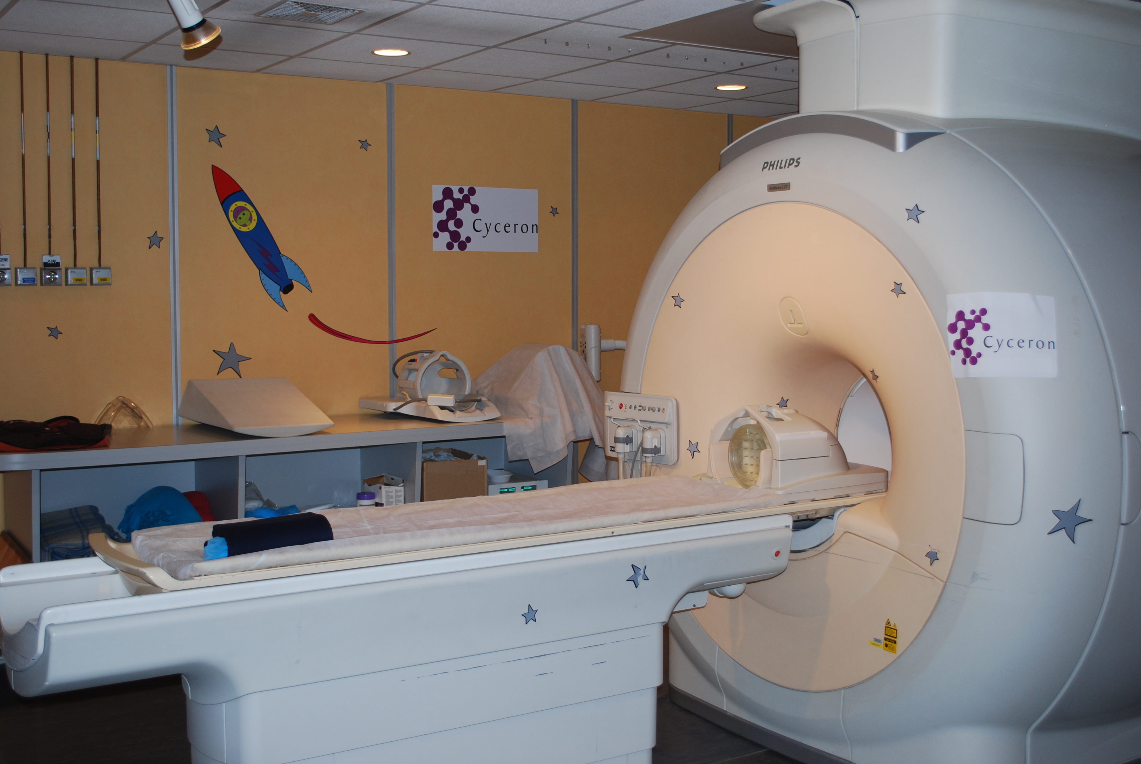
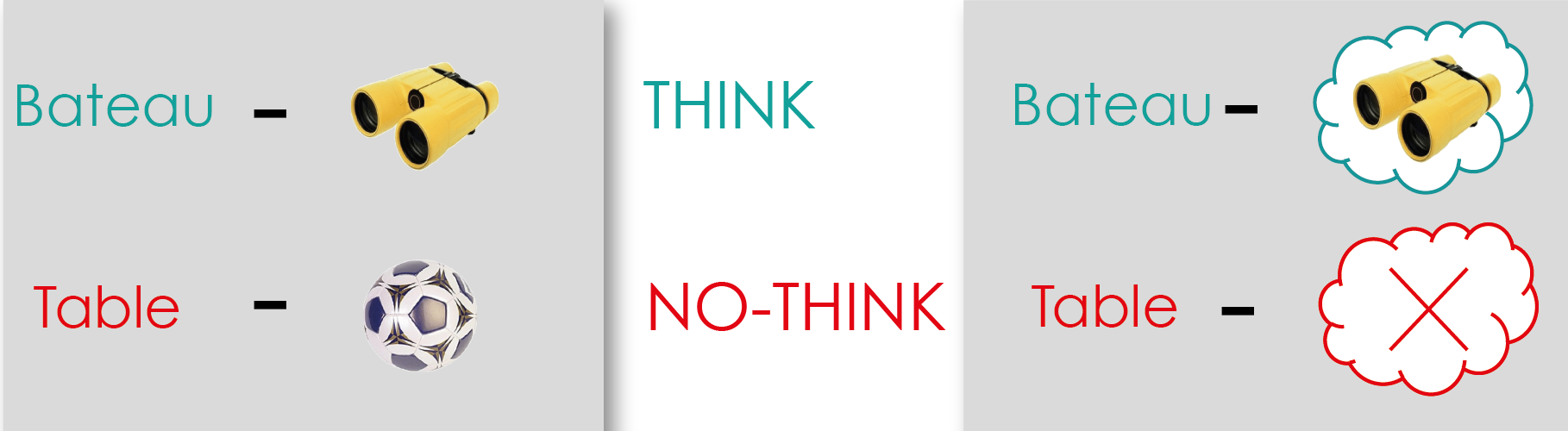
The participants had to perform the Think/No-Think task in the MRI machine.


Methods and results

Together with his colleagues, Gagnepain worked with 102 survivors of the Paris attacks, 55 of whom had PTSD. Also involved in the study were 73 people who had not been exposed to the attacks.

In order to model the resurgence of intrusive memories observed in the PTSD of these volunteers without putting them through the ordeal of viewing the shocking images of the attacks, the scientists opted for a brain imaging research protocol based on the Think/No-Think method (see box).

The aim of this method is to create associations between a cue word and an unrelated everyday object (for example, the word chair with the image of a ball), in order to reproduce the presence of an intrusion when confronted with the cue word. “Then we can study the capacity of the participants to drive and suppress from their mind the intrusive image that emerges against their will when confronted with the cue word”, says Alison Mary, researcher and co-author of the article.

The Think/No-Think method is used to obtain a model of memory intrusion (see box). Credits: Pierre Gagnepain


The Think/No-Think method

For the researchers, it was important not to expose survivors to new traumatizing images that could plunge them back into a state of distress. The Think/No-think paradigm models the situation of patients with PTSD, confronted with images and memories that frequently intrude on them, but without using potentially traumatic stimuli.

During the learning phase, the participants learn pairs of stimuli by heart (for example, the word chair associated with the image of a ball). The objective? When the word chair is subsequently presented to the participants, the image of the ball is automatically reactivated. The word chair acts as a cue for mental intrusion, triggering the associated memory of the ball. The emergence of this memory is spontaneous, simulating certain characteristics of the genuine intrusive memories of PTSD.

In the phase that follows, the brain activity of the participants is measured using functional MRI, according to two conditions:

·         Think: one of the words is presented written in green and the subject has to visualize the associated image precisely.

·         No-Think: one of the words is presented in red and the subject has to clear their mind and prevent the image from emerging, whilst maintaining their attention on the word. Here, researchers distinguish the situations in which the image does not emerge and those that lead to intrusion, even if the intrusion is brief. This means that they can observe the differences in brain activity in both cases, in order to closely study the memory control mechanisms used to repress an intrusive image.

Intrusive memory control and resilience

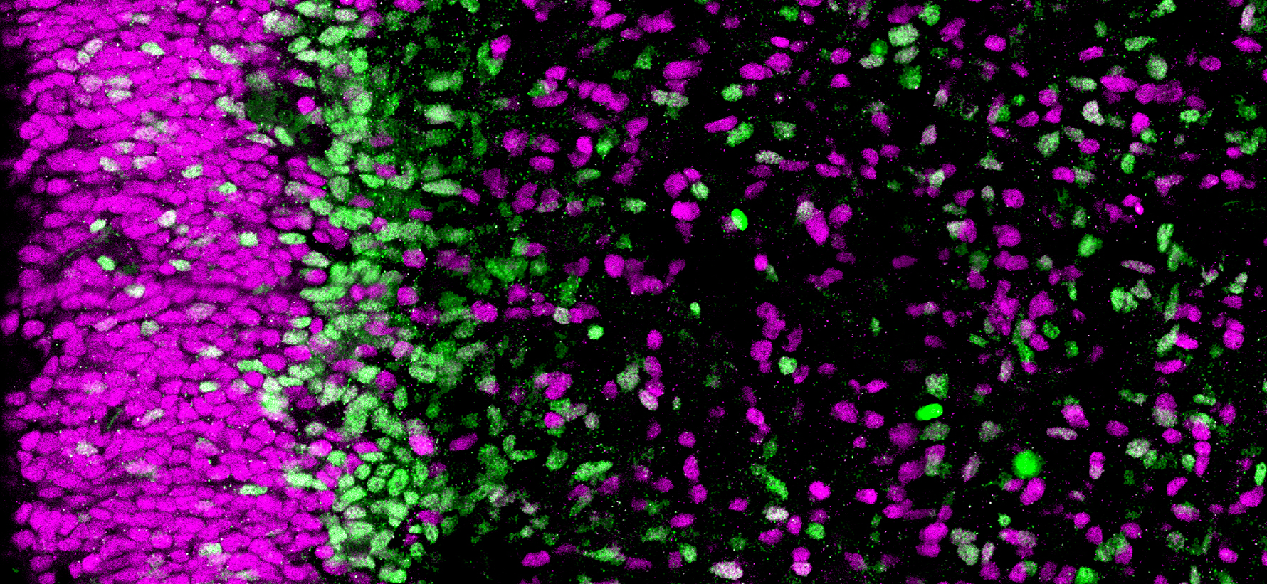
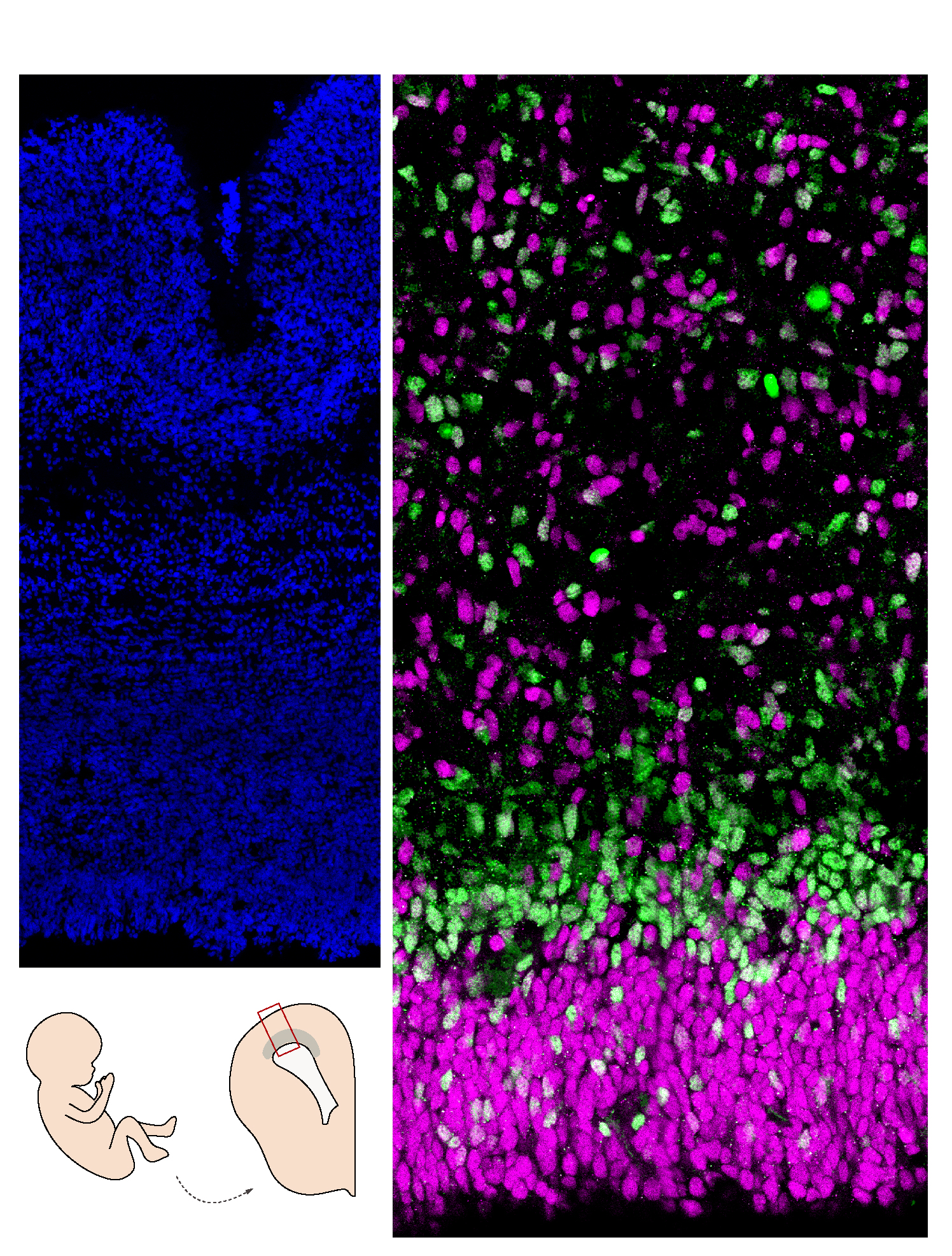
The researchers looked at the brain connections between the control regions located in the frontal cortex (at the front of the brain), and the memory regions, such as the hippocampus. They hoped to identify any differences among the three groups of participants (the first not exposed to the attacks, the second exposed but without PTSD, and the third also exposed but with PTSD).

The results show that the participants with PTSD present a deficiency of the mechanisms that suppress and regulate the activity of the memory regions during an intrusion (particularly the activity of the hippocampus).  

Conversely, the functioning of these mechanisms is to a large extent preserved in the individuals without PTSD, who are able to fight the intrusive memories. “In our study, we suggest that the memory suppression mechanism is neither intrinsically poor nor responsible for the intrusions, as was previously believed. However, its dysfunction is. If we use the analogy of a car’s brakes, it is not the act of applying them – or in our case the act of suppressing the memories – that poses a problem, but the fact that the braking system is faulty, which leads to their overuse”, explains Gagnepain.

  1. Implications of this research

These findings will make it possible to challenge traditional thinking on PTSD and devise new avenues for treatment.

Scientific implications

Firstly, the study highlights that the persistence of the traumatic memory is probably not solely linked to a dysfunction of memory but also to a dysfunction of the memory control mechanisms.

Secondly, although the memory suppression processes in victims of PTSD have long been considered problematic and ineffective (because they enable the traumatic memories to return in an even more violent way), the study shows that the problem is not this mechanism as such, but its poor implementation by the cerebral networks.

Patients with PTSD are actually considered to be in an ongoing state of memory “suppression”, even when there is no memory intrusion, in order to compensate this deficient memory control system. It remains to be determined whether these control difficulties arise after the trauma or were present before, rendering the individual more vulnerable.

Therapeutic implications

Many of the current PTSD therapies are aimed at recontextualizing the problematic memories, at making the patients aware that these memories belong in the past, and at reducing the feeling of fear that they generate.

Designing new interventions which are disconnected from the traumatic events and which stimulate the control mechanisms identified in this study could be a useful addition in training patients how to implement more effective mechanisms of suppression. “The current treatments all involve confronting the trauma, which is not always easy for the patients. It could be imagined that this type of task stimulates the mechanisms of suppression, thereby facilitating the processing of the traumatic memory in the traditional therapies”, the researchers point out.


Findings in interaction with the other components of 13-Novembre

The study also makes it possible to improve research into the brain function of the “resilient” survivors who have not developed PTSD. “This study goes further than all the others, which traditionally focus on military personnel exposed to traumatic situations. However, their exposure has rarely been to the same extent, to the same situations, or at the same frequency. So it is often difficult to study in parallel PTSD sufferers and resilient individuals who have been exposed to the same horrifying situation. We have that possibility here and I hope the progress made will have major positive impacts on trauma victims worldwide”, emphasizes Gagnepain.

Several studies are now emerging in order to supplement these findings. The researchers will in particular use the data obtained from the brain imaging sessions to take a closer look at specific alterations of the hippocampus, a key structure in the expression of intrusive memories. Since these participants have already been studied on two occasions separated by a two-year interval, the researchers will also be able to study their individual evolution and try to identify biomarkers to predict it. “It will be interesting to compare the data from 13-Novembre, which studies the collective and individual evolution of traumatic memories, with those of Remember, which studies the brain mechanisms used to fight this trauma. This research is original and unprecedented on the global scale”, concludes Gagnepain.


It is this ability to bring so many disciplines into dialogue and explore the entire complexity of human memory that makes the 13-Novembre program so rich.

Participant accounts*

Alice’s story

For Alice and the father of her children, November 13, 2015 was meant to be a special day – their first romantic meal out since the birth of their second son. Living in the XI arrondissement, they had chosen a local restaurant. Just after their meal was cut short by the babysitter because their youngest child was sick, and just after they decided that Alice would go and see to him, there was an explosion in the Comptoir Voltaire café across the road. Assuming a gas leak in one of the nearby buildings, Alice had no idea what was actually happening.

It was only when she was climbing the stairs to her apartment, whilst the news of the attacks was making the tour of the globe, that Alice began to realize what she had experienced. For her, the rest of the night was associated with a floating, anxious sensation while her husband, gone to join colleagues for a wrap party, found himself confined at the party location. This sensation was followed by incomprehension in the face of the horror throughout the weekend that followed.

The week following the attacks was hard. Alice had difficulty realizing what had happened, she cried a lot. Every day, she feared for the lives of her children, who were attending schools in the neighborhood. She was rapidly diagnosed with PTSD.

Participation in the studies

One year after the attacks, Alice heard of an enrolment campaign conducted at the INA, which was looking for volunteers to take part in the Inserm and CNRS 13-Novembre program. Encouraged by her psychologist, by a desire to better understand the events of that evening and by a wish to share her account with the scientific community, she went along. Everything then fell into place, with Alice also accepting to participate in the program’s brain imaging study sponsored by Inserm, Remember.

For Alice, taking part in a biomedical study has been a very intense but enriching human experience. “In Caen, the team welcomed me with a great deal of kindness. A team of doctors and psychologists was there to ask questions about my health and to explain the protocol and its objectives. I was exhausted when I left: learning the word list was not easy and doing the Think/No-Think task in the MRI machine was difficult because of the noise. But I was well looked after and happy to know that my participation has helped to improve knowledge of brain functioning in PTSD”, she explains.

Alice has continued to make progress with the support of a psychologist whom she met at a crisis unit at the XI arrondissement city hall. She is happy to have been able to participate in Remember and hopes that its findings will serve to improve the treatment of people living with PTSD, and to make the disorder less alarming.

Dominique’s story

It was at a meeting of an association for the victims of the attacks that Dominique first heard about the 13-Novembre program codirected by Francis Eustache and Denis Peschanski. A survivor of the Parisian café attacks, Dominique has always had a strong interest in science, particularly in the major challenge that represents exploring the human brain to elucidate its mysteries. The transdisciplinary aspect of the program – at the crossroads of social and neurosciences – as well as the researchers’ desire to study the construction of memory, aroused his curiosity.

As a survivor of the attacks, you feel a kind of guilt. I wanted to do something, I wanted to help move things forward after these events. Participating in a scientific study with such important themes as the collective memory of a country, the treatment of victims with PTSD, and coming together as a society after the attacks were important to me. And what is more, I felt capable of doing it”, he explains.

After participating in the 13-Novembre program filmed INA sessions, Dominique then accepted to go to Caen twice as a volunteer in Remember. An experience that was both difficult and highly enriching. “The discussions with the various researchers were fascinating, I had lots of questions about their work, about what they were looking for in the brain. However, the MRI part was a real challenge. While I am not claustrophobic, it is still a narrow space with little light, and the machine is very noisy, producing sounds that can evoke gunshots. It is very hard when you are trying to fight your intrusive memories. But I was well looked after by the team”, he specifies.

In order to conquer the MRI step during his second session in Caen, Dominique had practiced being in confined spaces by spending time under his bed at home. However, to his great surprise, the second session went without incident. He is ready to return to Caen next year for the third and final series of experiments in order to collect additional data for studying the longer-term evolution of the study participants, and to build on the study published in Science.

* Names were changed at the request of participants

For more information about the 13-Novembre program: Paris Attacks: What Will We Remember? (an interview with Francis Eustache and Denis Peschanski).


[1] Currently at the European Centre of Sociology and Political Science of the Sorbonne (CNRS/Université Paris 1 Panthéon-Sorbonne/EHESS)

[2] With the scientific component driven by the CNRS and Inserm and the administrative component by héSam Université, 13-Novembre is funded by the French Secretary General for Investment via the French Research Agency (ANR) as part of the Investments for the Future program (PIA). It has 31 partners and the support of 26 entities. It is a component of the facility of excellence, MATRICE. For more information: https://www.memoire13novembre.fr/partenaires-et-soutiens

Resilience after trauma: the role of memory suppression

Collective Memory Shapes the Construction of Personal Memories

Researchers analyzed media coverage of WWII in order to identify the shared collective representations associated with that period. Credits: Adobe Stock

For sociologists, our individual memories are shaped by the collective memory of our community. Until now, this phenomenon had never been studied at the neurobiological level. Inserm researchers Pierre Gagnepain and Francis Eustache (Inserm/Université de Caen-Normandie/Ecole Pratique des Hautes Etudes/Caen University Hospital/Cyceron Public Interest Group), in association with their colleagues from EQUIPEX MATRICE led by CNRS researcher Denis Peschanski, studied the collective representations of WWII in France, using brain imaging to show how collective memory shapes individual memory. Their findings have been published in Nature Human Behaviour.

In the last century, French sociologist Maurice Halbwachs declared that personal memories are influenced by their social contexts. From this perspective, the memory function of individuals cannot be understood without taking into account the group to which they belong and the social contexts related to collective memory.

Until now, these theories had never been tested by neuroscientists. Inserm researchers Pierre Gagnepain and Francis Eustache (Inserm/Université de Caen-Normandie/Ecole Pratique des Hautes Etudes/Caen University Hospital/Cyceron Public Interest Group), in association with their colleagues from EQUIPEX MATRICE (involving several French teams), led by CNRS historian Denis Peschanski, decided to take a closer look, using brain imaging techniques. They have for the first time revealed in the brain the link between collective memory and personal memories – innovative research which has been published in Nature Human Behaviour.

Collective memory is comprised of symbols, accounts, narratives and images that help to construct a community identity. For a better grasp of this concept, the researchers began by analyzing the media coverage of WWII in order to identify the shared collective representations associated with it. They studied the content of 30 years of WWII reports and documentaries, broadcast on French television between 1980 and 2010, and transcribed.

Using an algorithm, they analyzed this unprecedented corpus and identified groups of words regularly used when discussing major themes associated with our collective memory of WWII, such as the D-Day Landings. “Our algorithm automatically identified the central themes and the words repeatedly associated with them, thereby revealing our collective representations of this crucial period in our history”, states Gagnepain.

Visiting the Caen Memorial Museum

But what is the link between these collective representations of the war and individual memory? To answer that question, the researchers recruited 24 volunteers to visit the Caen Memorial Museum and asked them to view photos from that period, which were accompanied by captions.

Based on the words contained in the captions, the team was able to define the degree of association between the photos and the various collective memory themes identified previously. If words which had previously been associated with the theme of the D-Day Landings were found in the caption, for example, the photo was then considered to be linked to this theme in the collective memory. In this way, the researchers were able to establish proximity between each of the images: when two photos were linked to the same themes, they were considered to be “close” in the collective memory.

Gagnepain and his colleagues then turned their attention to the perception of these photos in the memory of the individuals. They tried to find out whether there was the same degree of proximity in the memories of the individuals as between the photos. The volunteers underwent an MRI examination during which they recalled the images seen at the Memorial Museum the day before. The researchers were especially interested in the activity of their median prefrontal cortex, a brain region linked to social cognition.

As such, the researchers compared the level of proximity between the photos by looking at the collective representations of WWII in the media and, via brain imaging, by looking at the individual memories that people had of these images following a visit to the Memorial Museum. The team showed that when photo A was considered close to photo B – because it was linked in the same way to the same collective theme – it also had a higher probability of triggering brain activity similar to photo B in the brains of the subjects.

This novel approach enabled indirect comparison between collective memory and individual memory. “Our data demonstrate that collective memory, which exists beyond the individual level, organizes and shapes personal memory. It constitutes a shared mental model making it possible to link the memories of individuals across time and space”, emphasizes Gagnepain.

Other research is ongoing in order to deepen our understanding of the interaction between collective and individual memory. Nevertheless, one lesson can already be learned from this study: the functioning of our memories cannot be researched without taking into account the social and cultural context within which we evolve as individuals.    

“Brain Map” paves the way for Personalized Medicine

Adobe Stock

Using cerebral imaging, connections between the various brain regions can be visualized. These connections form a veritable “map” of its structure, specific to each individual. A team led by Christophe Bernard, Inserm researcher, and Viktor Jirsa at the Institute of Systems Neuroscience (Inserm/Aix-Marseille Université), has shown that having the knowledge of these “maps” is enough to predict not just brain function but also the potential development and treatment of neurological diseases. Their findings have been published in PNAS.

Over the previous three decades, rapid progress in brain imaging has enabled major advances in the field of neurosciences. Magnetic resonance imaging (MRI), has paved the way for a deeper understanding of the brain and of the mechanisms of certain diseases.

Using MRI, researchers can access the general organization of the brain and more particularly the map of the neural connections between its different regions – a bit like a map of the roads that link various towns. “This map is unique to each individual and is more accurate than even a fingerprint”, highlights Inserm researcher Christophe Bernard. To pursue this analogy, neurological diseases such as Alzheimer’s and epilepsy are associated with a reorganization of the “maps”: the connections between brain regions are modified and some “roads” disappear.

While it is possible to accurately visualize the brain of each individual after using MRI to obtain a map of its connections, is it also possible, just from this map, to accurately predict its function and the potential development of diseases, as well as their treatment? Is it enough just to have a knowledge of this “map” to make these types of personalized predictions for patients?

The “Virtual Brain”

These are questions that Bernard, researcher at the Institute of Systems Neuroscience (Inserm/Aix-Marseille Université) and his colleagues have tried to answer in a new study published in PNAS. They began their research by using brain imaging techniques to visualize very precisely the connections between the brain regions of several mice.


Mapping the brain connections of a mouse makes it possible to predict its brain activity. Results obtained following virtualization of mouse brains using The Virtual Brain platform. Credits: Christophe Bernard

Based on these “maps”, they then created virtual models of the brain of each mouse using a technology called The Virtual Brain[1], in collaboration with researchers from Technion in Israel. In each of these virtual brains, the researchers generated electrical activity, mimicking what happens in a real brain.

This enabled them to study which brain regions communicate together – findings that were compared with the experimental data obtained using functional imaging from each mouse in a resting state. The researchers were thus able to show that having a knowledge of the “map” of each mouse is sufficient to explain the brain activity of that same mouse as seen using functional imaging. They were also able to show which connections make each brain unique.

Although these findings remain to be validated in humans, they are already paving the way for the personalized medicine of the future. “Our research validates the strategy of patient brain virtualization in order to explore, using a computer, the optimal therapeutic strategies prior to their personalized transfer. We can imagine that the ability to predict the development of certain diseases in an individual based on the unique map of their brain will enable us to envisage prevention strategies and personalized therapeutic options”, Bernard highlights.

[1] The Virtual Brain, a neuroinformatics platform developed by Viktor Jirsa at the Institute of Systems Neuroscience (Inserm/Aix-Marseille Université) in collaboration with Randy McIntosh (Baycrest Centre, Toronto) and Petra Ritter (Charité, Berlin), is used to create individual brain models. Currently being evaluated in patients with drug-resistant epilepsy, it can, for example – following virtualization of the patient’s brain – explore and predict the best form of neurosurgery to cure such epilepsy.

Developpemental Coordination Disorder, or Dyspraxia, a New Collective Expert Review by Inserm

©Frédérique Koulikoff/Inserm

Inserm has published a new Collective Expert Review, this time looking at developmental coordination disorder (DCD) – otherwise known as dyspraxia. In order to produce this document, which was commissioned by the French National Solidarity Fund for Autonomy (CNSA), a group of experts studied over 1400 scientific articles, exploring this relatively unknown disorder that affects around 5% of children. Its recommendations include guaranteeing access for everyone to professionals trained in DCD diagnosis and management, as well as enabling each child to get the most out of their education.


Developmental coordination disorder (DCD), also known as dyspraxia, is a commonly occurring condition that affects 5% of children on average. When a certain level of motor coordination is required, children with DCD do not perform as well as their peers in daily life activities (washing, dressing, eating, etc.) and at school (handwriting).

Inserm was commissioned by the French National Solidarity Fund for Autonomy (CNSA) to produce a Collective Expert Review of the scientific knowledge of DCD. Over a two-year period, Inserm’s Collective Expert Reviews Unit coordinated a dozen researchers and consulted ten specialists in order to review a scientific corpus of over 1400 international articles and issue recommendations on improving the diagnosis and management of children with DCD.

DCD varies markedly in its intensity and expression and is often associated with other neurodevelopmental disorders (of language, attention and learning), as well as a high risk of developing emotional, behavioral or anxiety disorders. These impact the child’s quality of life and participation in activities, particularly schoolwork. One of the key obstacles to their academic integration concerns handwriting.

To limit these impacts, the Expert Review states that spotting the signs is paramount when it comes to establishing rapid monitoring and individualized management for the child according to the severity of their condition, verbal competency, age, and any concomitant disorders.


Recommendations of the Inserm Collective Expert Review

The recommendations put forward by this Collective Expert Review can be summarized according to three main areas of focus.

The first consists of guaranteeing access to diagnosis for everyone, and as soon as possible following identification of the initial signs. In this respect, the Expert Review highlights the need to train professionals, emphasizing the importance of deepening the criteria and standardizing the tools needed to establish a diagnosis according to international standards.

Establishing such a diagnosis involves at the very least the participation of a doctor trained in developmental disorders, as well as that of a psychomotricity therapist or occupational therapist.

The second area of focus concerns what happens post-diagnosis, where there is no one standard intervention of unanimously recognized efficacy. Once diagnosed, it is therefore important to establish appropriate interventions that take into account the child’s profile, quality of life, and that of their family. The experts advise prescribing group sessions for the least affected children and one-on-one sessions for the others. They also recommend preferring interventions that focus on learning the skills needed for school and day-to-day life. Finally, these interventions must increase the involvement of the child’s family, teachers and anyone else interacting with them (sports coaches, etc.).

The objective of the third and final area of focus is to enable each child to get the right support at school. This requires that the school and its staff make the necessary additional arrangements for the child during their exams, in accordance with the French Disability Act (2005). This also involves raising the awareness of and training those involved in supervising and interacting with the child in everyday life, whether at home, school or during leisure

fermer