Menu

La thérapie génique, un espoir contre les maladies de Charcot-Marie-Tooth

Weightlessness: A Challenge for Both the Body and the Brain!

Etude des mouvements

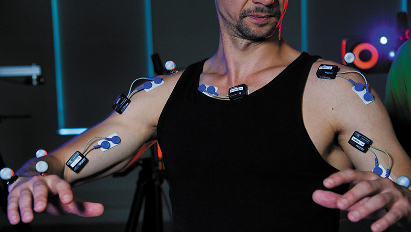
3D motion capture ©Inserm/Guénet, François


With one week to go before astronaut Thomas Pesquet sets out on his space mission Alpha, knowledge about how we adapt to gravity here on Earth is progressing. Researchers from Inserm and Université de Bourgogne within the Cognition, Action and Sensorimotor Plasticity (CAPS) laboratory are interested in how the movements that depend on this omnipresent force are carried out.

For the past 30 years, it was thought that the brain – responsible for motor control – worked to continuously compensate for the effects of gravity. In an initial study in 2016, the researchers had suggested that our brains use gravity to minimize the effort our muscles have to make. Those results were recently confirmed by new experiments conducted in collaboration with New York University on non-human primate models and humans, the findings of which have been published in Science Advances.


Our brain uses the effects of gravity to minimize the physical exertion required.

In order to perform our many activities, the movements of our limbs need to be precise. For a movement to be successful, the brain must generate muscle contractions by anticipating the environmental factors liable to affect that movement. One of the most important of these factors is gravity. The brain develops an internal representation of gravity that it can use to anticipate its effects on our body.


What is the purpose of this anticipation?

Initially, the researchers thought that the brain continuously compensated for the effects of gravity to achieve movements undisturbed by them. Recent studies by researchers from Université de Bourgogne and Inserm at the CAPS laboratory in collaboration with a team from New York University (Dora E. Angelaki, Professor of Neuroscience at the Tandon School of Engineering – New York) challenge this idea. The researchers hypothesized that anticipating the effects of gravity allows us to plan movements that use those effects on our bodies to minimize our muscular exertion.

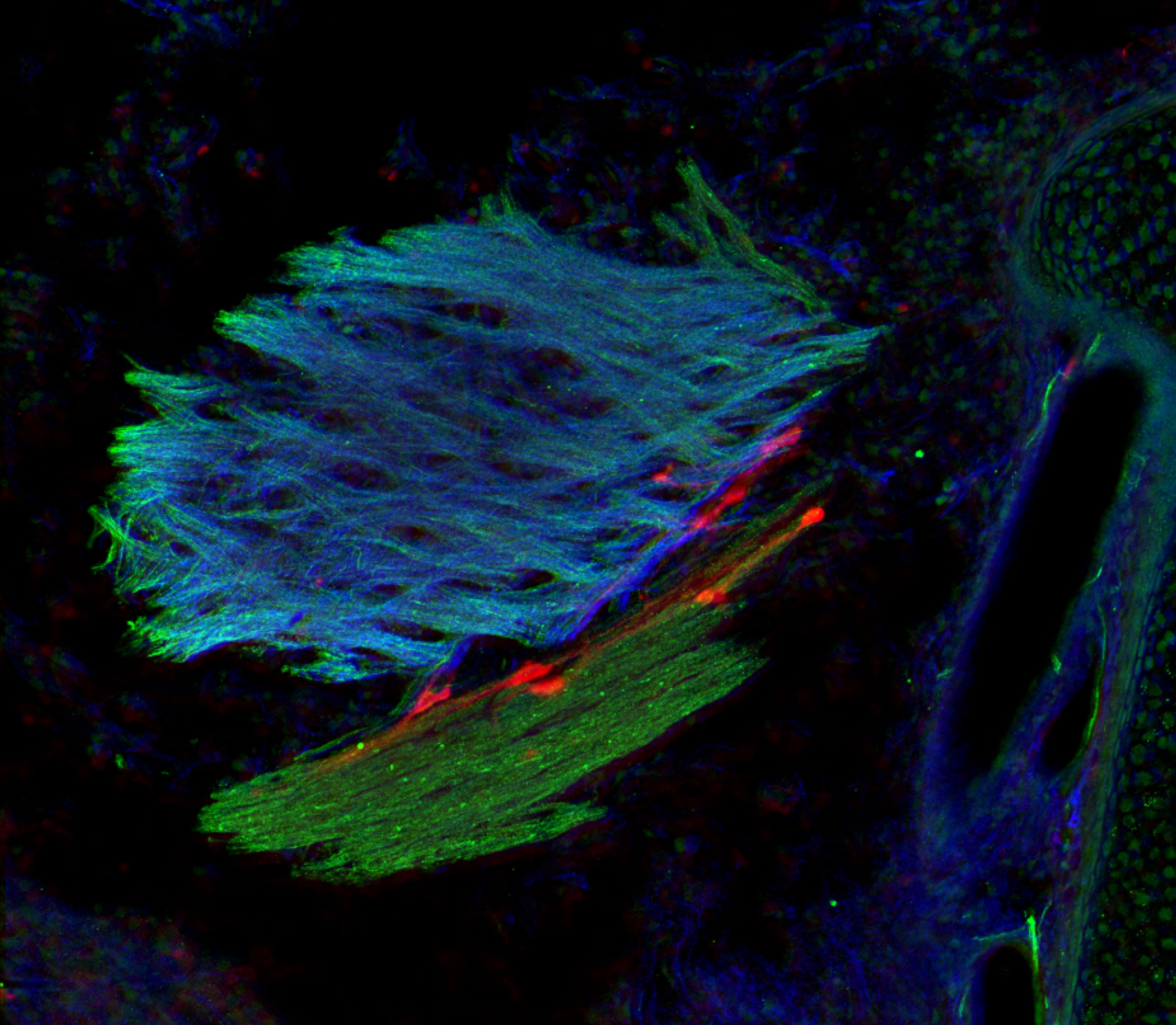
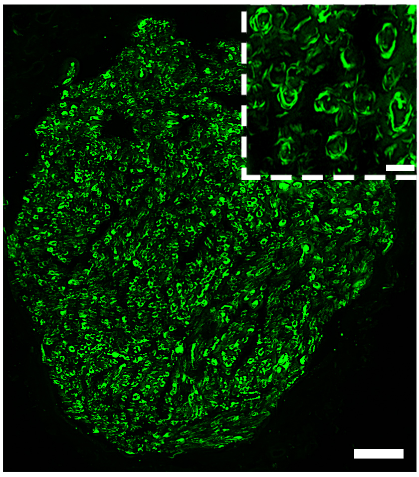
To confirm this theory, the research team recorded the activations that the brain sends to the muscles. These recordings were made in non-human primates and humans performing horizontal and vertical arm movements.

The information obtained show that the brain sends electrical commands that activate and deactivate the muscles in a very precise way – phenomena that last just a few milliseconds – in order to harness the effects of gravity to accelerate our downward movements and decelerate our upward movements. These findings were observed in both the non-human primates and humans.

This observation supports the hypothesis of profound nervous system adaptation to its environment. 

In the long term, this new knowledge could be put to use in various fields such as movement assistance for people with disabilities or the programming of humanoid robot movements.

The Vicious Circle of Anorexia Nervosa: Burning Calories May Well Be More Important Than Eating Less

Anorexie

Seeking a reward effect through physical exercise is thought to constitute a key component of the disease, influenced by genetics. © Bruno Nascimento on Unsplash.

In anorexia nervosa patients, the weight loss through lack of food is accompanied by fatigue and reduced physical capacity. Yet despite this they often continue to perform intense physical activity which contributes to that weight loss. Researchers from Inserm and Université de Paris at the Institute of Psychiatry and Neuroscience of Paris and the University Hospital Group Paris Psychiatry & Neurosciences show that physical exercise generates positive emotions not just in the patients (which was expected) but also – more surprisingly – in their relatives without the condition. However, this was not the case in the control subjects.

Seeking a reward effect through physical exercise is therefore thought to constitute a key component of the disease, influenced by genetics. This research, published in the International Journal of Eating Disorders, could make it possible to focus the management of anorexia nervosa patients on calories burned (sport) rather than exclusively on deficiencies (diet).

Anorexia nervosa is an eating disorder that affects mostly young women and girls between the ages of 15 and 25. Its lifetime prevalence in women is estimated to be just over 1%. Philip Gorwood and Laura Di Lodovico at the Institute of Psychiatry and Neuroscience of Paris (Inserm/Université de Paris) and the University Hospital Group Paris Psychiatry & Neurosciences have spent years trying to elucidate the disease and improve how it is managed.

In particular, their work has focused on the reward effect associated with not eating and the resultant weight loss. “We know that there is a vicious circle with anorexia nervosa, in which what makes a person lose weight is so rewarding in terms of how they feel, that they can overlook the dangers that they otherwise understand. This abnormality in the decision-making process is clearly related to the reward effect (the brain sends back messages that positively reinforce the maintenance of the disorder). But it is complicated to understand how a lack (a lack of food) can in itself be a ‘reinforcer’. That is why we have focused on the other dimension of weight loss – physical exercise,” explains Gorwood.

Based on these questions, the scientists showed in a previous study that anorexia nervosa is associated more with the pleasure of losing weight than with the fear of gaining it, and that this aspect is genetically influenced.

In their latest research, they continue to reflect on the clinical criteria of the disease and its heritability[1]by focusing on the concept of physical exercise. “This is an atypical approach because physical exertion is not considered a clinical manifestation of anorexia, even though many patients do a lot of sport, especially to manage their hunger and burn calories,” clarifies Gorwood.

The team felt that studying this aspect was all the more interesting given that yet again there is a contradiction: patients persist with exercising despite the fact that being underweight gradually reduces their physical capacities.

Patients, their relatives, and others

The protocol for this study was original because it allowed the researchers to focus not only on the emotions and perceptions of the patients following standardized physical exercise, but also on those of members of their family (particularly their mothers and sisters). Enrolled in this study were 88 female patients with anorexia nervosa, 30 of their relatives without the condition, and 89 healthy controls. Each was asked to perform standardized physical exercise and then answer questionnaires concerning the emotions they felt afterwards and the perception of their body image.

The scientists showed that for the same amounts of exertion, the emotions reported by the patients were more positive than those of the controls. “Doing sport sends the patients a message of positive reinforcement, making them continue this activity despite feeling tired or weak. The calories burned in association with this physical activity is a key factor that leads to its continuation,” explains Gorwood.

While this aspect was not found in the controls, it was present in the patients’ relatives. The study therefore suggests that this trait is shared within the families of people with anorexia.

Physical activity is associated with a rewarding effect, and this is thought to be involved in the heritability of the disease.

These findings have implications in terms of managing the condition, emphasizing the importance of focusing part of that management on physical exercise. The idea is to work with the patients so that they rediscover the pleasure of physical exercise (therefore in moderation) and lose their addiction to it, which is probably associated with the goal of weight loss. While specialized teams already considered this aspect of management to be important, the study provides firm scientific arguments in favor of pursuing this approach, legitimizing this care practice and putting it to widespread use.


[1] heritability refers to the proportion of variation in a population trait (in this case, anorexia nervosa) that can be attributed to the genetic factors we inherit.

Huntington’s Disease: Exploring the Avenue of a Potential Neuroprotective Treatment

huntington

Huntington’s disease is a rare and hereditary neurodegenerative disorder. A striatal neuron expresses the causative mutant huntingtin protein (red), which accumulates in the nucleus (blue) to form an aggregate of huntingtin and other proteins, including ubiquitin (yellow). © Frédéric Saudou


Huntington’s disease is a hereditary disorder that causes degeneration of the neurons involved in cognitive, motor and psychiatric functions. While existing treatments address the symptoms and relieve certain aspects of the disease, they cannot alter its course. Researchers from Inserm, Université Grenoble Alpes and Grenoble Alpes University Hospital at the Grenoble Institute of Neuroscience are hoping to remedy this. They are studying a new therapeutic approach in the hope of offering patients the first neuroprotective treatment – one that protects neurons – in the years to come. The therapeutic molecule in question has shown promising results in mice and is currently undergoing preclinical evaluation. Their research has been published in Science Advances.

Huntington’s disease is a rare and hereditary neurodegenerative disorder. It usually begins between the ages of 30 and 50 and manifests with cognitive disorders, psychiatric disorders and uncontrolled movements that worsen over time until death some 20 years later. In France, this condition affects around 18,000 people: 6,000 already have symptoms, whereas around 12,000 carry the mutated gene and will develop symptoms later. The team of Frédéric Saudou, Director of the Grenoble Institute of Neuroscience (Inserm/Université Grenoble Alpes/Grenoble Alpes University Hospital), is working on a new therapeutic approach in an attempt to provide solutions for these patients.

The disease is caused by an abnormality on the gene coding for the protein huntingtin, which interacts with and regulates the activity of at least 400 other proteins involved in various cell functions, including the transport of molecules. This abnormality leads to a reduction in the transport of a key molecule, BDNF, in the brain between the cortex and the striatum. The role of this molecule is to promote the survival of neurons and ensure the connections between them. This reduced transport therefore causes the death of neurons in these brain regions.

“Long before symptoms develop, a reduction in BDNF transport is observed. This molecule is essential for the survival of neurons and for neural connections between the cortex and striatum – two regions involved, among other things, in mood and movement control,” explains Saudou, a professor at Université Grenoble Alpes and Grenoble Alpes University Hospital.

The researcher and his colleagues therefore thought that restoring its circulation would at least afford the brain partial protection from neuron death.


A molecule to restore BDNF transport

In collaboration with Inserm Research Director Sandrine Humbert, Saudou and his team had previously shown that BDNF is transported within vesicles made up of numerous proteins, including huntingtin. In this new study, the researchers identified an enzyme that regulates the transport of these BDNF vesicles by controlling a cell mechanism known as “palmitoylation.” By increasing palmitoylation with the help of a molecule called ML348, they were able to restore the transport of BDNF vesicles.

Several in vitro experiments on human neurons and in vivo experiments on mice have shown that ML384 crosses the blood-brain barrier and restores BDNF traffic from the cortex to the striatum. When administered in a mouse model of the disease, it reversed the symptoms.

Injecting ML348 reduced the motor and psychiatric behavioral disorders, allowing the mice to regain activity close to that of their healthy counterparts,” explains Saudou. What is more, this molecule improves BDNF transport in human neurons derived from induced pluripotent stem cells (iPS cells) from Huntington’s patients, demonstrating that this molecule is potentially capable of having an effect in humans.

Following this proof of concept, the researcher and his team will move on to the preclinical testing phase to evaluate, using cell and animal models, the behavior of the molecule in the body, its safety, and identify effective doses. The ultimate goal is to develop a drug for patients. If these results are confirmed, this molecule could become the first “neuroprotective” treatment for Huntington’s disease, sparing certain neurons from degeneration and perhaps slowing its progression.

Charcot-Marie Tooth disease: A 100% French RNA-based therapeutic innovation

Maladie de Charcot

© Liliane Massade, Diseases and hormones of the nervous system (INSERM/Université Paris-Saclay)

  • Charcot-Marie Tooth disease causes progressive paralysis of the legs and hands. No treatment is available today.
  • After more than a decade of research, a French team has developed an RNA-based therapy that allows model mice to regain their mobility.

Charcot-Marie Tooth disease is the most common hereditary neurological disease in the world. It affects the peripheral nerves and causes progressive paralysis of the legs and hands. No treatment is currently available to fight this disease, which is due to the overexpression of a specific protein. Scientists from the CNRS, INSERM, the AP-HP and the Paris-Saclay and Paris universities have developed a therapy based on degrading the coding RNA for this protein in mice. Their work is patented and was published on 9 March 2021 in Communications Biology.

In molecular biology, transcription is when a DNA molecule is copied to make an RNA molecule. This RNA molecule is then “translated” into a protein, which can perform different functions within the body’s cells. When a specific protein called PMP22 is made twice as much as normal, it causes type 1A of genetic Charcot-Marie Tooth disease to develop. This overproduction leads to gradual paralysis of the legs and hands.

The challenge then, is to standardize the expression of this protein in people with Charcot-Marie Tooth disease. French scientists1 have developed a patented2therapy based on reducing RNA coding for the PMP22 protein. To achieve this they used other small RNA molecules capable of interfering with a specific RNA, here the one that encodes PMP22, and of degrading or reducing its translation into protein.

The difficulty in developing this therapy has been to stabilise these small RNAs, known as small interfering RNA or siRNA, which degrade very rapidly in biological environments.

Researchers have coupled them with another molecule called squalene, which is typically used in cosmetology and pharmacology. Biocompatible, biodegradable and forming nanoparticles in water, squalene protects siRNA from degradation. It also controls the size of the particles formed and the amount of siRNA injected.

These scientists then showed, in mice models for this disease, that injecting these interfering RNAs completely and rapidly restored of mouse locomotor activity and strength. The siRNAs penetrate the peripheral nerves, strengthen the myelin sheath3 around those nerves, and normalize the nerve signal velocity. The effect of treatment lasts for three weeks for severe forms and more than ten weeks for milder forms of the disease.

This therapeutic strategy for hereditary peripheral neuropathies, developed entirely in France, is proof of concept for a new precision medicine based on siRNA normalization of the expression of an overexpressed gene. It will now be developed in humans with pre-clinical and clinical studies.


1 At the Laboratoire Maladies et Hormones du Système Nerveux (INSERM/Université Paris-Saclay), the Laboratoire Aspects Métaboliques et Systémiques de l’Oncogénèse pour de Nouvelles Approches Thérapeutiques (CNRS/Université Paris-Saclay/Institut Gustave Roussy), the Institut Galien Paris-Saclay (CNRS/Université Paris-Saclay), the Service de Neurologie – Centre de Référence Neuropathies Périphériques Rares at the CHU de Limoges, the Service de Neurologie – Centre de Référence National des Neuropathies Amyloïdes Familiales et Autres Neuropathies Périphériques Rares at the CHU du Kremlin-Bicêtre (AP-HP/Université Paris-Saclay) and the Environmental, Toxicity, Therapeutic targets, Cellular Signaling and Biomarkers Laboratory (INSERM/Université de Paris). This work has received ANR funding under the Avenir programme (Labex NanoSaclay, reference ANR-10-LAM-0035).

2 References: WO/2020/064749, PCT/EP2019/075736

3 Myelin is a lipid-rich layer that isolates axons from neurons and allows for increased transmission of the nerve signal. Myelin dysfunction leads to very disabling nerve diseases. The PMP22 protein is present in myelin and is essential for nerve function.

Communiquer pendant nos rêves, c’est possible !

Better Access to Education Reduces Differences in Cognitive Capacity Between Men and Women

From one generation of women to the next, access to higher education is linked to an improvement in certain cognitive aspects. © Adobe Stock

Elderly women are currently at a higher risk than men of developing dementia, particularly Alzheimer’s disease. A phenomenon that could partially be explained by inequalities in access to education between the sexes during the first half of the 20th century. Researchers from Inserm and Université de Paris, in collaboration with University College London, have shown that certain cognitive capacities have improved in women over recent generations, and that this is linked to a larger number of women accessing higher education. In the longer term, they believe that sex-based inequalities in dementia risk could decrease. Their study has been published in The Lancet Public Health.

Elderly women currently have a 50% higher risk than their male counterparts of developing Alzheimer’s disease – a condition for which several risk factors are already well known, such as cardiovascular disease and level of education. It was the latter risk factor that Inserm researcher Séverine Sabia and the EpiAgeing team from the Center for Research in Epidemiology and Statistics at Université de Paris decided to study.

The current generation of very elderly people was born between 1920 and 1940, a time when few women had access to higher education.

However, from the 1960s, things began to change, with greater numbers of women accessing universities and on a more equal footing with men, to an extent that their educational attainment has more or less caught up with that of men in developed countries. A development that Sabia and her colleagues believe could reduce sex-based differences in dementia risk in the years to come.

To test this hypothesis, they compared the cognitive capacities during aging of women and men according to their education levels over several generations. These individuals were participants in two British cohorts – ELSA (English Longitudinal Study of Ageing) and Whitehall II – which had enrolled a total of 15,924 people from the general population, born between 1930 and 1955. The researchers divided them into three subgroups according to their year of birth: 1930-38, 1939-45, and 1946-55. The education level of the various participants was also known.

For each participant, two components of cognitive function were assessed several times over the 1997-2015 follow-up period. The first was short-term memory, which involves remembering a list of words just heard, followed by verbal fluency, which is the ease at which a person can produce words by naming as many animals (for example) as possible in one minute. “This is the first time to my knowledge that research of this type has focused on cognitive trajectories during aging in men and women combined with changes in education level in successive generations,” explains Sabia.

In both cohorts, the overall education level was higher in the youngest group (born between 1946 and 1955) than in the oldest group (born between 1930 and 1938). In addition, the proportion of women with a level equivalent to that of the high school diploma more than doubled, increasing from 14% to 33% (compared to 36% and 54% for men).

Data from this study suggest that women’s memory capacity has improved in recent years. They outperformed men in the tests at all ages, with the gap widening yet further among the “youngest” generation. In terms of verbal fluency, while men fared better than women in the oldest group born between 1930 and 1938, this gap narrowed for the participants born more recently, with the opposite effect being observed for the generation born between 1946 and 1955.

At the same level of education, women are at absolutely no disadvantage compared to men when it comes to memory or verbal fluency,” clarifies Mikaela Bloomberg, first author of the study.

“Although we studied just two of the components of cognitive function, the trend here is indicative of better cognitive reserve in women born more recently, thanks in part to improved access to higher education. This could ultimately result in a reduction in male/female differences in dementia risk in countries where access to education is similar regardless of sex.

These findings therefore underline the importance of access to education for all in order to promote aging in good health,” concludes Sabia.

Neuroscience Provides New Ways to Understand the Origin of Our Emotions

Emotions have long been considered as innate and universal biological experiences, quite distinct from each other. ©Adobe Stock

Are our emotions innate or are they the product of our culture and environment? This question has long been the subject of debate in the field of neuroscience. Researchers from Inserm, Université de Caen Normandie, Ecole Pratique des Hautes Etudes and the University Hospitals of Caen and Rennes provide robust clinical data in favor of the second hypothesis. Their work suggests that our ability to know and recognize emotions is built up gradually and depends on our knowledge of language. Their findings have been published in the journal Brain.

Throughout the history of neuroscience, the question of the origin of emotions has always intrigued scientists. Based on Charles Darwin’s theories, they have long considered emotional states as innate and universal biological experiences, quite distinct from each other.

Faced with the observation that emotions are not defined in the same way in all cultures, and that the boundaries between the categories (joy, sadness, anger, etc.) are not the same throughout the world, this viewpoint has nevertheless continued to evolve. A so-called “constructionist” hypothesis of emotions has thus developed over recent decades, postulating that emotions are not innate but rather concepts learned in childhood and associated with our physical sensations.

These concepts would be enriched throughout life, according to our experiences and environment. However, robust data from brain imaging and clinical practice were lacking to confirm this theory.

To decide between the two schools of thought, Inserm researcher Maxime Bertoux and the team of the Neuropsychology and Imaging of Human Memory laboratory (Inserm/Université de Caen Normandie/École Pratique des Hautes Etudes) in association with the University Hospitals of Caen and Rennes and GIP Cyceron studied 16 patients suffering from a rare neurodegenerative disease, known as “semantic dementia”.

This disease is characterized by a degradation of conceptual memory, i.e. a loss of knowledge about the world and language. “Patients have difficulty mobilizing what they have learned throughout their lives, for example, remembering that Paris is the capital of France. They are also unable to identify everyday objects and remember how they work or what they are used for, or understand the meaning of words. However, the degradation of conceptual knowledge associated with this disease should not impact patients’ ability to know and recognize emotions, if they are truly innate,” explains Bertoux.

Brain network identified

The participants were tested on their conceptual knowledge of four emotions: anger, pride, surprise and embarrassment. As part of the first step, they were asked to give a synonym for each of these emotions and then choose another similar word from a list. They were then asked to give an example of a context in which this emotion could be felt and then, from a list of situations, choose the one most likely to cause the emotional state in question. As part of the second step, the participants watched photos and videos of actors expressing emotions. They then had to recognize which emotion was represented.

Compared to the healthy participants, the conceptual memory of emotions was more impaired in the participants with semantic dementia. On average, these patients were, for example, less able to give or choose the correct synonym for a particular emotion, and also less able to select the appropriate context in which to expect to feel it. They also had greater difficulty recognizing emotional states expressed by others, whether positive or negative, presented in photographs or videos.

Based on these findings, the researchers reveal a strong correlation between the loss of conceptual knowledge memory and the difficulty in recognizing emotions and their positive or negative nature.

The researchers also used brain imaging techniques to identify the brain networks mobilized during the performance of these various exercises. Their results suggest that the same network is at work in both the tasks of recognizing facial emotions and those of mobilizing conceptual knowledge about emotions.

“Our study highlights the strong link between “affective” neurocognitive processes, linked to the recognition of emotions, and “conceptual” processes that were supposedly distinct. We show that our conceptual and linguistic knowledge play a decisive role in how we perceive emotions. This enables us to supply new elements to confirm the constructionist theory of emotions: that we culturally construct our emotions from childhood,” emphasizes Bertoux.

This work is also of interest in the clinical field. Indeed, many psychiatric and neurodegenerative diseases cause emotional disturbances.

“Our study supports the utility of cognitive, behavioral and emotional approaches in mental illness and neuroatypical states. Recognizing an emotion in others but also regulating our own emotions depends on our ability to have learned to name them and distinguish them conceptually,” concludes Bertoux.

A new cerebral canalopathy associating intellectual disability and abnormal movements

Brain scan, X-ray © Fotolia


Dysfunctions of ion channels – or channelopathies – in the brain are today associated with more than 30 neurological diseases such as epilepsy or cerebellar ataxias. Structures located on the membrane of cells allowing the passage of ions (for example sodium and potassium ions) between the interior of a cell and its external environment (extracellular environment), these channels make it possible in particular to generate and control d potentials. action in neurons. A study conducted at the Brain Institute (Sorbonne University / Inserm / AP-HP / CNRS) identified a new cerebral channelopathy originating from dominant mutations in the KCNN2 gene, encoding the SK2 ion channel. The results were published in Brain on November 27, 2020.


Pathogenic variants of the KCNN2 gene identified in patients and their location on the protein structure of the SK2 channel.

The variants in red are pathogenic variants truncating (introducing a stop codon into the protein sequence). Variants in black are pathogenic missense variants associated with loss of function. The variant in gray was classified of unknown significance because the channel with this variant did not show any particular deficit in electrophysiology.


Dr Fanny Mochel, geneticist in the genetics department of the Pitié-Salpêtrière hospital AP-HP and researcher at the Brain Institute (Sorbonne University / Inserm / AP-HP / CNRS) and Professor Christel Depienne, A geneticist at the Institute of Human Genetics at the University Hospital of Essen (Germany) and also a researcher at the Brain Institute have identified a new syndrome associated with mutations in the SK2 channel. The study published in the scientific journal Brainconcerns 10 patients, 6 men and 4 women aged 2 to 60 years with more or less severe intellectual delays associated, for some, with autism spectrum disorders or psychotic episodes. These cognitive disorders are in all cases associated with tremors, symptoms of cerebellar ataxia or even abnormal movements.

Thanks to a collaboration with Agnes Rastetter from the genotyping / sequencing platform of the Brain Institute (Sorbonne University / Inserm / AP-HP / CNRS), the genome of a first patient recruited at Pitié-Salpêtrière was analyzed at the search for genetic mutations at the origin of this syndrome. This analysis revealed a mutation in the KCNN2 gene interrupting its coding sequence, absent from the patient’s parents ( de novo mutation ). Brain imaging by MRI (magnetic resonance imaging) in this patient showed abnormalities in the structure and integrity of the white matter of the brain, that is, the cerebral sheath that protects the axons of neurons.

In addition, an international collaboration has enabled researchers to identify 9 other patients with mutations in the KCNN2 gene . The majority of these mutations had arisen de novo while a mutation was transmitted in a familial form of the same syndrome.

Finally, by working jointly with Carine Dalle from the electrophysiology cell exploration platform of the Brain Institute, the teams of Dr Mochel and Depienne have shown a deleterious role of these mutations on the function of the SK2 channel, i.e. that is to say a loss of function leading to a dysfunction of the ion channel SK2 and therefore a loss of regulation of the action potential, support of the nervous message.

The results of this new study have identified a new cerebral channelopathy originating from dominant mutations in the KCNN2 gene , encoding the SK2 ion channel. This new syndrome is characterized by the presence, on the one hand, of cognitive symptoms, in particular an intellectual disability, and, on the other hand, of motor symptoms such as abnormal movements.

This new pathology, the cause of which is now known, is very heterogeneous from a point of view of symptoms and requires multidisciplinary management at the border between genetics, for the search for mutations in the KCNN2 gene, pediatric neurology and neurology. for the management of cognitive and motor manifestations of patients.

Early Puberty: Is There a Potential Explanation for Some Cases?

GnRH (red cell) neurons that are born in the nose use olfactory fibers (green and blue marking) to migrate into the brain during fetal life. © European Research Council/Agence Nationale de la Recherche Médicale/Métropole Européenne de Lille

Until now the general consensus had been that puberty was triggered by the acceleration of growth. However, a research team from Inserm, Lille teaching hospital and Université de Lille working at the Lille Neuroscience and Cognition Laboratory has discovered in mice a mechanism associated with the prepubertal growth spurt and the triggering of early puberty. This mechanism is regulated by the GnRH neurons, which orchestrate fertility, through the expression of their protein Nrp1. The team’s findings, published in The EMBO Journal, challenge existing knowledge of the triggers of puberty and pave the way for the study of this mechanism in humans and its potential role in some cases of early puberty.

The general consensus is that a growth spurt at the beginning of puberty triggers the activation of GnRH neurons[1] in the brain and the release of sex hormones. During embryonic development, these GnRH neurons appear in the nose and migrate towards the hypothalamus in the brain – from where they later go on to orchestrate fertility.

This study, conducted by Inserm Research Director Vincent Prévot and his team within the Lille Neuroscience and Cognition laboratory (Inserm/Lille teaching hospital/Université de Lille), has challenged this scientific consensus by re-examining the roles of GnRH neurons in triggering puberty.

This study follows previous research in which Prévot and his team had shown that a protein, called Nrp1, which is present along the axons linking the nose to the olfactory system of the brain, is a component of the “rails” that enable GnRH neurons to migrate from the nose to the brain during embryonic development. However, Nrp1 is also expressed by the GnRH neurons themselves, which led the researchers to study its role in the migration and functioning of those neurons. In order to do that, they developed a mouse model in which Nrp1 was inactivated only in the GnRH neurons but continued to be expressed elsewhere. These neurons were labeled with fluorescence in order to observe them over a period of time.

What the research team observed in the mutated mice was an increase in the quantity of GnRH neurons in the nose, which is associated with migration towards the brain starting earlier. Physiologically, the authors observed weight gain and more rapid growth of the mutated mice in relation to the control animals, as well as an earlier start to puberty.

These observations suggest that the GnRH neurons could in reality control the prepubertal growth spurt and not the other way around as the researchers had hitherto thought.

“We will research connections and communications between the GnRH neurons and the appetite and growth regulation functions to explain this phenomenon, specifies Prévot. It is the first time to my knowledge that these GnRH neurons are attributed functions that differ from reproduction.”

The researchers also saw in the mutated mice that the neurons had colonized an unusual region of the brain, the olfactory bulb – the center of olfactory information processing -, when they are usually only concentrated in the hypothalamus. This observation led them to verify whether the perception of odors – known to play a role in sexual attraction – could be modified. They then observed that very young female mice presenting a lack of Nrp1 had, contrary to those of the control group, a preference for the odors of male mice versus those of members of their own sex. Early puberty in those mutated mice could therefore be accompanied by sexual attraction that also takes place earlier.

It remains to be verified whether these mechanisms occur in humans. “These findings suggest that a very early growth spurt could be associated with early GnRH neuron activation. A phenomenon that could also be associated with certain variants of the NRP1 gene. These results open up new avenues in preventing the risks of early puberty in children. We will now go on to explore the inhibition of GnRH neural activity with drugs that are already used in a clinical setting”, envisages Prévot.


[1] A group of cells in the hypothalamus secrete gonadotropin-releasing hormone (GnRH),  which controls puberty and fertility. GnRH stimulates secretion of the sex hormones LH and FSH that in turn stimulate the gonads. Acquiring these functions is a lengthy process that starts with the migration of the GnRH cells in the nose towards the brain during embryonic development, and then continues with their maturation and activation at puberty.

Research shows that treatment with growth hormone in children who have recovered from cancer does not increase the risk of a second tumor.

Growth hormone deficiency is a common complication of radiotherapy. © Adobe Stock


Teams from Bicêtre AP-HP hospital, Inserm, Gustave Roussy and the University of Paris-Saclay studied the influence of growth hormone treatment on the risk of a second tumor. in 2,852 adults recovered from childhood cancer. The data confirm that treatment with growth hormone in these children with growth hormone deficiency does not increase the risk of developing a second cancer. This study therefore provides reassuring data on the long-term fate of these children cured of cancer and treated with growth hormone to enable them to reach normal adult height. The results of this study were published in the European Journal of Endocrinology in September 2020.

Growth hormone deficiency is a common complication of brain radiation therapy. Children treated with radiation therapy need growth hormone treatment to reach normal adult height, but there have been concerns about a possible increased risk of another tumor developing in adulthood caused by it. growth hormone treatment.

Researchers from unit 1018 of the “Center for Research in Epidemiology and Population Health (CESP)” (Inserm / Université Paris-Saclay / Gustave Roussy) and from Bicêtre AP-HP hospital, analyzed data from a French cohort, Euro2k, which brings together 2,852 survivors of pediatric cancer diagnosed before the age of 18 before 1986. Among them, 196 had been treated in childhood with growth hormone.

The research team studied the influence of growth hormone treatment on the occurrence of second tumors with a follow-up of 26 years, taking into account the doses of radiation received by all the organs of the body. These were obtained by reconstituting the radiotherapy received for each child. 

In this cohort, 374 survivors developed a second tumor, 40 of whom received growth hormone treatment in childhood. Analysis of the data shows that treatment with growth hormone is not associated with an increased risk of second tumors. However, these researchers found in survivors who received growth hormone treatment for more than 4 years, a slight increase (x2) risk of meningioma, a benign tumor of the meninges favored by high doses of radiotherapy. This slight excess risk of meningioma in survivors who received more than 4 years of treatment with growth hormone is not significant, however, and there is no evidence that the treatment with growth hormone either. responsible.

“This study provides information on the long-term fate of the children whom we treat with growth hormone for a growth hormone deficiency secondary to the treatment of their cancer. These new data allow us to approach the treatment with growth hormone calmly. in these children cured of cancer, when necessary and to reassure families about the absence of an increased risk of second tumors during this treatment. concludes Dr Cécile Thomas-Teinturier, pediatrician-endocrinologist at the Bicêtre AP-HP hospital and first author of the study.

fermer