Menu

The creativity of the human mind rooted in errors ?

©AdobeStock

Why do some of our choices appear to be driven by a desire to explore the unknown? An Inserm team from École Normale Supérieure led by 2017 ERC grant-winner Valentin Wyart has shown that most of these choices are not motivated by curiosity but by errors caused by the brain mechanisms implicated in evaluating our options. These findings have been published in Nature Neuroscience.

Where shall we eat tonight – at our usual restaurant or one we have not tried before? Where shall we spend our next vacation – in the old family cabin or a rental halfway around the world? When we have various options to choose from, our decisions do not always lead us towards the safest one based on our previous experiences. This variability which is characteristic of human decisions is most often described in terms of curiosity: our choices are thought to be the reflection of a compromise between exploiting options that are known and exploring others whose outcomes are more uncertain. Curiosity is even considered to be an attribute of human intelligence – a source of creativity and unexpected discoveries. This interpretation is based on a very strong (albeit rarely explicitly stated) hypothesis, according to which we weigh up our options without ever making any errors.

An Inserm research team from the Laboratory for Cognitive and Computational Neuroscience (LNC2) funded by the European Research Council (ERC) wanted to test this implicit hypothesis upon which many findings are based. The researchers suspected that our capacity to weigh up and review our options is largely overestimated, based on findings published in 2016 in Neuron:

One of our researchers had previously shown that our capacity to make the right choice based on partial indicators is restricted by errors of reasoning when these indicators are combined, and not by hesitancy at the time of making the choice. So we asked ourselves whether these errors of reasoning could be responsible for some of the choices considered as being a matter of curiosity by current theories. “

To back up their suspicions, the researchers studied the behavior of around one hundred subjects using a slot machine-style game that consisted of choosing between two symbols associated with uncertain rewards. They analyzed the behavior of the participants using a new theoretical model developed by Charles Findling, postdoc in the team and co-lead author of the article, which takes into account evaluation errors of the symbols. 

As such, the authors discovered that over half of the choices usually considered as being a matter of curiosity were in reality due to errors of evaluation. “This finding is important, because it implies that many choices in favor of the unknown are made unbeknownst to us, without our being aware of it” explains team leader, Valentin Wyart.

Our participants have the impression of choosing the best symbol and not the most uncertain, but they do it on the basis of wrong information resulting from errors of reasoning. “

The participants played a slot machine-style game which involved choosing between two symbols associated with uncertain rewards. In this example, the participant has to choose between the left-hand symbol which won them money in the previous tests, and the right-hand symbol which has not been tried recently and whose outcome is therefore more uncertain. The current theories describe this type of choice as being the reflection of a compromise between exploiting known options (in this example, selecting the left-hand symbol) and exploring other more uncertain options (selecting the right-hand symbol).

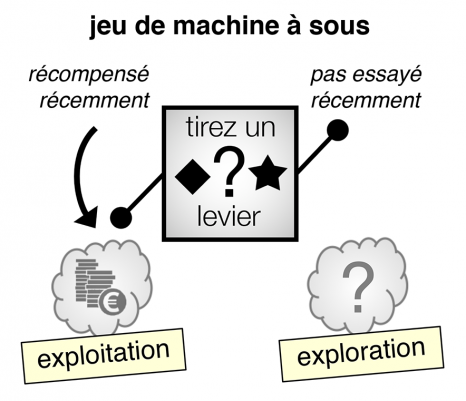
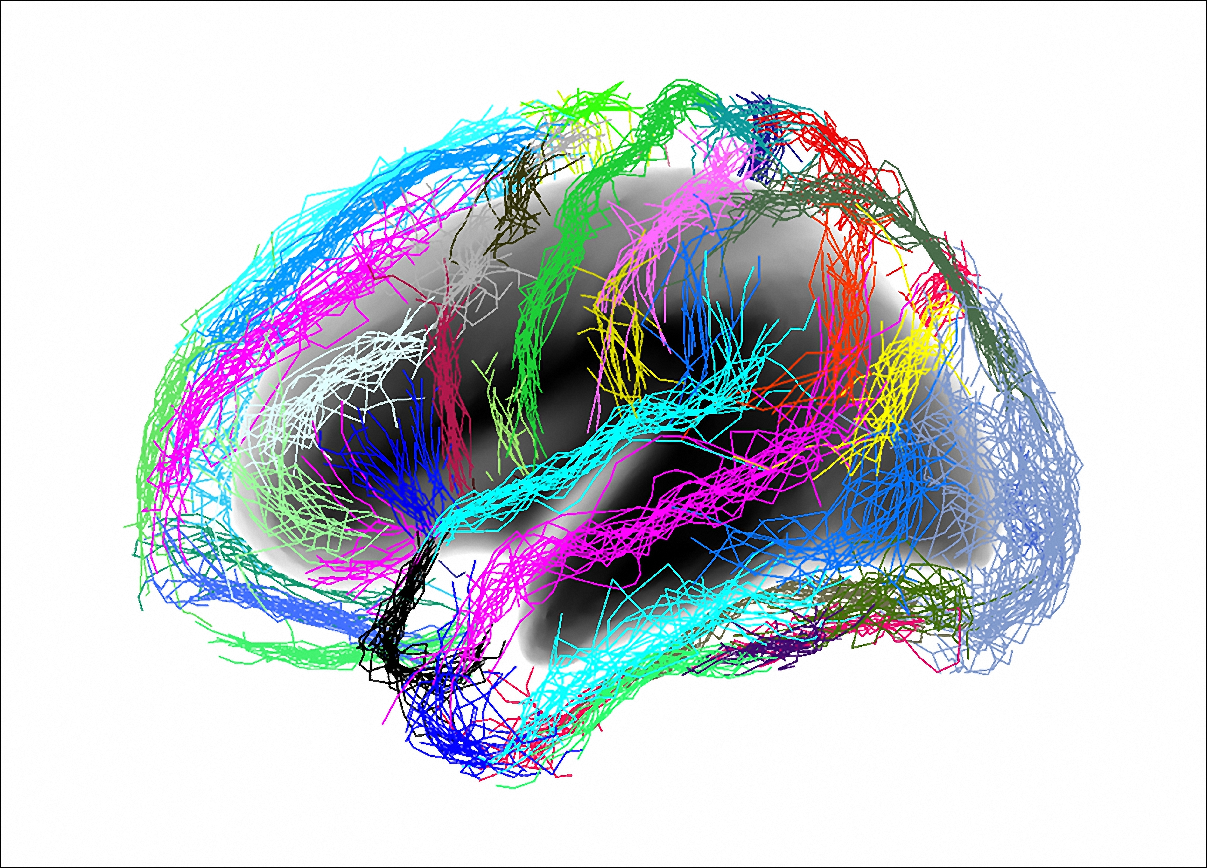
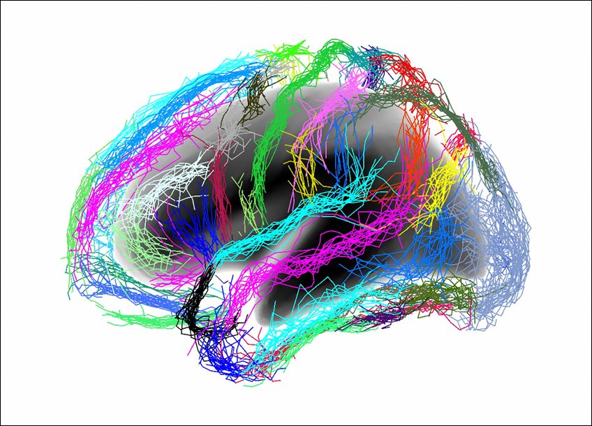
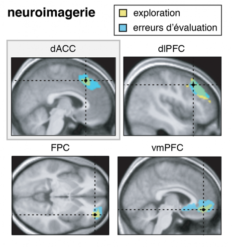
In order to understand where these errors come from, the researchers recorded the brain activity of some of the participants using functional magnetic resonance imaging. They discovered that the activity of the anterior cingulate cortex, a region involved in decision-making, fluctuates with the evaluation errors made by the participants. The greater the activity of this region when evaluating the options, the greater the evaluation errors. For Vasilisa Skvortsova, postdoc and co-lead author of the article, “these errors of evaluation could be regulated via the anterior cingulate cortex by the noradrenaline neuromodulator system, controlling the precision of the mental operations performed by the brain”. In other words, our brain is thought to use its own errors to produce choices in favor of the unknown, without relying on our curiosity. “This is a radically different vision from the current theories that consider these errors as negligible”, insists Wyart.

The researchers recorded the participants’ brain activity using functional magnetic resonance imaging. They discovered that the brain regions that activate when the participants explore uncertain symbols (shown in yellow) are the same ones that activate when the participants commit errors of reasoning. The brain regions shown are the dorsal anterior cingulate cortex (dACC), the dorsolateral prefrontal cortex (dlPFC), the frontopolar cortex (FPC) and the ventromedial prefrontal cortex (vmPFC).

Although these findings may appear surprising, are they really? Many major discoveries are the result of errors of reasoning: the discovery of America by Christopher Columbus, who believed he had reached the “East Indies” – a navigation error of 10,000 km, but also the discovery of radioactivity by Henri Becquerel, who initially thought that the radiation emitted by uranium was due to the re-emission of solar energy, or the discovery of the pacemaker by John Hopps when attempting to treat hypothermia with the help of radiofrequency. Going further, “the evolution of the species is also based on random variations of the genome, in other words genetic errors, some of which are retained by natural selection”, reiterates Wyart. So there is in fact nothing surprising about our brain taking advantage of its errors in order to think outside the box.

How People with Autism Might Avoid Socio-Emotional Situations

One hypothesis put forward to explain the repetitive behaviors of people with Autism Spectrum Disorder is a lack of cognitive flexibility. However, this may well not be the case. A recent study by a team of researchers from Inserm and Université de Tours used MRI to track the brain activity of autistic and non-autistic subjects faced with situations similar to those that cause problems in the daily lives of people with the disorder. Their findings, published in Brain and Cognition, suggest that the inflexibility of autistic individuals is actually the result of a strategy used to avoid socio-emotional situations. This research, which suggests now considering the cognitive and socio-emotional domains as closely linked rather than dissociated, opens up new avenues in the understanding and management of autism.

In their daily activities, people with Autism Spectrum Disorder (ASD) experience difficulty adapting their behavior to environmental changes. ASD is characterized by two main diagnostic criteria: the individual experiences persistent difficulties in social communication and is locked into repetitive behavioral patterns, restricted interests and/or activities. But while both criteria need to be present in order to diagnose ASD, little attention has been paid to how they interact.

In a study led by Marie Gomot, Inserm researcher at the Imaging and Brain laboratory (Inserm/Université de Tours), this question was explored by comparing the cognitive management of socio-emotional information and that of non-social information in people with ASD.

While the processes responsible for ASD symptoms have not yet been fully elucidated, one current hypothesis is a lack of cognitive flexibility – in other words, difficulty in alternating between multiple tasks and in analyzing one’s environment in order to adapt to these changes.

To evaluate this flexibility, the researchers used MRI to track the brain activity of ASD and non-ASD participants who underwent a test simulating situations similar to those that cause problems in the daily lives of people with the disorder.

The research team used a modified version of a test traditionally used in neuropsychology in order to test cognitive flexibility while processing non-social or socio-emotional information. Five cards were presented, each illustrated with a different face. The participants were asked to match the central card with one of four surrounding cards, according to one of the following three rules: frame color (non-social information), facial identity (social information) or facial expression (socio-emotional information). In order to evaluate their cognitive flexibility, the participants were asked throughout the test to make different matches (same color, same identity or same facial expression) by changing or maintaining one of the three rules.

The research team saw no significant difference between the ASD and non-ASD participants when it came to the behavioral parameters measuring cognitive flexibility alone – namely the capacity to adopt a new rule. However, the study did reveal the importance of information type for these cognitive flexibility processes in ASD. While the ASD participants needed more attempts than the non-ASD participants in order to adopt the rule linked to socio-emotional information, they had no particular difficulty in adopting those involving the processing of non-emotional information.

In parallel, the MRI revealed a significantly higher level of brain activity in the ASD participants when they were required to demonstrate cognitive flexibility. This brain activity only stabilized when the ASD participants received confirmation that they had found the correct rule to apply, thereby suggesting that people with ASD require a higher level of certainty in order to adapt to a new situation.

“These findings are important because they suggest that the implementation of routines and repetitive behaviors by people with ASD might not be due to a genuine lack of cognitive flexibility but rather to avoid being confronted with certain socio-emotional situations, specifies Gomot. The need for a high level of certainty combined with a poor understanding of the codes that govern socio-emotional interactions would thereby lead to the avoidance of tasks with a socio-emotional component.” And to conclude, “this research confirms the close link between cognitive and emotional dysfunction in ASD and the need for future studies to take them into joint consideration more often.

A new discovery! How our memories stabilise while we sleep

Photo by Kinga Cichewicz on Unsplash

Scientists at the Center for Interdisciplinary Research in Biology (CNRS/Collège de France/INSERM)1 have shown that delta waves emitted while we sleep are not generalized periods of silence during which the cortex rests, as has been described for decades in the scientific literature. Instead, they isolate assemblies of neurons that play an essential role in long-term memory formation. These results were published on 18 October 2019 in Science.

While we sleep, the hippocampus reactivates itself spontaneously by generating activity similar to that while we are awake. It sends information to the cortex, which reacts in turn. This exchange is often followed by a period of silence called a ‘delta wave’ then by rhythmic activity called a ‘sleep spindle’. This is when the cortical circuits reorganize to form stable memories. However, the role of delta waves in the formation of new memories is still a puzzle: why does a period of silence interrupt the sequence of information exchanges between the hippocampus and the cortex, and the functional reorganisation of the cortex?

The authors here looked more closely at what happens during delta waves themselves. They discovered, surprisingly, that the cortex is not entirely silent but that a few neurons remain active and form assemblies, i.e. small, coactive sets that code information. This unexpected observation suggests that the small number of neurons that activate when all the others stay quiet can carry out important calculations while protected from possible disturbances. And the discoveries from this work go even further! Spontaneous reactivations of the hippocampus determine which cortical neurons remain active during the delta waves and reveal transmission of information between the two cerebral structures. In addition, the assemblies activated during the delta waves are formed of neurons that have participated in learning a spatial memory task during the day. Together these elements suggest that these processes are involved in memory consolidation. To demonstrate it, in rats the scientists caused artificial delta waves to isolate either neurons associated with reactivations in the hippocampus, or random neurons.

Result: when the right neurons were isolated, the rats managed to stabilise their memories and succeed at the spatial test the next day.

These results substantially change how we understand the cortex. Delta waves are therefore a means of selectively isolating assemblies of chosen neurons, which send crucial information between the periods of hippocampo-cortical dialog and the reorganisation of cortical circuits, to form long-term memories.

1 An associate member of the Université PSL, since 2009 the Collège de France has had a deliberate policy of welcoming independent teams who can benefit from shared technical and scientific services and an exceptional multidisciplinary environment. Twenty-two teams are currently housed in the Center for Interdisciplinary Research in Biology et de Physique of the Collège de France. Supported in particular by the CNRS, this facility is open to researchers in France and elsewhere. It helps to make Paris an attractive place to conduct research.

Science serves gender equality

© Tim Mossholder on Unsplash

In a commentary published in the journal Nature Human Behavior, Inserm researcher Violetta Zujovic and her colleagues from the XX initiative committee at the Brain & Spine Institute (Inserm/CNRS/Sorbonne Université) at the AP-HP Pitié-Salpêtrière Hospital explain how a neuroscientific approach can be used to more effectively combat gender inequality. The actions taken by the researchers have increased the number of female speakers invited to take the floor at the research center from 25 to 44% in the space of a year and a half, and have increased the number of women in senior roles from 25 to 31%. Their neuroscience-based approach now constitutes an important mechanism for changing attitudes and behaviors. Among other things, their future work will focus on using “nudges”—a technique originating in neuroscience that tries to influence our behavior in our own interest—to improve gender equality. An example for others to follow.

Although internationally recognized as a country that supports women’s career progression through suitable infrastructure and specific legislation, France performs poorly when it comes to offering equal opportunities to men and women. This is the paradox highlighted by the XX initiative committee, made up of a dozen scientists, in a commentary published in Nature Human Behavior. If the entire community, including the scientific community, recognizes these inequalities, why do gender-related behaviors persist? The published commentary warns of the impact of cognitive biases, which are beyond our control but deeply rooted in our attitudes. Unconscious prejudices and stereotypes have a powerful influence over almost every decision we make.

Despite the norms of equality, and despite the evidence that teams are more successful when they include both men and women, social prejudices continue to silently pull the strings, for example by creating sexist or racist biases. The neuroscientific community is at the forefront of this issue, not only in raising awareness of such forms of unconscious bias, but also because it has the tools at its disposal to understand their cognitive origins and break societal stereotypes.

The XX initiative committee at the Brain & Spine Institute (ICM), which includes male and female neuroscientists from Inserm, the CNRS, and Sorbonne Université, have proposed a roadmap in several stages. Their recommendations highlight a crucial first stage: realization by each individual of their own implicit assumptions derived from existing prejudices, and the impact these have on their decision-making.

This first stage was put into practice by collecting comprehensive data on the male to female ratio by level of responsibility and job title. The internal ICM data gathered in April 2017 showed that just 26% of women held a management or joint management post within the institute, whose staff is 63% female.

Other figures showed that only 25% of invited speakers at the institute’s weekly scientific seminars were female.

The simple fact of presenting the findings of this investigation triggered a collective reassessment process that has resulted in the number of women heading up a research group increasing from 26 to 31%. The entire community has also been involved in efforts to showcase more female researchers, which has had the practical result of the number of female scientists taking the floor at the institute increasing from 25 to 44%.

“Thanks to the collective drive to combat gender inequality, we were also able to revise various institutional forms to remove all gender-specific information,”explains Violetta Zujovic, the Inserm researcher who led this work.

In this societal and economic context, in which we are trapped by unconscious prejudices and gender stereotypes, one of the key recommendations proposed by the researchers is to inform men and women and train them to overcome these forms of bias. The committee is holding a meeting on “Gender Bias: Science and Practice” on April 3, 2020, which is open to all, along with practical workshops providing individuals with the tools to combat their own prejudices and understand how to assess their own values and skills.

The impact of this committee, and the neuroscientific approach it instigated around eighteen months ago, highlights the importance of combining national policies with practical measures. To that end, the neuroscientists are already working on developing “nudges” that will cleverly encourage rather than impose male/female equality. Overall, these measures are designed to enable individuals to understand and recognize the unconscious gender-related prejudices that we perpetuate and pass on, and to individually strive for cultural change.

In May 2019, Inserm also set up a professional parity and equality mission that will shortly set out practical measures for advancing professional equality. These will be incorporated into a multi-year plan and integrated into the institution’s strategy.

Sport Has Its Benefits but Do Not Overdo It

©Braden Collum/Unsplash

Sport is good for health, we are always being told. However, in top athletes, excess physical activity can be harmful, as the cases of “overtraining syndrome” show. Responsible for major fatigue and reduced sporting performance, it is a phenomenon that intrigues scientists. A study performed by Mathias Pessiglione, Inserm Research Director at the Brain & Spine Institute (Inserm/CNRS/Sorbonne Université) in conjunction with the French Institute of Sport, Expertise and Performance (INSEP) and the French Anti-Doping Agency (AFLD) shows that intensive physical training can harm brain capacity, particularly cognitive control. The full results have been published in Current Biology.

Inserm researcher Mathias Pessiglione and his team were interested in identifying the causes of a common phenomenon in top athletes, known as “overtraining syndrome”. This is expressed by reduced sporting performance and intense fatigue. Athletes suffering from this syndrome may be tempted by products likely to restore their performance, hence the involvement of AFLD in the project.

The primary hypothesis of the researchers was clear: the fatigue caused by overtraining is similar to that caused by mental effort and is thought to be linked to the same brain mechanisms. Another recent study had already shown that mental fatigue affects cognitive control and leads to impulsive decisions.

To test their hypothesis, the team spent nine weeks working with 37 triathletes, who were split into two groups. The first underwent the “usual” high-level training whereas the second had additional training during the last three weeks of the experiment, with sessions lasting 40% longer, on average. The participants were all monitored at the Brain & Spine Institute, both behaviorally and via functional MRI.

Training sessions 40% longer, on average

From this, the researchers were able to identify similarities between overly intensive physical training and excessive mental work. This excessive physical activity leads to reduced activity of the lateral prefrontal cortex (a key region for cognitive control), similar to that observed during mental effort. A reduction in brain activity expressed by impulsive decisions, in which short-term gratification is prioritized over long-term goals. In the case of top athletes, such impulsiveness can, for example, lead to the decision to stop right in the middle of a sporting performance or abandon a race in order to end the pain felt during physical exertion.

Beyond these top athletes, the researchers consider that, clinically, fatigue and reduced cognitive control could constitute the first stage in the development of burnout syndrome, which affects many people across various professional domains.

The next step for the researchers is to propose and test interventions in order to avoid the onset of actual burnout – namely the total exhaustion of the individual.

A novel, more effective method for diagnosing Alzheimer’s disease


Inserm/Auzias, Guillaume/Baillet, Sylvie/Colliot, Olivier

Currently, anatomical analysis of the cerebral cortex by MRI can be used to diagnose 80% of patients with Alzheimer’s disease. But what if analysis of another brain structure could provide better results? This was the finding of a collaborative team of researchers from Inserm, the Université de Paris, and the CEA (French Alternative Energies and Atomic Energy Commission), led by Maxime Bertoux, now an Inserm researcher within Unit 1171, Degenerative and Vascular Cognitive Disorders (Inserm / Université de Lille / Lille University Hospital). According to their study, published in Neurobiology of Aging, morphological analysis of the cortical sulci could make it possible to identify Alzheimer’s disease in 91% of cases. The size of these sulci also appears to be associated with the stage of disease progression and cognitive decline. Their research suggests the potential value of this method in diagnosis and patient follow-up.

Anatomical analysis of the brain using MRI (magnetic resonance imaging) typically consists of measuring the thickness of the cerebral cortex (the tissue also known as “gray matter” that covers the two cerebral hemispheres) and the volume of various areas of the brain such as the hippocampus, the atrophy of which is one of the early signs of Alzheimer’s disease. This method can be used to correctly detect the disease in around 80% of patients. The team led by Inserm researcher Maxime Bertoux has shown that MRI analysis of the cortical sulci is even more effective.

The sulci are the grooves of the brain, which tend to get wider with age. This change in the sulci is accompanied by a decrease in the thickness of the surrounding cortex. The acceleration of this process in Alzheimer’s disease had been shown by the same research group in previous studies. This time, the group wanted to find out whether morphological analysis of the sulci could be used as a diagnostic marker for the disease and its stage of progression.

The researchers performed a brain MRI scan of 51 patients with Alzheimer’s disease, some at an early stage and others at an advanced stage, along with 29 control participants without the disease. Diagnosis was made based on a laboratory work-up, which included both a lumbar puncture to test for the presence of disease biomarkers, and positron emission imaging (a PET scan) to show amyloid deposits, the build-up of protein in the form of plaques characteristic of some neurodegenerative diseases.

The researchers then used the Morphologist software program recently developed at NeuroSpin (the Center for Neuroimaging at the CEA), which allowed them to digitally recreate a “mold” of the brain in negative from an MRI scan. The software then extracted a mean value for the width of each sulcus and the thickness of the surrounding cortex from 18 areas of each cerebral hemisphere. Meanwhile, the researchers took standard measurements of the volume of various areas of the brain and of the cortical thickness, in order to compare the two methods.

An algorithm was then used to correlate the health status of each participant (control or patient) to the measurements found. The researchers found that the width of a group of several sulci, in particular in the frontal and temporal lobes, was associated with Alzheimer’s disease. This measure could be used to determine the health status of participants in 91% of cases, compared to only 80% for the standard anatomical measurements. The morphology of the sulci also appears to change with the stages of the disease: in patients with more advanced cognitive decline, they were wider.

“These measures indicating disease progression appear to be correlated with cognitive performance,” explains Bertoux, “which may be very useful for clinical trials investigating the efficacy of a potential drug. In addition, these measures require only an MRI machine and largely automated analysis, meaning they can be taken in a large number of health institutions. The method still requires validation in larger groups of patients, but it could have great clinical potential,” he concludes. Bertoux is already using this new approach to identify the specific characteristics of other neurodegenerative diseases, including frontotemporal dementia.

No rest for neurons during sleep

Adobe Stock

An Inserm team has provided the first description of the behavior and language of the neurons responsible for memory consolidation during sleep. The researchers have shown that, far from being organized in a static and linear way as was previously thought, neurons vary in their role over time, and that information pathways are continually changing. Their research has been published in Science Advances.

Brain cells are continually exchanging information. During sleep, this plays a particularly important role in consolidating memory. But little is currently known about how these exchanges take place. Electroencephalograms, which measure overall electrical brain activity, show regular waves that vary in speed depending on the phase of sleep, but do not provide information about how information is processed at the neuronal level. This is what the team led by Christophe Bernard (Institute of Systems Neuroscience – Inserm U1106) have succeeded in revealing. To do so, the team used electrodes to record electrical activity from around a hundred neurons concentrated in a particular region. It is these electrical signals that carry information. Three areas known to be involved in memory were recorded in rats during sleep: the hippocampus, prefrontal cortex, and entorhinal cortex.

“Because of the regularity of waves seen on the encephalogram, we thought that neurons worked in a very precise and repetitive way to transmit or store information (rather like a fine-tuned machine). But the recordings show that this is not at all the case,” notes Christophe Bernard.

Groups of neurons organize themselves for very short periods to store and transmit information, and continually take turns over time. And within each group, only a few neurons play a dominant role. “So there’s a sequence of substates, with ultimately around half of the neurons in these three regions playing a key role in processing information from one moment to the next. In other words, there is no hierarchy among the neurons, but rather an even distribution of roles,” explains Christophe Bernard.

Free-flowing circulation

The other major discovery is that, during a given substate, the information does not always follow the same pathway. “This came as a surprise, as the dominant theory was that information transfer followed a set pathway. But we found that this is not the case. In the brain, the partners with which a neuron exchanges information fluctuate from one moment to the next. It works a bit like the internet,” illustrates the researcher.

“An email sent from Paris to Sydney passes through servers located in various countries during its journey, and these servers vary over the course of the day depending on traffic. It’s the same in the brain: even if the information is the same, it does not follow a set pathway, and the partners are never the same.”

Finally, this research has made it possible to decode the type of language spoken by the neurons. If a substate corresponds to a “word,” then the sequence of substates constitutes a sentence. But while the meaning of the words and sentences still evades the researchers, they have been able to establish that the neurons speak a complex language, which makes it possible to optimize information processing. A simple language contains very few words; it is easy to understand but struggles to convey complex concepts. A chaotic system of language contains a word for every possible situation, and is impossible to learn. The language of neurons is complex, like human languages, and its complexity is notably greater in paradoxical sleep (during dreams) than in deep sleep.

The researchers will now look at what happens during the waking state, when performing specific tasks, or in disease. This will include studying the possible link between memory loss in subjects with epilepsy and the complexity of neuronal language.

A New Therapeutic Target for Treating Spinocerebellar Ataxias?

Cellules de Purkinje dans une coupe horizontale du cervelet de souris exprimant une protéine fluorescente (GFP) sous le contrôle du promoteur des récepteurs dopaminergiques D2. Ces cellules dégénèrent chez les patients atteints d’ataxie spinocérébelleuse SCA3. ©Inserm/Valjent, Emmanuel

Spinocerebellar ataxias are neurodegenerative genetic diseases of the cerebellum and brain stem that lead to numerous motor disorders. The most well-known of these ataxias is SCA3, which is also called Machado-Joseph disease. In her research published on June 14 in Acta Neuropathologica, Nathalie Cartier-Lacave, Inserm researcher at the Brain and Spine Institute, discovered with her team the crucial role of an enzyme that can improve symptoms of this disease in mice.

Certain neurodegenerative diseases are caused by a mutation that leads to the production of malformed proteins in possession of excess amino acids (polyglutamine expansion). This occurs with Huntington’s disease and some forms of spinocerebellar ataxia.

In this study, a team from the Brain and Spine Institute (Inserm/Sorbonne Université/APHP) led by Nathalie Cartier-Lacave looked at another group of diseases that presents this polyglutamine-expanded protein production – spinocerebellar ataxias – and more specifically SCA3. In this disease which affects 1-2 in every 100,000 people, the ataxin 3 protein mutates and aggregates in neurons, leading to their death and the subsequent onset of motor disorders. The researchers were able to show that supplying a key enzyme of brain cholesterol metabolism, CYP46A1, to the regions affected by the disease improves symptoms. A strategy that could also be effective in the other ataxias linked to polyglutamine expansion.

The researchers began by studying the cholesterol metabolism in mice with SCA3, revealing an imbalance in this metabolism and decreased levels of the enzyme CYP46A1.
These initial findings led the researchers to test whether or not restoring the expression of this enzyme in SCA3 mice could be beneficial. They performed a single injection of a gene therapy vector carrying gene CYP46A1 into the cerebellum of SCA3 mice, revealing reduced degeneration of the Purkinje neurons of the cerebellum, an improvement in the motor disorders, and decreased ataxin 3 aggregates when compared with untreated mice with the disease.

“These findings show that CYP46A1 is an important therapeutic target for restoring this metabolism, decreasing
toxic mutated protein aggregates and thereby improving the symptoms of the disease”, explains Inserm Research Director Cartier-Lacave.

To further elucidate the phenomenon, the researchers revealed that the pathway used to evacuate the malformed or mutated proteins – the autophagy pathway – is disrupted in SCA3 mice. This led them to conclude that ataxin 3 proteins aggregate as a result of dysfunction of this pathway. However, if normal CYP46A1 levels are reinstated, autophagy is restored, and the disease symptoms attenuated.

Interestingly, the researchers also observed improved evacuation of the ataxin 2 aggregates during overexpression of the enzyme, leading to hopes for treatment, with one product having the potential to be effective in multiple severe and rare diseases.

A European program (Erare) coordinated by Inserm at the Brain and Spine Institute (N. Cartier, A. Durr) is in progress to confirm these results on other models of ataxia and to evaluate the feasibility and tolerance of a potential therapeutic application in patients with these severe genetic diseases.

Une étude montre des effets bénéfiques particulièrement importants du café dans le traitement d’une maladie neurologique orpheline


When narcolepsy makes creative

©Adi Goldstein/ Unsplash

Sleeping we make it more creative? The study of narcolepsy, which enjoy privileged access to REM sleep, could provide key information to understand this phenomenon. A team including doctors from the hospital Pitié-Salpêtrière AP-HP and researchers from Inserm, CNRS and Université Sorbonne within the Institute for Brain and Spinal Cord Disorders, in collaboration with a team University of Bologna in Italy, revealed the existence of a greater creativity in patients with narcolepsy. The results of the study suggest a link between a particular phase of sleep, REM sleep, and creative abilities. This important advance, published in the journal Brain May 29, 2019, opens new avenues in understanding the cognitive functions of sleep and mechanisms of creative thinking.

Narcolepsy is a rare sleep disorder that affects approximately 0.02% of the general population. It is characterized by uncontrollable sleep phases. These sleepiness have the distinction of often begin immediately by a particular phase of sleep, REM sleep, a situation not to encounter in normal times.

Indeed, our sleep consists of several stages and REM sleep is always preceded by a slow phase of sleep. So it usually sleep at least an hour before accessing this particular sleep. Narcoleptic people therefore have privileged access to REM sleep. They also have many parallels symptoms associated with REM sleep, as if they existed in them a porous barrier between wakefulness and that sleep phase. For example, the majority of them are lucid dreamers, that is to say conscious dreaming when they are dreaming and can sometimes influence the dream scenario. If more than half of the adult population reported having made a lucid dream at least once in his life, regular lucid dreamers (several times a week) are very rare.

Data from the current literature suggest that either nap including REM sleep is followed by an increased period of greater mental flexibility to solve problems. Narcoleptic individuals with privileged access to this sleep phase, would there be a long-term effect on their creativity?

By meeting regularly with narcoleptic patients in my service, I noticed they seemed more change in creative activities than average; not only in their careers but also in their leisure or their thinking. “Says Dr. Isabelle Arnulf, head of the Sleep pathology department at the Pitié-Salpêtrière, AP-HP. From this observation was born the idea of exploring the creative capabilities of these patients with regard to their particular access to REM sleep.

A study by Celia Lacaux, a researcher at the Sorbonne University, and Delphine Oudiette researcher at Inserm, within the department of sleep pathologies of the Pitié-Salpêtrière Hospital AP-HP led by Prof. Isabelle Arnulf to ICM, tested in collaboration with a team from the University of Bologna in Italy, the creative capacities of 185 narcoleptic individuals and 126 control individuals.

Defining and measuring creativity is not an easy task. In neuroscience, it can be defined as the ability to produce something both original and adapted to the constraints. To evaluate and obtain the fullest possible extent, the researchers used two methods:

  • A “subjective” measure based on creativity questionnaires in 185 narcoleptic subjects and 126 control subjects: a test “creative profiles” focused on the personality and creative profile, and a test of “creative fulfillment” on personal achievements participants in various fields of the arts and sciences, cinema writing, through humor, cooking or architecture.


  • A measure ‘objective’ creative performance through a “paper and pencil” test for two hours, called EPOC (Evaluation of the Creative Potential) in 30 patients and 30 controls. It assesses the two main dimensions of creativity: divergent thinking which demand from a stimulus to generate the most possible responses; and convergent thinking, which requires the integration of several objects in a single generation, coherent and original.

Narcoleptic individuals generally received higher scores than the control subjects, both objective measures and subjective. ”  If narcoleptic subjects had higher scores than control subjects, only some of them really stood out in terms of creative fulfillment. This suggests that we really encourage narcoleptic people realize their potential. “Said Delphine Oudiette, Inserm researcher at the MHI, who led the study. ”  Moreover, among people with narcolepsy, the subgroup of lucid dreamers obtenaitles highest scores of creative profiles test, suggesting a role of dreams in the creative abilities. 

This increased creativity could be explained by the privileged access to REM sleep and dreams enjoyed narcoleptic people and gives them the opportunity to “incubate” their ideas during brief naps during the day.

”  This is a strong argument to say that regular access to REM sleep and dreams promotes creativity. Sleep on it, you will find a solution! It is also the first time we show that narcoleptic subjects are better than average in an area as important as creativity, bringing the same positive note to this difficult disease to live with. “Celia Lacaux concludes, first author of the study. Further work will be needed to confirm this but these early results provide important clues to understanding the functions of REM sleep and dreams.

Hypertension: A New Drug Coming Soon?

Prise de la tension artérielle chez un patient ©Inserm/Depardieu, Michel

Firibastat is the first in a new class of antihypertensive drugs targeting the renin-angiotensin system in the brain. A phase IIa placebo-controlled clinical trial provided initial data on its efficacy in hypertensive patients and was coordinated by Michel Azizi from Inserm/Hôpital Européen Georges-Pompidou AP-HP Clinical Investigation Center 1418 and the Hypertension Department of the same hospital and by Catherine Llorens-Cortes, Inserm Research Director at Unit 1050 “Center for Interdisciplinary Research in Biology” at Collège de France. On the basis of these results, published in The Journal of Hypertension, the phase IIb trial was launched in the USA.

Hypertension is the most common chronic disease in France. Linked to an abnormally high pressure of the blood on the vessel walls, its general lack of symptoms can make it appear harmless. Yet if not brought under control, it constitutes one of the main causes of cardiovascular, cerebrovascular and neurodegenerative complications (myocardial infarction, stroke, Alzheimer’s disease…). Hypertension can be normalized by means of lifestyle measures, either alone or more often than not in combination with medication. However, up to 30% of patients either do not respond – or respond poorly – to the treatments currently available. To remedy this, interventional approaches and new therapeutic targets linked to the pathophysiology of the disease are being studied.

The development of the drug firibastat[1] in the treatment of hypertension continues. It is the first in a new therapeutic class targeting the brain renin-angiotensin system controlled by angiotensin III, a peptide that in various hypertension models exerts a stimulating effect on blood pressure via three mechanisms. It increases the activity of the neurons that favor vasoconstriction, it inhibits the reflex that adjusts the intensity of heart contractions to the blood pressure level and, finally, it contributes to the increased secretion of the anti-diuretic hormone vasopressin in the blood, reducing the volume of urine produced by the kidneys.

Firibastat counters these various mechanisms by specifically inhibiting aminopeptidase A, an enzyme in the brain that produces angiotensin III. This drug, taken orally, becomes active in the brain after crossing the blood-brain barrier. Its safety was already tested in healthy subjects in two phase I clinical trials. The recently-published results of the phase IIa trial confirm the safety data and provide the initial efficacy elements. Enrolled in this trial were 34 patients (73% of whom male) with a daytime ambulatory blood pressure of between 135/85 and 170/105 mmHg. They had an average age of 57 and were not obese (average BMI 26.8 kg/m2). One half of the patients received firibastat for four weeks followed by placebo for another four weeks and the other half received the same treatment but in the reverse order: placebo followed by firibastat.

The results demonstrate better control of systolic blood pressure (SBP) with firibastat after four weeks with an average -4.7 mmHg reduction in SBP versus +0.1 mmHg with placebo. A difference which, however, is not statistically significant. “This can be explained not just by the small number of subjects but also by the fact that they had moderate hypertension, explains Inserm Research Director Catherine Llorens-Cortes. Firibastat is an antihypertensive agent and not a hypotensive agent, meaning that it can act on hypertension but will have no effect on normal blood pressure. “Its efficacy should therefore increase with the severity of the hypertension”, clarifies the researcher. A phenomenon which appeared to be confirmed in this trial because the decrease in ambulatory SBP reached -9.4 mmHg in the event of strong baseline hypertension whereas the benefit was less marked for lower baseline SBPs. The authors also verified the safety of firibastat and observed that it did not interfere with the systemic renin-angiotensin system, controlled by angiotensin II.

“These encouraging findings gave the green light to the phase IIb study which has recently been completed in the USA. It has confirmed the efficacy of firibastat in 254 hypertensive overweight high-cardiovascular-risk patients after two months of treatment, including in African-American patients whose hypertension is most often resistant to the treatments currently available.” clarifies Llorens Cortes.

[1] In partnership with the company Quantum Genomics, and with the support of the French National Research Agency (ANR): ANR RPIB CLINAPAI and LabCom CARDIOBAPAI

fermer