Menu

Dimly lit working environments : correcting your body clock is possible!

Researchers at Inserm, led by Claude Gronfier (Inserm Unit 846: Stem Cell and Brain Institute), have, for the first time, conducted a study under real conditions on the body clocks of members of the international polar research station Concordia. The researchers have shown that a particular kind of artificial light is capable of ensuring that their biological rhythms are correctly synchronised despite the absence of sunlight. The full significance of this result can be appreciated with the knowledge that disturbance to this biological clock causes problems with sleep, alertness, cardiovascular problems and even depression.

These results, published in Plos-One, could be rapidly transformed into practical applications for working environments that are dimly to moderately lighted (polar research stations, thermal and nuclear power stations, space missions, offices with no windows, etc.). They could enable the design of lighting strategies intended to maintain the health, productivity and safety of staff.


Horloge biologique, travail et luminosité

Fotolia


The system that allows our body to regulate a certain number of vital functions over a period of about 24 hours is called the body clock (or circadian rhythm). Located deep within the brain, it consists of 20,000 neurons whose pulsatile activity controls the sleep/wake cycle, body temperature, heart rate, the release of hormones, etc. The cycle determined by the internal clock lasts spontaneously between 23.5 to 24.5 hours, depending on the individual. In order to function correctly, it refers to the signals that it receives from the external world and that it interprets as indicators for the purpose of constantly resynchronising itself every 24 hours.
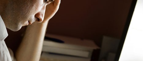
This is why the intake of food, physical exercise and the external temperature, for example, are said to be ‘time setters’. The most important ‘time setter’, however, is light. After inappropriate exposure to light, your entire body clock is thrown out of order with consequences for cognitive functions, sleep, alertness, memory, cardiovascular functions, etc.


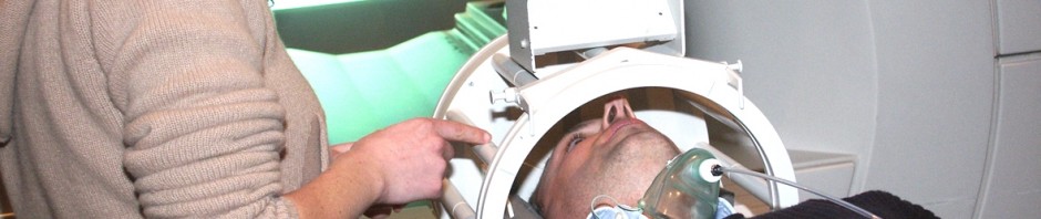
For the first time, scientists have been able to study under real conditions how various types of artificial light influence the way the biological clock behaves in situations where the natural light is insufficient. For nine weeks of the polar winter (no sunlight during the day), the staff of the international polar station Concordia were alternately exposed to a standard white light and a white light enriched with blue wavelengths (a particular kind of fluorescent light that is perceived as white by the visual system). For the purposes of the study, the researchers asked the staff not to change their day-to-day habits, particularly the times they got up and went to bed.

Once a week, samples of saliva were taken in order to measure the rates of melatonin (central hormone) secreted by each of the individuals.


The details of the results show that an increase in sleep, better reactions and more motivation were observed during the ‘blue’ weeks. Moreover, while the circadian rhythm tended to shift during the ‘white’ weeks, no disturbance in rhythm was observed during the ‘blue’ weeks. In addition, the effects did not disappear with the passage of time.

On a general level, the study shows that an optimised light spectrum enriched with short wavelengths (blue) can enable the circadian system to synchronise correctly and non-visual functions to be activated in extreme situations where sunlight is not available for long stretches of time.

The effectiveness of such lighting is due to the activation of melanopsin-containing ganglion cells discovered in 2002 in the retina. These photoreceptor cells are basically essential to the transmission of light information to a large number of so-called ‘non-visual’ centres in the brain.

‘Although the benefits of “blue light” for the biological clock have already been demonstrated in the past, all the studies were conducted under conditions that are difficult to reproduce under real conditions’, explained Claude Gronfier, the main author of this work.


These results could quickly lead to practical applications. In working environments where the intensity of the light is not sufficient (polar research stations, thermal and nuclear power stations, space missions, offices with no windows, etc.), they could enable the design of lighting strategies intended to maintain the health, productivity and safety of staff.

‘Beyond a professional context, we envisage this strategy more broadly as a practical approach to the treatment of problems with the circadian rhythms of sleep and non-visual functions in conditions where the lighting is not optimal.’

What should be remembered from this work is as follows:

  • White light enriched with blue is more effective than the standard white light that is found in offices and homes for the purpose of synchronising the biological clock and activating the non-visual functions that are essential to the correct functioning of the body. It is thus not necessary to use blue lights or even LEDs to obtain positive effects.

  • The effectiveness of this light does not require high levels of illumination as is the case in the photic approaches to the treatment of problems with the circadian rhythms of sleep or seasonal affective disorder (5,000 to 10,000 lux are recommended in these approaches.)

  • Due to its effectiveness, this light does not require sessions of exposure to it (between 30 minutes and two hours are recommended in the photic approaches previously mentioned). In this study, the light comes from the lighting of the rooms being used.

  • The effects of this lighting approach do not disappear with the passage of time. This study shows that the effects are the same from the first to the ninth week of observation.

Gronfier 1 (2)Gronfier 1


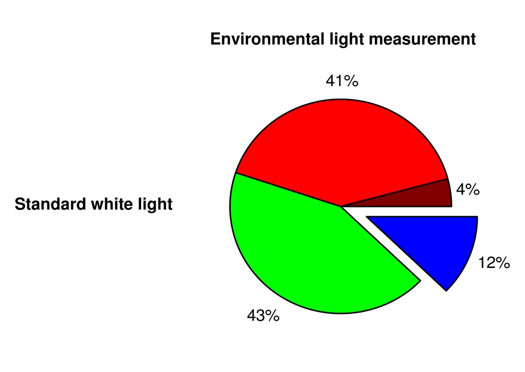
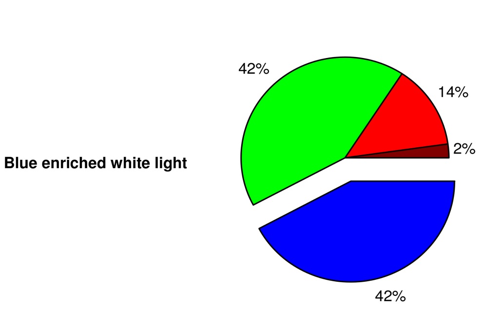
Composition of standard white light and light enriched with blue

On the left, the spectrum of the white light consists of roughly equal parts of red and green (about 40%), then blue (12%) and infra-red waves (4%). On the right, the proportions are different (42% blue and 14% red). Nevertheless, with the naked eye, a human will perceive a white light in both cases.

Antiphospholipid syndrome : discovery of a promising route to improving patient care

Teams of the Nephrology–Adult Transplant department of the Necker Hospital-Sick Children (AP-HP, department led by Prof Christophe Legendre, Paris Descartes University) and of Dr Fabiola Terzi, Inserm research director of the ‘Therapeutic mechanisms and strategies for chronic renal diseases’ team, have just discovered, in part, the molecular mechanisms underlying the development of vascular lesions in the course of antiphospholipid syndrome. By combining fundamental research and monitoring a single cohort of kidney-transplant patients with antiphospholipid syndrome, the researchers have highlighted a beneficial effect of sirolimus, commonly used as an immunosuppressor in organ transplants, to prevent recurrence of vascular lesions on the transplanted kidney. This study was published in the New England Journal of Medicine on Thursday 24 July.

A rare and still poorly-described disease.

Antiphospholipid syndrome is a rare auto-immune disease with an estimated prevalence between 2% to 5% of the general population. It is characterised by the presence of clots that form repeatedly in the arteries or veins, and in pregnant women is accompanied by repeated episodes of miscarriage. Making the diagnosis requires, in addition to clinical signs, identifying the presence of antiphospholipid antibodies in the blood.

Alongside clinical signs, there is a still poorly-described form of this disease characterised by thickening of the intima[1], the wall of blood vessels, and resulting in a problem of the vessels supplying downstream organs (vessel problem called vasculopathy). These lesions are particularly well described in the kidneys where they lead progressively to terminal kidney failure. The physiopathological mechanisms of this thickening were unknown until now and anticoagulant treatments are ineffective against this phenomenon.

Nephrologists know this syndrome well and find themselves powerless against the rapid advance of the disease and the development of terminal kidney failure requiring dialysis. In an initial study we had shown that, unfortunately, these vascular lesions associated with antiphospholipid syndromes reappeared rapidly on the grafted kidney when these patients were transplanted and very significantly reduced the survival of the grafts. This observation was the basis for our thoughts” explains Dr Guillaume Canaud (Necker Hospital-Sick Children, AP-HP, Inserm Unit 1151, Paris Descartes University)

The origin of this advance: the role of the AKT/mTORC pathway

Work done by the teams Nephrology–Adult Transplant department of the Necker Hospital-Sick Children (AP-HP, department led by Prof Christophe Legendre) and Inserm research unit 1151 shows that the development of vascular lesions during antiphospholipid syndrome is largely associated with activation of the AKT/mTORC pathway in endothelial cells by antiphospholipid antibodies.

When this pathway is activated, whatever the cell type, it induces proliferation and growth of the cell” explains Dr Canaud. “First of all we observed in man that this pathway was highly activated in the vessels of patients with a kidney disorder linked to antiphospholipid syndrome and in vessels of the kidney graft after transplant in patients affected by this syndrome. It is also activated in other vascular regions for an extremely severe form of this syndrome“.

The researchers confirmed in vitro that antiphospholipid antibodies were capable of activating the AKT/mTORC pathway in endothelial cells in cultures. They were later able to test in vitro the impact of different AKT/mTORC pathway inhibitors, including sirolimus, on endothelial cells exposed to antiphospholipid antibodies.

Backed by these laboratory results, the researchers observed the impact of sirolimus treatment in a group of transplant patients with antiphospholipid syndrome.

Among 37 patients with antiphospholipid syndrome, transplanted at the Necker Hospital from 2001 to 2009, 10 received sirolimus as an immunosuppressor. These ten patients, compared with the 27 patients that did not receive sirolimus but another class of immunosuppressor, were spared the recurrence of vascular lesions and saw the survival of their transplant very significantly improved.

For Dr Canaud,For the first time, this study describes the mechanisms that lead to thickening of vessel walls in these patients.

Inhibition of the AKT/mTORC pathway, using sirolimus, makes it possible to prevent the reappearance of vascular lesions after the graft and so improve renal survival (12 years post-transplant).

This observation opens a promising therapeutic route in transplant patients with antiphospholipid syndrome, or even in patients carrying this syndrome but not transplanted”.


[1] The tunica intima is the inner layer of blood vessels, corresponding to the endothelial cells and the underlying connective tissue.

New resistance mechanisms to melanoma targeted therapies : contribution of the translation of RNAs into proteins

French investigators have discovered new resistance mechanisms to targeted therapies used for less than three years in the treatment of melanoma. This discovery enables us not only to better understand why these treatments become ineffective but also to reveal new avenues for the management of these aggressive tumours. These studies have been published in the review Nature and have the benefit of an early on-line publication.


The treatment of metastatic melanoma remains a major problem in oncology. Half of the patients suffering from this disorder have a mutation of a protein called BRAF. Medicines targeting this mutated protein, vemurafenib (Zelboraf®) and dabrafenib (Tafinlar), enable the progression of this type of skin cancer to be significantly delayed. Unfortunately, over time, these anti-BRAF compounds loose their efficacy.

Investigators from the Predictive Biomarkers and new molecular strategies in anti-cancer therapy laboratory (Inserm/Gustave Roussy/Paris-Sud University) have shown that the mechanisms used by tumours to resist these treatments involves a protein complex called eIF4F which regulates the synthesis of proteins from RNA. 

 From the biopsies of tumours from patients, investigators also showed that the formation of this complex was diminished in tumours which responded to anti-BRAF and was increased in resistant metastases.

They have also shown that compounds developed by a pharmacochemistry team of the CNRS and by the University of Strasbourg which inhibit the elF4F complex bring about an improvement in the efficacy of vemurafenib in cellular and murine models.

Mélanome

Inserm/Dantchev, Dimitri

These results offer new prospects for the prediction of the efficacy of melanoma treatments using medicines targeting the BRAF protein.

Moreover, over the long term they may result in more effective new treatments emerging to treat not only this fearsome type of cancer, but also certain types of thyroid, colon, lung and brain cancers.

These studies have been conducted by Stéphan Vagner (Inserm U981/Gustave Roussy/Université Paris-Sud, Villejuif; Current address: CNRS UMR3348/Institut Curie, Orsay) and by Caroline Robert (Inserm U981/Gustave Roussy, Dermatology Department/Paris-Sud University, Villejuif) in collaboration with Laurent Désaubry (Therapeutic Innovation Laboratory, CNRS UMR 7200/University of Strasbourg, Illkirch).

The team of Drs Caroline Robert and Stéphan Vagner was supported by PAIR melanoma (Fondation ARC, The league Against Cancer and by INCa), Cancéropôle Ile de France and the group “Ensemble contre le mélanome” (Together against Cancer). This study has also benefited from the support of AAREC Filia Research, from the Wenner-Gren Foundation and from the Swedish Society of Medicine.


HIV : male circumcision also benefits women

Having proven effective in reducing the number of new HIV infections in men, circumcision also appears to play a role in the reduction of HIV incidence in women. These findings are from the study ANRS 12126 coordinated by Professor Bertran Auvert (Inserm U1018, Université de Versailles-Saint Quentin, Hôpital Ambroise Paré) and conducted in the township of Orange Farm in South Africa. They will be presented as an oral communication by Kévin Jean (Inserm U1018) at the 20th International AIDS Conference organized by the International AIDS Society and held at Melbourne from 20 to 25 July 2014

The World Health Organization (WHO) and the Joint United Nations Programme on HIV/AIDS (UNAIDS) have, since 2007, recommended considering circumcision as a complementary strategy in the prevention of HIV infection in men. These recommendations are based on the study ANRS 12165, begun in 2002 in Orange Farm in South Africa by Professor Bertran Auvert (Inserm U1018, Université de Versailles-Saint Quentin, Hôpital Ambroise Paré), the results of which have been confirmed by two other trials conducted in Kenya and in Uganda. This strategy has been deployed principally in regions of South and East Africa where the rate of HIV infection by heterosexual transmission is high and the rate of circumcision is low.

In the framework of a previous study (ANRS 12126), Professor Bertran Auvert’s team has shown that circumcision seems to be well accepted by the male population when it is offered in real-life conditions. In Orange Farm, many men are willing to undergo circumcision (the rate of circumcision has increased from 12% to 53%) and the number of new infections in circumcised men has decreased (half as many new infections as in uncircumcised men). But what about women?

Kévin Jean and Bertran Auvert, together with their colleagues at the NICD/NHLS in Johannesburg, present, for the first time, on the occasion of the 20th International AIDS Conference of the International AIDS Society, the first data on the indirect effect of male circumcision on women.

In women who have intercourse only with circumcised men, circumcision appears to reduce the prevalence (proportion of people infected) and the incidence (rate of new infections) of HIV infection, and does not seem to promote risky behavior (increase in number of sexual partners, non-use of condoms).

The data are from the combination of three cross-sectional studies conducted in 2007, 2010 and 2012 among 2452 women aged 15 to 29. Blood test results, data on sexual behavior, and circumcision status of male sexual partners were recorded. First, the scientists compared the rate of HIV prevalence in women who only had circumcised sexual partners with the rate of HIV prevalence in women who had uncircumcised sexual partners. Over 30% of women reported having had sexual relations only with circumcised men. The rate of HIV prevalence in these women was 17.8%, whereas it was almost twice as high (30.4%) in women who had sexual relations with uncircumcised men.

Second, they used a mathematical model to estimate the HIV incidence rate in these two groups of women for the same period. The rate of new infections in women who only had circumcised sexual partners was 20% lower than in the other women.

These results confirm the importance of voluntary circumcision programs in national HIV/AIDS prevention programs.

The study does not stop here. It is being continued with a new survey among 3000 seronegative and circumcised adult males. Sixteen months after their inclusion in the cohort ANRS 12285, the scientists will examine their sexual behaviors (condom use, risky behavior…) and measure the number of seroconversions. This new study will determine the effect in real-life situations of a large-scale circumcision program on HIV incidence in men and, by extension, will predict the effects of the spread of the male circumcision programs that are currently under way in South Africa and in some East African countries.

Drug users : a new strategy to reduce infectious risk

The risk of transmission of HIV and of hepatitis C virus in intravenous drug users can be reduced significantly by means of support and educational sessions delivered by their peers.
This type of community intervention, which is easily transposable, has been evaluated in the framework of ANRS AERLI, a study conducted jointly by the nonprofit organizations AIDES, Médecins du Monde and by Inserm U912 (Marseille). The results were presented in an oral communication at the 20th International AIDS Conference organized by the International Aids Society and held in Melbourne (Australia) from 20 to 25 July 2014.


Reduction of the risk of HIV and hepatitis C virus infection in intravenous drug users is a public health priority. The ANRS AERLI study on support and education sessions on the risks associated with injecting drugs has assessed an innovative intervention: offer intravenous drug users individual guidance on safe injection practices. Given by trained peers using a standardized protocol, these sessions were based on an educational intervention tailored to each drug user’s practices and questions.

In all, 288 sessions were conducted among 113 drug users recruited in 8 centers specialized in offering help in risk reduction for drug users (CAARUD). A control group of 127 drug users, comparable in terms of drug injection history, age and sex, was set up in 9 other centers. All participants were questioned, at inclusion and again 6 and 12 months later, on injection behaviors involving a risk of transmission of hepatitis C virus and of complications at the injection site.

Data analysis revealed a significant benefit of the educational intervention. In the “Intervention” group, there was:

– a 43% decrease in injection practices involving a risk of transmission of hepatitis C virus (44% of drug users reported at least one unsafe practice before the intervention versus 25% 6 months later);

– a 41% decrease in complications at the injection site (66% of drug users reported complications before the intervention versus 39% 12 months later).

In comparison with the control group, we observed a major effect of peer-to-peer education on practices that run the risk of infection transmission,” commented Patrizia Carrieri (Inserm U912). “This is all the more valuable as a large proportion of these drug users remain on the margins of the health care system.”

This type of intervention has two other advantages: it is inexpensive and is easily implemented in centers for drug users.

“This trial also demonstrates the relevance of community health care interventions, including in research, because the intervention model tested here derives directly from the peer support practices used by nonprofit organizations like AIDES and Médecins du Monde” emphasize Jean-Marie Le Gall, AIDES and Marie Debrus, Médecins du Monde.

Given the observed benefits, said Professor Jean-François Delfraissy, ANRS (France REcherche Nord&sud Sida-hiv Hépatites) Director, it is now conceivable to set up the community intervention on a larger scale, thus enhancing risk reduction among drug users.

 This type of intervention could also be very useful in countries where the drug user population is large and greatly exposed to the risk of infection by HIV and hepatitis C.”

Repeated home-based HIV screening in South Africa : a strategy well accepted on a large scale

The first results of the pilot phase of the trial ANRS 12249 TasP show that the repeated offer of screening for HIV has been well accepted by a rural population in South Africa. The challenge remains of bringing infected individuals into health care facilities for treatment of the infection. These results were presented as an oral communication at the 20th International AIDS Conference organized by the International AIDS Society and held at Melbourne (Australia) from 20 to 25 July 2014.


Does initiation of triple antiretroviral therapy (ART) immediately after diagnosis of seropositivity reduce the transmission of HIV in the population and thereby also the acquisition of new infections (incidence)? This is a pivotal question in development of strategies to fight the HIV epidemic.

Launched in March 2012, the ANRS 12249 TasP (Treatment as Prevention) Trial is one of four international randomized trials designed to assess the efficacy of the TasP strategy in a large population. It is conducted the province of KwaZulu-Natal, South Africa, one of the highest prevalence areas in the world, and the highest in South Africa (16.9% in 2012 according to the latest national survey of the general population).

In this trial, 22 geographical zones (“clusters”), each of approximately 1000 inhabitants, were defined and randomly divided into two groups (an intervention group and a control group comprising 11 clusters). All inhabitants were routinely offering repeated (every six months) rapid HIV testing in their homes. In the intervention group, people found to be seropositive were offered immediate antiretroviral treatment, whatever their CD4 cell count. In the comparison group, treatment was offered according to currently recommended South African Department of Health guidelines. Mobile clinics worked in each cluster to expedite access to care.

Results from the pilot phase of the trial, were presented at the international HIV meeting in Melbourne. This pilot phase was conducted in 10 geographic clusters containing over 12,000 residents above 16 years of age, and followed up for between 12 and 18 months. HIV status was determined in close to 9,000 of these individuals, either because they reported that they were seropositive or because they agreed to rapid HIV testing.

The initial findings are as follows:

HIV status was determined for 82% of those contacted, meaning that home-based testing was acceptable to this population. Among people who were found to be seronegative at the first home-based contact, 85% agreed to a second HIV test at the next home visit.

– Scientists also found a prevalence of HIV infection of 31%, much higher than the initial estimate.

– Around 25% of the 2,570 HIV infected individuals learned of their HIV status through the study.

– In respect to those diagnosed with HIV infected individuals and not already receiving therapy only 48% presented to a clinic within six months.  63% were linked within a year.

– In the intervention group, 80% of those with CD4 >350 cells/mm3 received therapy.

“These initial results are very important,” pointed out Professor François Dabis (Institut de Santé Publique, Epidémiologie et Développement, Inserm U 897, Bordeaux), one of the co-investigators of the trial, “because they validate the feasibility of the TasP strategy, which aims to test and treat the whole adult population so as to curb transmission.

 Rapid home-based HIV testing is very well accepted and we observed no major hindrance likely to call the intervention into question. We noted simply that people newly diagnosed as seropositive need time to engage with care, notably when they feel in good health. However, once in care, ART uptake is good. We are enhancing our strategies to encourage linkage and retention in care for all trial participants”.


“Validation of phase I of this trial is essential,”
explained Professor Jean-François Delfraissy, Director of ANRS (France REcherche Nord&sud Sida-hiv Hépatites). “We can therefore pursue the trial.” The study entered its second phase in June 2014. The first results on the effect of TasP on the incidence of HIV infection in the population should be known by the end of 2016.

The ANRS 12249 TasP Trial is coordinated by Professor François Dabis, Professor Marie-Louise Newell (University of Southampton, United Kingdom) and Professor Deenan Pillay (Director of the Africa Center for Health and Population Studies, Mtubatuba, South Africa, and University College London, United Kingdom). The trial is run in partnership with the Department of Health, KwaZulu Natal, South Africa, GTZ (German government agency operating in the field of technical cooperation) and the Wellcome Trust (United Kingdom). The International Initiative for Impact Evaluation (3ie) is providing funding for the second phase of the study.

Radiotherapy ‘flashes’ to reduce side effects

Treating hard and fast seems to be a good way to limit the side effects of radiotherapy. This is the discovery made by researchers at the Curie Institute, Inserm and the Vaud University Hospital, published in Science Translational Medicine on 16 July.
Radiotherapy remains one of the benchmark local treatments for cancer patients: increasingly accurate, it consists of irradiating cancer cells to destroy them while preserving neighbouring healthy tissues and organs as much as possible. By increasing the intensity of the irradiation 1,000 times over a very short time, the researchers have shown that the efficacy remains the same, but healthy tissues are better protected.


“Eradicating the tumour, while limiting side effects, has always been the aim of radiotherapists”
, emphasises Vincent Favaudon, researcher at the Curie Institute. Radiotherapy is still one of the most effective approaches for treating cancers. It is offered to more than half of patients, combined with surgery and/or chemotherapy. For more than 20 years, developments in imaging, data processing, dosimetry and accelerators have made it possible to ‘sculpt’ the irradiation volume increasingly accurately, depending on the location and shape of the tumour. Despite everything, side effects due to irradiation of healthy tissues remains a crucial problem.

An effect for each mode of administration

In collaboration with Marie-Catherine Vozenin (Inserm and Vaud University Hospital, Lausanne, Switzerland), the radiobiologist Vincent Favaudon, eminent Inserm Research Director, studied the effects of radiotherapy on healthy and tumorous tissues depending on its mode of administration. “The Curie Institute laboratories on the Orsay site has an experimental electron linear accelerator that can deliver high radiation doses in a very short time, as a flash”, he explains. “To give an idea of scale, this accelerator delivers a radiation dose-rate 1,000 to 10,000 times more intense than conventional radiotherapy”.

The researchers wondered if this modified the effects on the tissues. “In our tumour models, a conventionally-administered 15 Gy dose to treat a lung tumour certainly leads to the occurrence of a pulmonary fibrosis between 8 weeks and 6 months after irradiation, while when using a ‘flash’ irradiation no fibrosis appears below 20 Gy”, explains the radiobiologist. This protective effect was also observed on apoptosis (programmed cell death produced following unrepaired damage to the DNA), blood capillaries and skin lesions.

“On the other hand, anti-tumour efficacy remained the same on all the tumour models we tested”, notes Marie-Catherine Vozenin, Inserm researcher and Head of the radiobiology laboratory in the Radio-oncology Department Vaud University Hospital. ‘Flash’ irradiation therefore protects healthy tissues very selectively from side effects arising.

The equipment currently used in most radiotherapy departments, which operate using X-rays, is not efficient enough to generate the dose-rates needed for ‘flash’ irradiation. A major technological advance would be required to achieve it”, continues Vincent Favaudon. “However, the ‘Pencil Beam Scanning’ system currently being installed at the Curie Institute Protontherapy Centre will be capable of such performance and the medical team, assisted by the researchers, is planning to undertake a pre-clinical trial very quickly”.

Pencil Beam soon at the Curie Institute Protontherapy Centre

Since spring 2013, the Curie Institute Protontherapy Centre (Orsay) has been preparing to commission the technology known as ‘Pencil Beam Scanning (PBS)’, which will enable a proton beam to sweep over the tumour.

Institut de Recherche en Cancerologie de Montpellier (IRCM)

Plateforme de radiothérapie clinique du (CRLC) centre régional de lutte contre le cancer. Val d’Aurelle-Paul Lamarque, Montpellier. Inserm/ P Latron

Installed in the treatment room with an isocentric arm – with which it is possible to orientate the beam around the patient depending on all effects – this cutting edge technology will enable the indications for protontherapy to be extended even further.

In this way we will be able to treat new locations, particularly extra-cranial tumours with complex volumes, by ensuring very good conformation to the volume of the tumour while improving the protection of neighbouring healthy tissues and organs“, rejoices Dr Remi Dendale, the centre’s medical director.

 “This is what we call Intensity-Modulated Protontherapy or IMPT, which will enable us to simplify preparation of treatments, by avoiding the need to manufacture compensators (used to adjust dose distribution at depth) and some of the collimators (used to modulate the outline of energy distribution in the lateral plane)“, says physicist Nathalie Fournier-Bidoz.

Images of tissue sections

Effect of irradiation with 17 Gy administered on 0.28 s on healthy lung tissue, equivalent to a dose-rate of 60 Gy/s (middle picture) and in 548 s, equivalent to a dose-rate of 0.031 Gy/s (right picture). Tissue irradiated with a very high dose-rate looks the same as non-irradiated tissue, while tissue irradiated at low dose-rate is completely altered.

Tissu pulmonaire


Working toward improved management of cranial trauma

Under normal conditions and because it cannot store oxygen, the brain cannot withstand being deprived of oxygen for more than a few minutes without risking serious consequences. After an accident (cranial trauma or stroke), emergency teams therefore try to restore cerebral oxygenation as quickly as possible. The faster and more precisely physicians work, the greater the chances of recovery. A multi-disciplinary team at the Grenoble Institute of Neuroscience (GIN, Inserm/ Grenoble-Alps University/Grenoble teaching hospital) comprising physicists, biologists and physicians (neurologists and anaesthetists) has developed a new method for measuring cerebral oxygenation using MRI. Besides being non-invasive, this technique identifies the least oxygenated areas of the brain with precision. Ultimately, it could be used to guide therapeutic interventions and make them more precise, less risky and more effective.
These results are published in the Journal of Cerebral Blood Flow and Metabolism.

Although the brain accounts for just 2% of bodyweight, it uses 20% of the total oxygen consumed by the body. It is unable to create ‘reserves’ and the slightest shortage of oxygen can result in serious consequences (e.g. loss of language functions and motor skills) and even death if not restored very quickly. Ideally in a trauma situation, it should be possible to immediately identify the areas of the brain most affected by a lack of oxygen and where possible avoid physically operating on already weakened bodies.

After an accident, it is vital to monitor oxygenation of the brain. Nowadays, standardized international methods, notably the installation of a probe in the brain, allow brain oxygenation to be estimated locally. This operation entails a neurosurgical procedure and only allows a highly localized measurement of cerebral oxygenation. The only means of mapping the brain’s oxygen levels is by a radioactive measurement of oxygen using functional imaging (a technique developed in the 80s). However, this method also has its limits as it is expensive and requires PET (positron emission tomography) equipment which is scarce in France. Finally, while the brain’s total oxygen consumption remains constant, it varies between the various regions of the brain.

The new MRI method developed by Inserm researchers measures brain tissue oxygen saturation or the quantity of oxygen present in the tissue microvascularization (expressed as StO2).

Thanks to this technique, researchers have observed a major reduction in cerebral oxygenation in an animal model ischemic stroke and, to a lesser extent, following cranial trauma. By mapping cerebral oxygenation, it has been possible to identify regional heterogeneity of StO2 in strokes, cranial trauma and a model brain tumour.

This innovative approach offers a number of benefits including the possibility of immediate use with humans. Unlike positron emission imaging (or PET imaging), which is the gold standard technique, no radioactive tracer needs to be injected when performing an MRI examination. StO2 maps with enhanced spatial resolution can also be produced by MRI. Moreover, MRI examinations are less expensive than PET examinations and MRI imaging devices are more widespread in France than PET imaging devices.

The results of this preclinical trial match those achieved with the two gold standard methods (measurement of blood gas levels and histological mapping of oxygen deprived areas) for different oxygenation levels.

CP_E_Barbier_StO2

From a clinical perspective, several applications may be foreseen, which are illustrated in this project by 3 situations:

  • In patients suffering from cranial trauma, this map could help identify the least oxygenated areas of the brain. Follow-up and therapeutic strategy could therefore be adapted to optimal effect.
  • In stroke patients, this technique should enable more effective identification of the area of ischemic brain tissue which is likely to recover after treatment.
  • Finally, certain malignant brain tumours contain poorly oxygenated areas which are known to be resistant to treatments such as chemotherapy and radiotherapy. It is strongly suspected that these poorly oxygenated areas contain tumour stem cells which cause tumour recurrence. By mapping them more effectively, it should be possible to adapt and customize treatments.


Although the reliability of this system has now been established, it remains to be seen to what extent it is capable of guiding the work of neurologists and anaesthetists. “In several years’ time, it is possible that a precise map of brain oxygenation will enable us to supply drugs to the right place or more effectively configure surgical operations to reduce intracranial hypertension” explains Emmanuel Barbier, Inserm research director.

This work was carried out using the IRMaGe platform, which is a platform of the ‘France Life Imaging’ national life imaging infrastructure. It was funded notably by the Agence Nationale pour la Recherche [French National Research Agency] (IMOXY project).

What are the risks of post-traumatic stress disorder after an accident?

Many patients continue to suffer from symptoms (headaches, pain) several months after an accident, which can pose a real handicap to their lives. The team of Emmanuel Lagarde, research director at Inserm’s Research Centre for Epidemiology and Biostatistics (Inserm/University of Bordeaux) has studied the subsequent development of 1,300 people who were admitted to A&R between 2007 and 2009 for trauma. The researchers demonstrate that it is possible to identify people who will develop post-traumatic stress disorder, which generally occurs when the individual’s life was put in danger. This will enable their care to be adjusted accordingly. Their work also reveals that post-concussion syndrome, which has wrongly been defined as the consequence of cranial trauma, is only one part of post-traumatic stress disorder.

The results of the study have been published in the journal JAMA Psychiatry.

Every year, one in ten people in France are taken to A&E with a trauma following an accident. The large majority of the victims have only mild injuries and are discharged from hospital quickly. However, a number of them continue to suffer long after their direct injuries have healed. They may, for example, have headaches, uncontrollable fear or maladies of various kinds, vision problems, balancing problems or be irritable. When the symptoms occur simultaneously in a single context, they constitute what is called a syndrome.

Following a trauma, two syndromes are described: post-concussion syndrome (PCS), which occurs after a mild cranial trauma, and post-traumatic stress disorder (PTSD), which is encountered in people who have been exposed to a stressful situation wherein their life, or that of another person, was put in danger. Post-traumatic stress disorder was initially described in soldiers who, after exposure to combat or an explosion, complain of nightmares or obsessive thoughts which they are unable to get rid of. The two syndromes have been described for several years in the successive editions of the Diagnostic and Statistical Manual of Mental Disorders (DSM) of the American Psychiatric Association, which is a current standard reference in the area of diagnosis in mental health.

In this study, 1,300 people who were admitted to A&E at the Bordeaux Hospital Centre between 2007 and 2009 were contacted three months after their accident. Over 500 were suffering from a mild cranial trauma when they were admitted to hospital while the others had various injuries, all either mild or moderate in severity. The researchers measured the occurrence of 36 symptoms which are included in the definitions of PCS and PTSD.

‘Post-concussion syndrome (PCS) does not deserve its name because, on the one hand, the symptoms that constitute it are not specific to cranial trauma and, on the other, they do not occur simultaneously. It seems that PCS is, in reality, only one part of post-traumatic stress disorder’,

explained Emmanuel Lagarde, research director at Inserm.

Post-traumatic stress disorder in the general population

The results obtained also make it possible to have a better understanding of post-traumatic stress disorder, which is still insufficiently described for non-military contexts. In the general population, this disorder occurs in 2% of injured people but this figure rises to 9% when the trauma is cranial. However, it is more frequent among women and people who have been in a road accident or have been attacked. The occurrence of PTSD is also influenced by the state of the victim’s physical and mental health before the accident. All this information can enable doctors to determine if early treatment should be provided.

This study puts the classification of post-traumatic complaints into question because it also questions the very existence of post-concussion syndrome, which should be seen as only one part of post-traumatic stress disorder. These results do not, however, question the reality of the suffering of a significant number of people affected by this disorder, for whom the symptoms continue to persist and considerably impact the quality of their lives.

‘This is why it is necessary to describe these syndromes and their origin more accurately, particularly because identifying them also has important consequences in terms of insurance, compensation and the care and rehabilitation policies of patients’,

stressed Emmanuel Lagarde, who is the main author of this work.

100,000 women with breast implants monitored for 10 years

In 2013 an estimated 346,000 women in France had breast implants. Some 30,000 of these are thought to have received an implant produced by the company PIP (some of which have since been removed). Beyond determining that these breast implants are defective, it appears that the only way to document their potential adverse effects is to conduct a large-scale epidemiological study. At the request of the Agence Nationale de Sécurité du Médicament et des produits de santé (ANSM) [French National Agency of Medicine and Health Products Safety], a group of researchers managed by Florent de Vathaire in Inserm Unit 1018 “Centre for epidemiology and population health research” is launching a study entitled LUCIE. The aim of this 10-year study is to monitor some 100,000 women who have or have had breast implants of any brand. The results will enable conclusions to be drawn with regard to the potential medium and long-term adverse events in women with PIP implants.

In 2013, ANSM estimated that 346,000 women in France had silicone breast implants (either for aesthetic or medical reasons) including 30,000 with PIP brand implants. In explantations performed between 2001 and 2013, one in every four PIP implants explanted was considered defective.

There are still uncertainties with regard to the long-term effects of these implants.


In order to examine the potential risk of these implants to the women who carry them, Inserm backed by the Agence Nationale de Sécurité du Médicament et des produits de santé (ANSM) is launching a national epidemiology cohort study entitled LUCIE with women who have or have had breast implants and have undergone surgery in France. The Inserm team managed by Florent de Vathaire of Unit 1018 “Centre for Research in Epidemiology and Population Health” will manage this study.

This cohort has been set up to evaluate and describe the incidence of adverse effects linked to silicone gel breast implants manufactured by PIP. The data collected will be used to determine the state of health of women who have had silicone gel breast implants and examine whether there is a link between any health events declared (breast cancer or other adverse effects such as allergic skin reactions or burst implants) and PIP breast implants.

The aim is to include approximately 100,000 women who have undergone breast implant surgery including 30,000 who currently have or have had a PIP implant (with the remaining implants produced by other manufacturers).


To achieve this goal, the cohort will be recruited in two ways:

1) Through a transfer of records to Inserm researchers by hospitals and clinics which have implanted or explanted breast implants since 2001, regardless of the manufacturer.

2) Through voluntary registration of women after they have signed an on-line consent form on a “live” website on which they will be asked to complete an on-line questionnaire.

People who give their consent to take part will be contacted by e-mail every two to five years and asked to answer further on-line questionnaires (main health events which have occurred since they were last contacted, lifestyle etc.). The women are scheduled to be monitored for at least ten years.

Together, Inserm and ANSM are working to improve scientific knowledge of the problems associated with breast implants and therefore ensure that these products are used safely.

cohorte img2

To help recruit and retain participants, Inserm researchers have taken the following steps to promote the study:

– A website to inform the public of progress with the study and disseminate results. Participants can access the on-line medical questionnaire via this website alarecherchedelucie.fr

– A campaign on social media (Twitter,Facebook,LinkedIn)
Twitter account @EtudeLucie
Page Facebook
On  LinkedIn

Any woman who has or has had breast implants can take part in this study even if she has not experienced any health problems. This is important for ensuring that the study is representative of people with silicone gel breast implants and will ensure that questions raised regarding breast implants are answered.

Women requiring further information can e-mail contact ten.eicul-etrohoc@tcatnoc or call the following number from 15 July:

cohorte img

fermer