Menu

Phage therapy : synergy between bacteriophages and the immune system is essential

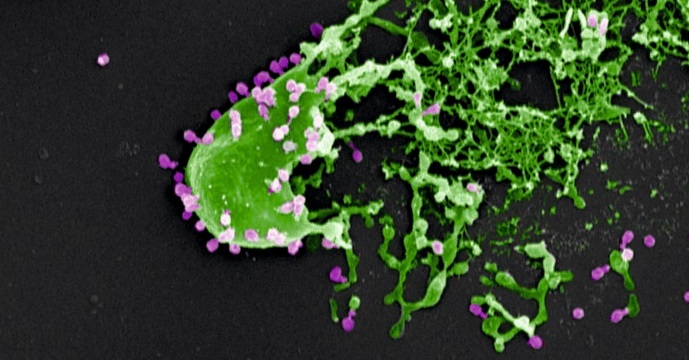
Bacteria (in green) assaulted and killed by bacteriophages (in purple). Electron microscopy image provided courtesy of M. Rohde and C. Rohde (Helmholtz Centre for Infection Research, Braunschweig/Leibniz Institute DSMZ, Braunschweig, Germany) and colorized by Dwayne Roach (Institut Pasteur).
© M. Rohde and C. Rohde

Phage therapy involves the use of bacteriophages, or phages, for treating bacterial infections. Phages are viruses that specifically attack bacteria and are harmless to humans. A significant decline in the use of this therapeutic strategy introduced 100 years ago was seen in the West following the development of antibiotics. However, there is now new interest in phage therapy, especially in Europe, given the alarming increase in the number of antibiotic-resistant bacterial infections.

Until now, there has been insufficient scientific data to understand how phage therapy works in vivo. While most in vitro studies have proven that phages specifically target and kill bacteria, none of these studies took account of the importance of the host’s response to this activity.

Two Institut Pasteur teams (Laurent Debarbieux’s Bacteriophage-Bacteria Interactions in Animals Group and the Innate Immunity Unit led by James di Santo (Inserm U1223)) in partnership with Joshua Weitz’s team at the Georgia Institute of Technology (Atlanta, U.S.), recently showed the importance of patients’ immune status in terms of the chances of phage therapy success. This finding is the result of an original dual approach combining an animal model and mathematical modeling.

In order to evaluate the efficacy of treatment with a single phage species, the researchers focused on the bacterium Pseudomonas aeruginosa, which is involved in respiratory infections such as pneumonia. This bacterium, which is resistant to carbapenems, or ‘antibiotics of last resort’, was ranked by WHO as one of the four biggest global threats in terms of antibiotic resistance.

The researchers demonstrated that phage therapy is effective in animals with a healthy immune system (known as ‘immunocompetent’). The innate immune system can be triggered quickly and phages initially act in tandem with it to fight off infection. Then, after 24 to 48 hours, some bacteria naturally develop resistance to the phages which consequently cease to function. The innate immune system then takes over to destroy the bacteria. Of all the immune cells involved, neutrophils (white blood cells originating in the bone marrow) play a predominant role.

In parallel, in silico simulations have shown that the innate response needs to destroy 20-50% of the bacteria in order for phage therapy to be effective, regardless of whether phage resistance is observed. Thus, in the model studied, the researchers proved that there are no circumstances under which phages are capable of eradicating a P. aeruginosa infection alone.

These findings are particularly significant since they suggest that patients’ immune status should be considered when undertaking phage therapy. Laurent Debarbieux explains: “In terms of clinical consequences, one could reconsider the selection of patients likely to benefit from phage therapy. It may not be appropriate or recommended for people with severe immunodeficiency”.

The researchers are now planning to decipher the exact immune processes involved and the underlying mechanisms. At the same time, clinical trials are ongoing, notably including the Phagoburn trial on skin infections in burn patients funded by the European Union’s 7th Framework Programme.

What life expectancy in good health?

Copyright Fotolia

Life expectancy has been on the increase for several decades in Western countries. But what about life expectancy in good health?

It is clear that the increase in life expectancy is a new source of inequality between men and women in terms of disability.

The work of Jean-Marie Robine (Inserm unit 1198 “Molecular mechanisms in neurodegenerative dementias”) and Claudine Berr (Inserm unit 1061 “Neuropsychiatry: epidemiological and clinical research” in Montpellier) shows that from 2004 to 2015, disability-free life expectancy (DFLE) had increased by 1.1 years for men, going from 61.5 years in 2004 to 62.6 years in 2015. For women, DFLE had virtually stagnated over this period, going from 64.2 years in 2004 to 64.4 in 2015.

These are the findings of a study published in the latest Weekly Epidemiological Bulletin (BEH) published on July 11, which draw on the work of Inserm researchers on aging.

Mobilized on the subject, Inserm researchers are available to answer your questions.

An antidiabetic drug moves a step forward

In humans, apelin is able to regulate blood sugar levels and increase the sensitivity of cells to insulin. These two observations have paved the way for a clinical trial led by Inserm researchers from Toulouse, and represent a promising step forward for the development of a new treatment for diabetes, in particular type 2 diabetes.

This work has been published in the journal Diabetes, Obesity and Metabolism


 It is a journey that began over 10 years ago. A classic research story that demonstrates the long road between discovering a therapeutic molecule and its possible use in humans. The potential use of apelin was demonstrated in 2008 by university professor Philippe Valet and his Inserm team. This ubiquitous molecule (it is found throughout the body) can, if necessary, regulate the body’s blood sugar level instead and in place of insulin. However, this rescue pathway is only activated if the main pathway does not function properly.

Normally, sugar from food is stored in the liver, muscle, and adipose tissue, and is released as and when the body requires. This process is however dependent on the action of insulin, which “captures” sugar for storage. If insulin does not function properly, it leads to diabetes (increased blood sugar levels). Either it is not produced by the body at all: this is type 1 diabetes. Or the insulin receptors located on the surface of the liver, muscle, and adipose tissue cells become desensitized: this is type 2 diabetes. This results in two problems: the levels of circulating glucose are too high, and in time this becomes harmful to the body.

After discovering this alternative pathway, which enables another way of absorbing sugar, the researchers soon had the idea of stimulating this natural pathway and producing synthetic apelin.

Today, the researchers report the positive results of a clinical trial in 16 patients that was carried out within the Diabetology Department headed by Professor Pierre Gourdy. Healthy but overweight men were recruited to take part in a study that aimed to prove the efficacy and tolerability of two different doses of intravenously-administered apelin. The first group received a dose equivalent to 9 nmol/kg, and the second group received 30 nmol/kg. The patients’ glycemia was measured before and after the injection.

The results show that the injection of the smallest dose led to better absorption of circulating blood glucose, while administration of the highest dose also led to a demonstrable increase in cell insulin sensitivity. No side effects were observed.

“This is what we call a ‘proof of concept’ study”, explains Philippe Valet. “Although the sample is a small one, the results that we have just obtained encourage us to move on to larger studies in order to confirm them on a larger scale and be able to consider proper marketing authorization.”

This work could notably contribute to research into the treatment of diabetes, which affects over 400 million people around the world.

Physical activity does not protect against the onset of dementia

 
© fotolia

Physical activity in adulthood is not associated with a reduced risk of developing dementia. However, a decline in this activity is observed during the decade preceding its diagnosis. If this decline in activity cannot be considered as an early sign of dementia, it could be one sign – among others – to be taken into account by the attending physician. This is demonstrated by a study conducted by an Inserm research team of the Center for Research in Epidemiology and Public Health (Unit 1018 Inserm / Université Paris-Saclay). The results were published in the British Medical Journal on June 22, 2017.

Over the past forty years in France, life expectancy in good health has increased by 10 years, from 72 years in 1970 to 82 years in 2010. This increase in life expectancy can be explained by the improvement in quality of life but above all by the progress made in the field of medicine. However, living longer also leads to a higher probability of developing dementia (reduction of cognitive abilities leading to difficulties in everyday activities). The World Health Organization (WHO) estimated in 2015 that 47 million people worldwide were suffering from this disease and this number is expected to triple by 2050.

Until very recently, many studies suggested that physical activity could be a neuroprotective factor, thus delaying the onset of cerebral pathologies. In order to study this hypothesis, a longitudinal study conducted by a team of Inserm researchers from the Center for Research in Epidemiology and Public Health (CESP), in collaboration with University College London, followed more than 10,300 people aged 35 to 55 at the time of inclusion, between 1985 and 2012 (i.e. 27 years). Every 4 years the researchers measured the physical activity of each subject and made them take numerous cognitive tests.

This study showed that physical activity in adulthood would not have a protective effect on the risk of dementia. Indeed, it could not be demonstrated that there was any association between the practice of physical activity (whether mild or moderate to intense) and the onset or not of dementia. Moreover, people following public health recommendations in terms of physical activity, i.e. more than 2 hours 30 min of moderate to vigorous physical activity, showed a decline in cognitive functions, such as memory or thinking skills, similar to people not following these recommendations. As stressed by Séverine Sabia, Inserm researcher heading this study, “These results are corroborated by two recent intervention trials1,2 and an expert report published on June 22, 2017 that concludes there is a lack of sufficient evidence of a protective effect of increased physical activity on the risk of dementia”.

Nevertheless, researchers observed a decrease in physical activity (up to 2 hours / week less) in the 9 years preceding diagnosis in subjects with dementia. These findings suggest that the decline in physical activity in these individuals may be part of the changes that occur during the preclinical phase of dementia such as Alzheimer’s disease. Even today, it is very complicated to diagnose this disease before certain clinical signs such as memory problems or loss of autonomy appear. It therefore remains to be seen whether maintaining a good level of physical activity during this preclinical phase of dementia could slow down the disease process.

While physical activity appears to have no protective effect on the onset of dementia, it is important to remember that practicing a sport is beneficial for the cardiovascular system and the prevention of obesity and type 2 diabetes. It is therefore necessary to maintain good regular physical activity, even of moderate intensity, as it is a major determinant of a person’s state of health at all ages of life.

Gut bacteria can help to predict how the body will respond to fatty foods

Scientists have found that certain compounds, produced by microbes in the guts of mice, could be used to show which animals are at greater risk of becoming obese, or developing health conditions such as diabetes or cardiovascular disease.

The group, led by scientists at Imperial College London and INSERM UMRS 1138 in Paris, tested the urine of mice for a number of these microbial compounds, finding that certain key chemical signatures could accurately predict how the animals would respond to a high-fat diet before they received it.

High-fat diets are a major driver of obesity and related health conditions, such as diabetes and cardiovascular disease. However, evidence from previous studies suggests different people eating the same high-fat diet may have different outcomes, making it hard to define a one-size-fits-all ‘healthy diet’.

Previous research has shown that the hundreds of species of bacteria and other microbes which inhabit our gut work with our own cells to carry out a number of roles, and that this microbial garden can be shaped by what we eat or medicines we take, such as antibiotics.

In the latest study, published in Cell Reports, researchers used genetically similar mice to highlight the role that gut bacteria played in how the body responds to changes in diet and the impacts on health.

Before animals switched diets, their urine was screened for compounds produced by their gut bacteria using magnetic resonance spectroscopy, giving the mice a profile of chemical signatures, generated by metabolites from their microbiomes.

The team found that once the mice were switched to the same high-fat diet, they had a range of outcomes, with some animals gaining more weight than others, or becoming less tolerant to glucose – one of the early warning signs of diabetes.

Analysis revealed that key chemical signatures in their urine were predictive of some outcomes, such as changes in behaviour, weight gain and tolerance to glucose. One compound in particular, trimethylamine-N-oxide (TMAO), was shown to be predictive of glucose tolerance.

“We know that our environment and genetics can influence our risk of obesity and disease, but the effects of these communities of bacteria living inside us are less well understood,” said Dr Marc-Emmanuel Dumas, from the Department of Surgery & Cancer at Imperial, who led the research. “By using a group of mice with the same genetic makeup, we were able to zoom in on the variability in animals switched to a high-fat diet.”

“This study shows that value of a diet is determined not only by your genes, but also the genes of your gut microbes. This work has implications in lots of different areas, which is why it’s so exciting.” Senior investigator on the study, Dr Dominique Gauguier, from INSERM-Paris and a visiting professor at Imperial, said: “Our results illustrate the strong capacity of an organism’s gut microbiome to drive the adaptation to environmental challenges regardless of genetic variation and underline the need of deeper physiological and molecular phenotyping of individuals in large scale genetic studies.”

The findings will be explored further as part of an ongoing large clinical trial of 2,000 patients, where details of their lifestyle, diet, medication and other factors, as well as their microbiomes being characterised. Pulling together all of these data, and building on previous findings, they will be able to reveal how people react to different diets, and how their microbiomes influence the outcome.

According to the researchers, the hope is that in future, a patient’s profile could be generated from urine and blood samples and used to predict which diet they will respond to best.

“Our findings reveal that measuring metabolites in urine before the diet switch, we can predict which animals will get fat and become intolerant to glucose and which ones won’t,” added Dr Gauguier. “These findings open up really strong perspectives into designing personalised diets and harnessing our gut bacteria to promote health.”

A major advance towards a treatment for accelerated ageing

In a study published today in the journal EMBO Molecular Medicine1, the team led by Prof. Nicolas Lévy identifies the mechanism associated with the accumulation of progerin, a toxic protein produced in the course of ageing, and demonstrates the therapeutic potential of a new drug – MG132 – to treat progeria, a rare syndrome involving premature and accelerated ageing. Nicolas Lévy and his team have demonstrated the ability of this drug to considerably reduce progerin production and simultaneously degrade it. This drug, along with other compounds from the same family, is undergoing evaluation for the treatment of other rare diseases, as well as more common diseases including certain types of cancer.

This work, supported by Inserm, Aix-Marseille University, the A*Midex foundation and AFM-Téléthon, paves the way to a therapeutic trial and the development of compounds to reduce the effects of accelerated and physiological ageing.

 Hutchinson Gilford progeria syndrome (HGPS) is an extremely rare and severe genetic disease that causes precocious and accelerated ageing in children. Although it spares the brain functions, it progressively leads to ageing in the vast majority of the organs, with particularly dramatic consequences being observed in the skin, adipose tissue, cardiovascular system and bones. Constantly fatal, death usually occurs around the age of 13 years. This disease, which affects 1 birth per 10–20 million worldwide, is caused by a mutation in the LMNA gene taht leads to the production  and  accumulation  of  a toxic protein, progerin, in cells nuclei. Progerin causes serious cellular dysfunctions (defects in DNA breaks repair, failure of cell proliferation    and    differentiation, etc…). Progeria is thus a unique model for understanding major mechanisms involved in natural ageing. Since 2003, Nicolas Lévy and his team have identified the gene and mechanism inducing progeria and other premature ageing diseases, developed therapeutic approaches, and conducted the first European trial in 12 children affected with the disease.

In the study published today, Nicolas Lévy’s team – UMR_S910, Aix-Marseille University/Inserm – has identified the mechanism whereby progerin accumulates without being degraded, and has identified a family of drugs that not only allow a tremendous reduction in its initial production, but also the simultaneous elimination of the remaining produced progerin. This study, using cells from children affected with progeria as well as a mouse model developed within this same team£, paves the way for a clinical trial for  progeria and other severe diseases of accelerated ageing. It will also be exploited in order to define the potential of each drug identified in the family, with respect to rare genetic diseases, cancers and natural ageing. For Dr Karim Harhouri, first author of the study, “These 5 years of work have enabled us to discover the real mechanism whereby progerin accumulates without being degraded, and a class of drugs that had not been exploited before, with a seemingly major therapeutic potential.”

“This work is part of the main thrust of our research in the area of rare genetic diseases, continuously aimed at translating knowledge of fundamental mechanisms into the most efficient possible treatments for our patients. This could not have been achieved without the convergence of talents, human skills and expertises to reach a common ambition, that of expanding effective treatments for our patients while reducing the access time; this is the philosophy we should be adopting, that of integrated research on care-related problems, and which we are upholding with the creation of the GIPTIS Institute*,” explains Nicolas Lévy, principal investigator, senior author of the study and proponent of the GIPTIS Institute*, which should open its doore in Marseille in 2020.

This work is the subject of a joint patent application – WO2016/113357 – holded by Aix- Marseille University, Inserm, AFM-Téléthon, CNRS and the ProGeLife** biotech company.

*GIPTIS : Genetics Institute for Patients, Therapies, Innovation and Science (www.giptis.com)

** www.progelife.com

A Tool to Predict Cognitive Decline Within 10 Years of Parkinson’S Disease Onset

©Fotolia

An international study with the participation of physicians from the AP-HP Paris public hospitals network and researchers from Inserm, UPMC and CNRS within the Brain & Spine Institute (ICM) has identified a clinical-genetic score to predict cognitive impairment in Parkinson’s disease sufferers. Cognitive impairment is one of the most debilitating characteristics to manifest in certain patients with the disease. The ability to predict its emergence within ten years of the onset of Parkinson’s is of major importance for their treatment and for the set-up of targeted clinical trials.

This study, published in The Lancet Neurology and funded notably by the National Institutes of Health (NIH), brings together U.S. teams from Harvard Medical School and Brigham and Women’s Hospital (Boston).


After several years of living with Parkinson’s, patients can suffer cognitive impairment in addition to the movement disorders characteristic of the disease. In their study, the researchers built an algorithm to identify those patients most subject to this impairment. It was developed using clinical and genetic data from 9 cohorts of patients with Parkinson’s from Europe and North America, i.e. roughly 3,200 patients who were followed over a 30-year period, from 1986 to 2016. 

In France, the DIG-PD cohort, sponsored by AP-HP and coordinated by Prof. Jean-Christophe Corvol from the Department of Neurology and Head of the Clinical Investigation Center at Pitié Salpêtrière Hospital, is part of the project, which is called “Drug Interaction With Genes in Parkinson’s Disease – DIG-PD”. In the French cohort, over 400 patients were followed annually for 6 years.

A number of factors were taken into account and analyzed. Age at disease onset, motor and cognitive severity, education level, sex, depression, as well as β-glucocerebrosidase gene mutation status turned out to be the biggest predictors of cognitive impairment and were included in the prediction model developed by the researchers. The study also revealed that education could play a role in the onset of impairment and that this factor could be associated with potential patient “cognitive reserve”.

Based on this data, the clinical score developed by the researchers precisely and reproducibly predicts the onset of cognitive disorders within 10 years of that of Parkinson’s disease. It was developed thanks to the clinical and genetic analysis of the 9 cohorts, i.e. the analysis of over 25,000 associated elements of data.

This tool is of considerable importance for the prognosis of cognitive impairment in patients with Parkinson’s disease. It could also be used to identify more precisely those at high risk of developing such disorders to enable them to anticipate treatment or participate in targeted clinical trials.

Opening of medically assisted procreation to female couples and single women

©Inserm/Lasalle, Bruno – Human egg fertilized

Today, the National Consultative Ethics Committee (CCNE) has decided to open up medically assisted procreation (MAP) to female couples and single women in France.

In a report released on June 19, the National Academy of Medicine has advocated egg preservation for women who wish to do so.

Medically assisted procreation (MAP) involves manipulating an egg and/or sperm to produce fertilization. This makes it possible to overcome certain difficulties in conceiving, without necessarily treating the causes of infertility.

According to the National Institute of Statistics and Economic Studies (Insee), more than 25,000 babies were born by MAP in France in 2014.

It is estimated that one in seven couples consults a doctor because they are having difficulty conceiving a child, according to the Biomedicine Agency. Around one in ten couples is treated for infertility problems, especially by MAP.

Regarding in vitro fertilization, artificial insemination, etc. Inserm researchers are at your disposal to answer your questions.

Read the Inserm information file on MAP (in French)

Read the “Fertility: our future generations in danger” article published in issue no. 36 of  Science&Santé.

FGF 19: a New Factor to Fight Skeletal Muscle Wasting

Skeletal muscle wasting, a process commonly associated with aging but also seen with various chronic diseases (obesity, cancer, kidney failure), situations of immobilization (accidents, post-operative periods) or prolonged weightlessness (astronauts), strongly impacts quality of life. Researchers from the Cardiovascular, Metabolism, Diabetology and Nutrition (CarMeN) laboratory (Inserm/Inra/Université Claude Bernard Lyon 1/Insa Lyon) in Lyon (France), led by Hubert Vidal, Inserm Research Director, in collaboration with the team of Dr. Jérome Ruzzin from the Department of Biology of the University of Bergen (Norway), have discovered that a hormone produced by the intestine called fibroblast growth factor 19 (FGF19), is able to increase skeletal muscle mass in mice and increase the size of human muscle cells in culture. The researchers have also shown that FGF19 protects from muscle wasting in various experimental mouse models, highlighting its potential therapeutic value. These results were published in Nature Medicine on June 26, 2017.


Fibroblast growth factor 19 (FGF19), an enterokine (hormone secreted by the intestine) well known for its effects on biliary acid metabolism in the liver, is also able to target other tissues and play a glucose-regulation and lipid-homeostasis role. When investigating the therapeutic potential of FGF19 in metabolic diseases such as type 2 diabetes and obesity, the researchers showed that mice treated with FGF19 for 7 days gained less weight and adipose tissue despite eating more than the untreated mice. Under these conditions, the researchers have shown that skeletal muscle mass and muscle strength increases in the treated animals, thereby identifying for the first time a new role of FGF19.

At the molecular level, the researchers have identified a signaling pathway that mediates the hypertrophic effects of FGF19 in muscle and show that this effect is the consequence of enlarged muscle fiber size, irrespective of the type of fiber.

The researchers then demonstrated the therapeutic potential of FGF19 using various mouse models of muscle wasting, including glucocorticoid-treated mice, genetically-obese mice and aged mice. In each model, they demonstrated the capacity for treatment with FGF19 to maintain or increase muscle mass and strength. “For the first time, this demonstrates a potential use of FGF19 to fight skeletal muscle wasting and possibly also in agronomics to increase livestock muscle mass,” concludes Hubert Vidal and his collaborators. They envisage setting up clinical trials to validate these observations in humans.

Omega-3 Essential for the Brain from Adolescence into Adulthood

© Fotolia

Omega-3 fatty acids are crucial for the brain. Deficiency in these acids can lead to depressive mood. A new study led by Inserm and Inra researchers from Unit 901, “Mediterranean Institute of Neurobiology” (Inserm/Université d’Aix-Marseille) and UMR 1256, “Nutrition and Integrated Neurobiology” (Inra/Université de Bordeaux) reveals the mechanisms of disease that develop in adult mice on a low omega-3 diet since adolescence. It also demonstrates therapeutic approaches. The results have been published in The Journal of Neuroscience.

The rapid growth of western societies has been associated with significant dietary changes. The western diet is low in omega-3 essential fatty acids, which are found in large quantities in oily fish such as salmon as well as in chia seeds, nuts, and soy. This type of diet is a risk factor for mental health disorders such as depression and stress. It is important therefore to gain a better understanding of the mechanisms linking imbalanced diet to mental health disorders.

An Inserm team based at Marseille’s Mediterranean Institute of Neurobiology, in collaboration with an Inra team from Bordeaux, has developed a mouse model of omega-3 deficiency lasting from adolescence into adulthood. The researchers observed that introducing this low omega-3 diet from adolescence onward reduces the level of fatty acids in both the prefrontal cortex (which is involved in complex cognitive functions such as decision-making, executive control, and reasoning) and in the nucleus accumbens (which is involved in the reward system and the regulation of emotions), resulting in anxious behaviors and reduced cognitive function in adulthood.

The researchers then investigated the mechanisms underlying these results, and discovered that two primary forms of neuronal learning (in the synapses, the communication zones between neurons) are altered in the prefrontal cortex and the nucleus accumbens in omega-3-deficientmice.

With a view to developing innovative therapeutic solutions, the scientists showed that two methods were effective in completely restoring both the brain function and the emotional and cognitive behavior of omega-3-deficient adult mice. “We simply had to enhance the capacity of mGlu5 (the receptor for glutamate, the most important neurotransmitter in the central nervous system) in the neurons in order to re-establish communication, or inhibit the degradation of the main cannabinoid naturally secreted by the brain, which controls synaptic memory,” explained Olivier Manzoni and Sophie Layé, the researchers who led the study.
These results suggest that nutrition is a key environmental factor that influences brain function and behavior until adulthood, long after the end of the perinatal period. This study identifies nutritional risk factors for neuropsychiatric disorders, and points toward new therapeutic options for behavioral disorders associated with omega-3 deficiency.

Discovery of a new mechanism involved in the migration of cancer cells

A team of young researchers under the supervision of Guillaume Montagnac, Inserm research leader at Gustave Roussy, in collaboration with the Institut Curie and the Institut de Myologie (Myology Institute), has discovered a new mechanism which facilitates cell migration. On the surface of its membrane, the cell develops multiple small hooks which help it to attach to fibres outside the cell and move along them. This action helps us to understand better how a cell escapes from the tumour mass and moves around the body to form a new focus. This research is published in the 16th June issue of the American journal Science.

Cell migration is a normal process which is essential to life. In oncology it is involved in the formation of new metastases.

“Up till the present, we knew that the cell relied on certain structures to anchor itself within its environment. We have now identified new cell structures known as ‘clathrin-coated pits’, already known to be important for other cell functions. The cancer cell uses them as hooks to attach to other structures in order to move around, These novel structures underlie some 50% of cell adhesion to surrounding structures,” declared  Guillaume Montagnac, Leader of the ATIP-Avenir team, attached to Inserm Unit 1170, Normal and abnormal haematopoiesis”, at Gustave Roussy.  

Recognised in 1964, these clathrin pits are small invaginations of the cell membrane which allow it to renew itself or to help molecules to enter the cells. The cell uses them particularly to supply itself with nutritional material (iron, cholesterol, etc.).

Using fluorescence methods, the researchers succeeded in demonstrating with an aggressive human breast cancer line, known for its marked propensity to metastasise, that the clathrin pits adhere to collagen fibres and surround them. The pit squeezes the fibre, so strengthening its hold and allowing it to move.  

“Our Gustave Roussy team is one of the few with an interest in cell membrane dynamics when the cell is placed in 3D matrices under conditions close to normal ones. By studying these clathrin pits in 3D we were able to see the phenomenon when we were not expecting it,” concluded Guillaume Montagnac.

A breast cancer cell with actin (engine of migration) in red, the clathrin pits (cell hooks) in green and collagen fibres in blue

fermer