Menu

Resistance to Antidepressants: the Ability of Neurons to Self-regulate

Why are some depressed patients more or less totally resistant to the most commonly-prescribed antidepressants? This question was addressed by researchers from Inserm and Sorbonne Université at the Fer à Moulin Institute who were able to reveal the major role of neurons that secrete serotonin – the preferred target for antidepressants – in regulating their own activity. Implicated is a serotonin receptor carried by these neurons whose deficiency could be decisive in the absence of response to the most commonly prescribed antidepressants. This research, published in Neurospychopharmacology, will help elucidate the role of serotonin in psychiatric disorders.

Serotonin is a neurotransmitter – a chemical substance produced by some neurons in order to activate others – which is implicated in a number of psychiatric disorders, such as depression, addiction, impulsiveness and psychosis. It is secreted by specific neurons known as serotonergic neurons.

Releasing serotonin outside the neuronal cell activates neurons which possess receptors specific to this neurotransmitter. When these receptors detect sufficient serotonin in the extracellular environment, they send a message to activate or inhibit the neuron that expresses them. The serotonergic neurons also possess several types of serotonin receptors – called autoreceptors – making it possible to self-regulate their activity.

Researchers from Inserm and Sorbonne Universités/UPMC at the Fer à Moulin Institute (Inserm, UPMC) studied the role of one of the serotonergic neuron autoreceptors – known as 5-HT2B – in the regulation of their activity, in order to elucidate the lack of efficacy of some antidepressant treatments.

Usually, when a serotonergic neuron secretes serotonin in the extracellular environment, it is capable of recapturing some of that serotonin which it will release again later on.  This mechanism, ensured by a specific transporter, enables it to regulate the level of serotonin present in the extracellular environment. This transporter is the preferred target of the antidepressant drugs used to treat psychiatric disorders involving serotonin.  These drugs are called selective serotonin reuptake inhibitors (SSRIs) because they prevent this recapture by the transporter. In the context of depression in which serotonin secretion is too reduced, SSRIs make it possible to maintain normal levels of serotonin in the extracellular environment.

The research team took as their starting point the observation that, in the mouse, when the serotonergic neuron does not carry any 5-HT2B autoreceptors, there is lower than usual serotonergic neuron activity and that the molecules blocking the activity of the transporter, such as SSRIs, have no effect on extracellular serotonin levels. The researchers therefore showed that in order to have an effect, these molecules needed the presence and normal expression of the 5-HT2B serotonin receptor.

They also discovered that when a neuron secretes serotonin, its 5-HT2B autoreceptor detects the quantity present in the extracellular environment and sends a signal to the neuron for it to secrete more serotonin.  To avoid the excessive secretion of serotonin, the serotonergic neuron possesses a negative regulator: the 5-HT1A autoreceptor which also detects the level of extracellular serotonin and sends a signal to the serotonergic neuron to inhibit the secretion. In order to maintain normal neuronal activity, 5-HT2B makes it possible to maintain a certain level of activity by acting as a positive self-regulator.

These findings, which remain to be confirmed in human subjects, reveal a fine serotonergic neuron self-regulation mechanism balanced between the activator autoreceptors and the inhibitor autoreceptors.  They constitute a step forward in identifying new drug targets, in elucidating the role of serotonin in certain psychiatric disorders and in understanding the inefficacy of certain antidepressants.
In Mice, Exposure to Chlordecone has Transgenerational Effects on Sperm Production

A study coordinated by Inserm researchers at the Research Institute for Environmental and Occupational Health in Rennes shows that exposing pregnant mice to chlordecone[1] affects the third generation of their male progeny. The number of germ cells – from which spermatozoa originate – decreases, their differentiation is affected, and there is also a decline in the number of mature spermatozoa. This research has been published in Scientific Reports.

During embryonic development, maternal exposure to certain environmental factors can have an impact on the unborn fetus. Early exposure to certain endocrine disruptors is suspected to lead to effects on reproductive function. The objective of this new study was to test, in animals, the consequences on multiple generations following gestational exposure to the known endocrine disruptor, chlordecone.

It has indeed been firmly established that exposure to high doses of chlordecone in adulthood has adverse effects on the production and quality of sperm in laboratory animals and in humans. Previous epidemiological studies conducted by Inserm in the French West Indies have shown that current levels of environmental exposure to chlordecone do not cause changes in sperm characteristics when exposed in adulthood. But given the ability of chlordecone to cross the placental barrier, the question of this substance having an effect during exposure in utero remained unanswered.

To address this question, pregnant mice were exposed by oral route to a daily dose of chlordecone which is not known to induce harmful effects in this species (100 μg per kg of bodyweight). The selected exposure period (from embryonic day 6 to 15) is not just a critical window for the transmission of epigenetic information to the next generations but also a vulnerable time when it comes to germ-cell development.

The principal findings?

Exposing the pregnant females to chlordecone affects the third generation of their male progeny (the first was not directly exposed): the number of germ cells – or spermatogonia – decreases, their differentiation is affected, and there is also a decline in the number of mature spermatozoa.

In other words, explains Fatima Smagulova, Inserm researcher and scientific manager of this study and an ATIP/Avenir team: “the entire germline in the male is affected, whether quantitatively or qualitatively, after two generations.

These modifications appear to be correlated to changes in the location of certain epigenetic marks (notably histone methylation and acetylation) situated in the promoters of genes coding for transcription factors, some of which are regulated by estrogen receptor 1 (ESR1).

Finally, modifications in the expression of 377 genes coding for proteins implicated in essential cell functions (chromosomal segregation, cell division and DNA repair) were observed.

This rodent study shows that prenatal exposure to chlordecone at low doses leads to transgenerational effects on sperm production and suggests that the hormone properties of the compound could be implicated in the mechanisms behind these effects. However, the actual impact on the fertility of men living in the West Indies following prenatal exposure to chlordecone remains to be elucidated.

[1] Chlordecone is an organochlorine insecticide which was used from 1973 to 1993 in plantations in the French West Indies to combat the banana weevil. Its continued presence in the environment is responsible for contaminating local foodstuffs, whether plant or animal, terrestrial or aquatic. The local populations – including pregnant women – are exposed, as shown by impregnation studies previously conducted by Inserm, with current exposures occurring mainly through the consumption of contaminated foods.

When a Gut Bacterium Aggravates Metabolic Syndrome and a Probiotic Relieves It

©AdobeStock

While it is evident that obesity, type 2 diabetes and other metabolic complications are current public health issues, their prevalence is much less clear. A team of researchers from Inra, Danone, the Paris public hospital system (AP-HP), Inserm and Sorbonne Université have recently revealed, in an in vivo preclinical study, that the metabolic disorders linked to a high fat diet are aggravated by the proliferation of Bilophila wadsworthia, a pro-inflammatory gut bacterium which contributes to the deterioration of the intestinal barrier. These effects are attenuated by a probiotic bacterium known as Lactobacillus rhamnosus CNCM I-3690. The findings of this study pave the way for the development of nutritional approaches and probiotics which target the microbiota. They were published on July 18, 2018 in Nature Communications.

The name Bilophila wadsworthia probably does not ring any bells for the vast majority of us. It is true that, in a healthy individual, it represents less than 0.1 ‰ of the gut microbiota. However, it is significantly more abundant in individuals consuming a high fat diet. Yet, changes in the composition of the microbiota are commonly associated with metabolic dysfunction without the underlying mechanism of this relationship being properly understood.

In an in vivo preclinical study, researchers from Inra, Danone, the Paris public hospital system (AP-HP), Inserm, Sorbonne Université and their colleagues showed that a high fat diet creates conditions conducive to the proliferation of gut bacteria such as B. wadsworthia. This proliferation is accompanied by an aggravation of the various parameters which characterize metabolic syndrome (e.g. impaired glucose tolerance, reduced sensitivity to insulin and increased blood or hepatic lipids). It is also linked to gut inflammation, intestinal barrier dysfunction as well as bile salt metabolism disorders, favorable to the development of this bacterium.

The scientists then explored the therapeutic potential of a probiotic bacterium, Lactobacillus rhamnosus, revealing the utility of a specific strain known as CNCM I-3690. This strain limits the proliferation of B. wadsworthia, protects the intestinal barrier from its pro-inflammatory effects and improves the parameters of glucose regulation.

This research highlights the role of a gut bacterium, B. wadsworthia, in aggravating the metabolic effects of a high-fat diet. These findings, if they are confirmed in humans, pave the way for the preventive and therapeutic use of probiotic strains likely to ward off the specter of inflammatory and metabolic diseases such as type 2 diabetes and obesity, by restoring the functions ensured by a balanced gut microbiota and by contributing to improve dietary quality.

Obesity: Using the Microbiota to Fight the Harmful Effects of Fatty Liver

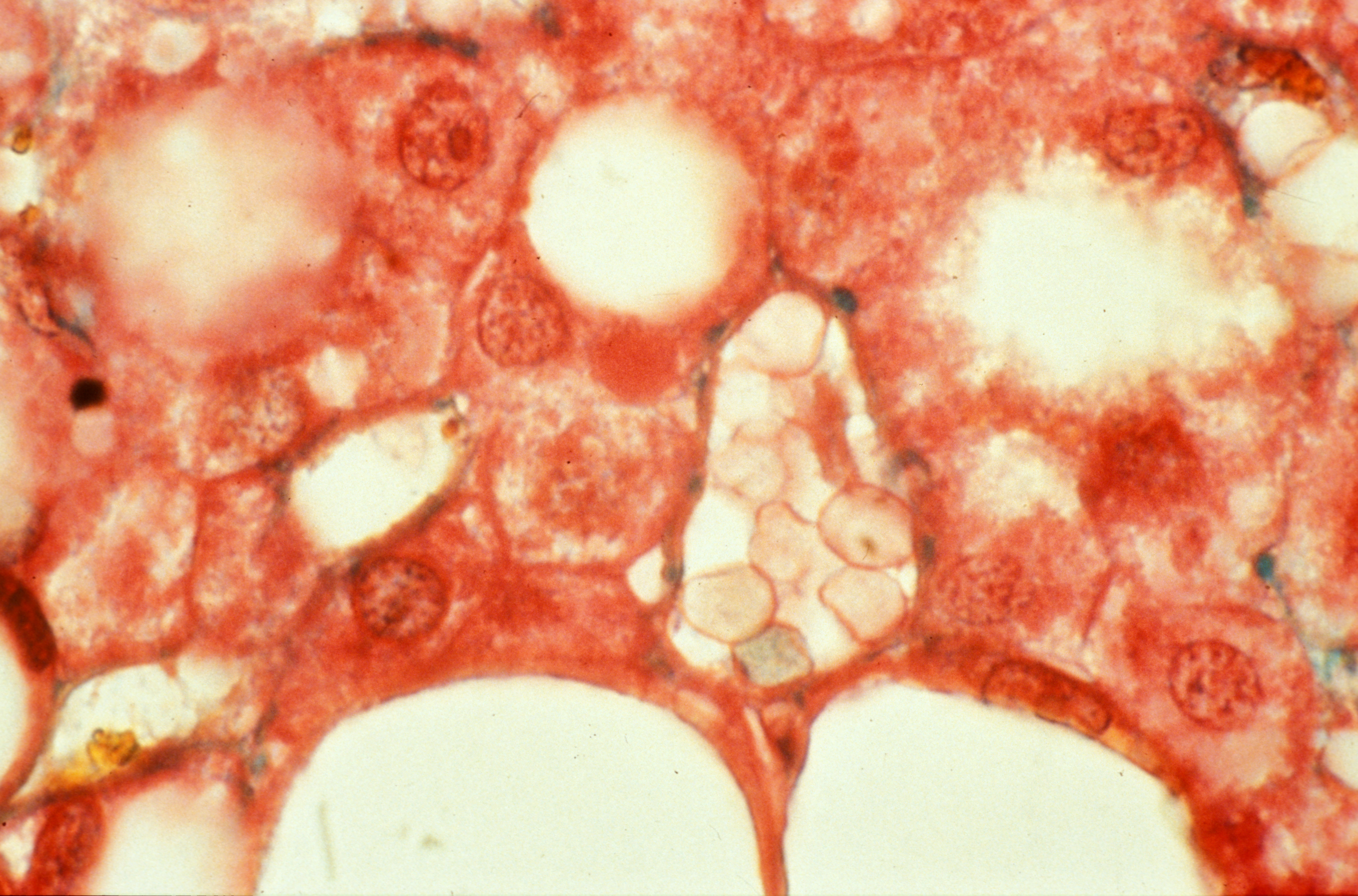
Detail of a stéatose, an accumulation of a fat, a triglyceride, in the hepatic cell. Copyright: Inserm/Hadchouel, Michelle

Day after day, the microbiota continues to yield its secrets. In a new study published in Nature Medicine, Rémy Burcelin, Inserm Research Director at the Institute of Cardiovascular and Metabolic Diseases (Inserm/UPS) along with researchers from Inserm Paris, Imperial College London, Girona Hospital and University of Rome Tor Vergata, show how certain gut bacteria cause the accumulation of fat in the liver and play a major role in fatty liver disease (hepatic steatosis). This research could ultimately lead to the availability of biomarkers to predict the disease and proposed therapeutic solutions based on nutritional, and pharmacological approaches and a new generation of probiotics.

Seventy to 80% of people with obesity and type 2 diabetes have “fatty liver”. Fat accumulated in this organ rapidly, when associated with inflammation, leads to the development of liver failure, a diminished ability to filter environmental and dietary toxins resulting in the risk of cancer.

No drug treatments exist at present. The only solutions involve diet and, in the most extreme cases, liver transplant.

Faced with this therapeutic dead-end, FLORINASH, a European consortium of researchers from France (Inserm), Italy and the UK decided to create and compile data from two large cohorts of 800 obese men and women, separating the groups according to the presence or absence of fatty liver. Various medical data were collected from the participants. Then, in a smaller subgroup of around 100 obese women, molecular analyses were carried out on liver biopsies and urine, plasma and fecal samples.

The objectives of this new study were to discover the biological pathway through which liver failure manifests, reveal biological markers to predict the risk of developing the disease in obese individuals, and find therapeutic solutions.

Existing findings from Inserm published in 2007 and 2011 had explained the process through which gut bacteria (microbiota) become harmful, leading to obesity and type 2 diabetes. The researchers were therefore wondering whether the microbiota also plays a role in the onset of the hepatic complications of type 2 diabetes and obesity.

Using the female patients’ biopsies and clinical data, the researchers applied a big data approach. An immense database containing the various molecular details of the microbiota composition, liver genes, plasma proteins and urine proteins was constituted. The researchers then developed algorithms able to identify a logical link between these data.

In other words, explains Rémy Burcelin, Inserm Research Director and study coordinator: “We wanted to see if we could identify, step by step, from the microbiota, the succession of mechanisms responsible for hepatic disease.”

Through this large-scale work in which the algorithms screened over three million bacterial genes, the researchers made two findings. The first is that the more the disease progresses,the greater the fall in microbial gene diversity, suggesting diminished microbiota composition even before the first symptoms appear. The second is that a specific compound of the microbiota, phenylacetic acid, potentiates fat accumulation in the liver while lipopolysaccharides trigger inflammation.

In order to prove the causal link between observation and effect, the researchers continued their work, this time in animals and human liver cells. This involved transferring into healthy mice the microbiota of human donors with fatty liver disease, leading to a drastic increase in their liver triglycerides. It was also shown that when phenylacetic acid was administered to mice, fat built up in their livers.

According to the team, it would be possible in the long-term, by manipulating intestinal bacteria, to prevent the hepatic complications of obesity. The idea is also to be able to lead to the development of a new generation of probiotics, food supplements and to a pharmacological strategy which interferes with the bacterial mechanisms responsible for the liver disease.

Chronic liver disease: discovery of the role of T cell mucosa-associated invariant (MAIT) in inflammation and fibrosis

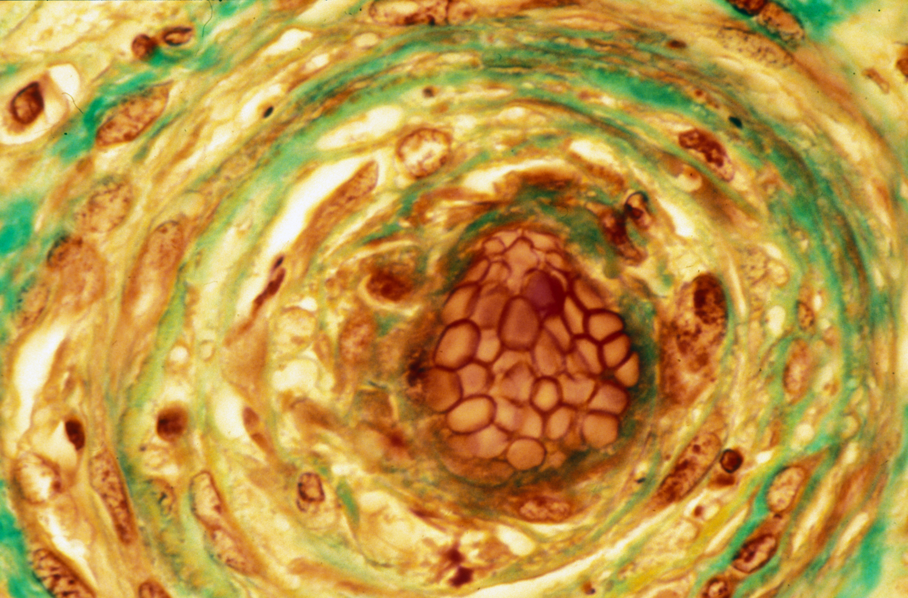
Crédits: Inserm/Hadchouel, Michelle

In collaboration with the team “Inflammation and stress in liver disease” of the Research Center for Inflammation (UMR 1149-Inserm-Université Paris Diderot), teams of Hepatology Services, anesthesia and resuscitation ‘pathology Beaujon hospital AP-HP, Paris Diderot University and the Cochin Institute (1016 UMR Inserm-University Paris Descartes), demonstrated that a specific population of T cells, called “MAIT “played a major role in inflammation and fibrosis associated with chronic liver disease.

These cells could represent an interesting antifibrogénique strategy to develop new therapeutic approaches to chronic liver disease. This study was the 1 st June 2018 to be published in the journal Nature Communications.

Cirrhosis is the latest evolutionary stage of fibrosis associated with chronic liver diseases regardless of their cause (mainly alcohol abuse, chronic viral hepatitis and metabolic steatopathy in France). It is estimated that in France 200 000 to 500 000 individuals with cirrhosis and more than 170,000 deaths per year are linked to the disease in Europe. Eventually, cirrhosis leads to liver failure whose only cure is liver transplantation. Indeed, there is to date no molecule which antifibrotic effect has been validated in clinical practice.

Chronic liver disease is characterized by persistent inflammation that contributes to their progression to more severe stages. They can progress to liver fibrosis and cirrhosis, and then require a liver transplant. A treatment for a regulation of this inflammatory response could be an interesting anti-fibrogenic approach.

The team of Dr. Sophie Lotersztajn (center for research on inflammation Inserm-Université Paris Diderot), in collaboration with anaesthesiologists services (Dr Emmanuel Weiss), pathology (Prof. Valerie Paradise) and hepatology (Pr Pierre-Emmanuel Rautou) Beaujon hospital AP-HP and a team from the Institut Cochin – Paris Descartes University (Dr Agnes LEHUEN) have focused on the role of invariant T cells associated with mucosa (MAIT) which function just beginning to be decrypted.

This work shows that during cirrhosis, MAIT cells in the liver and blood are activated and they accumulate in the liver in contact with the fibrogenic cells in the fibrous septa. In a mouse model enriched MAIT cells, fibrosis is exacerbated.

Conversely, deficient mice are resistant MAIT fibrotic process. Finally, in vitro studies demonstrate that MAIT cells interact with macrophages by increasing inflammatory and hepatic myofibrolastes stimulating their profibrogéniques properties.

This study highlights the role of MAIT cells in inflammation and fibrosis associated with chronic liver diseases and suggests that targeting these cells could be an innovative therapeutic approach antifibrogénique.

Cost-effective universal screening for hepatitis C in France

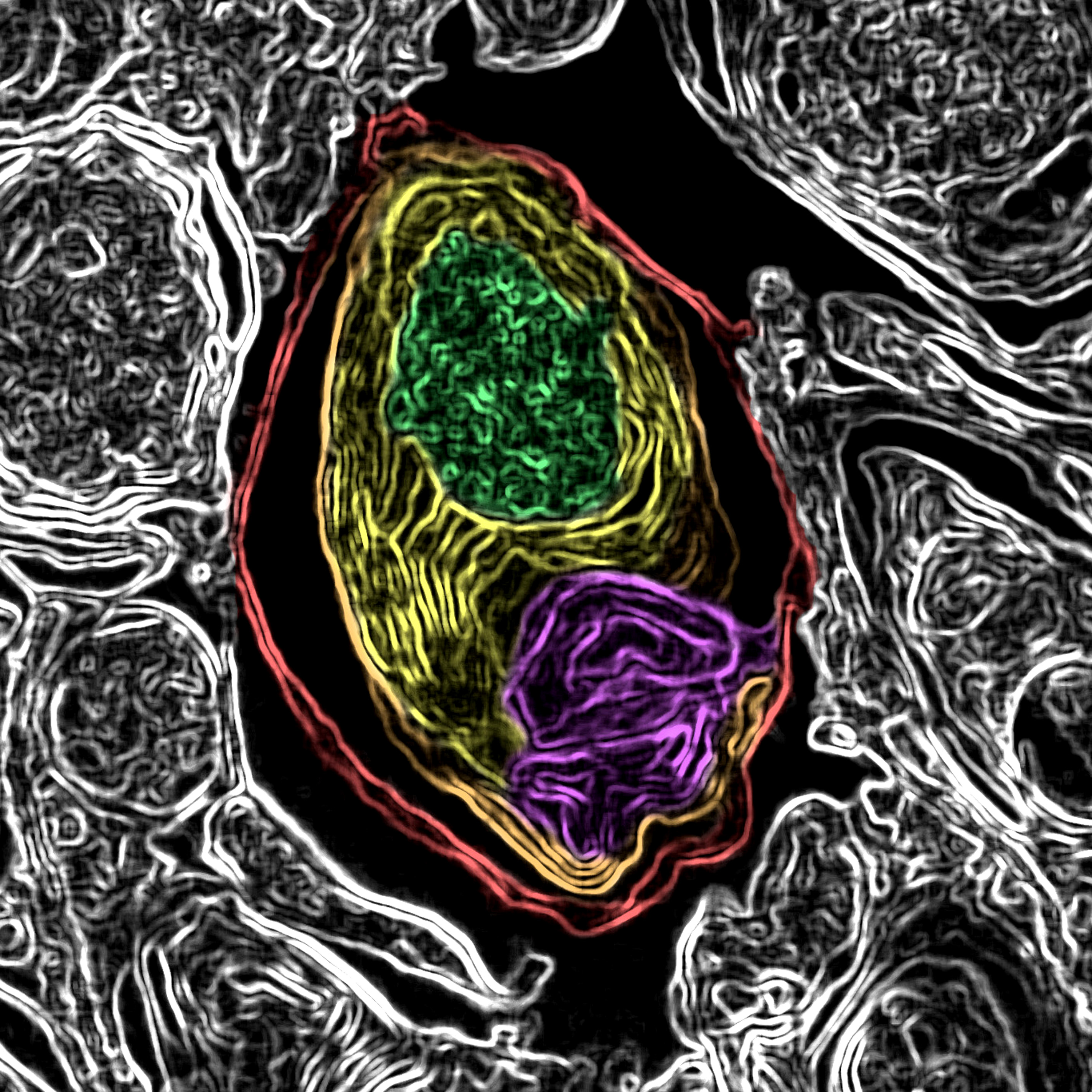
Crédits: Inserm/Jammart, Baptiste

An estimated 75 000 people in France are unaware they are infected by hepatitis C virus. An ANRS-funded study by Sylvie Deuffic-Burban, a research associate at IAME (Infection, Antimicrobials, Modeling, Evolution) (Inserm – Université Paris Diderot – Université Paris 13), and her team shows that a universal screening strategy applied to hepatitis C is cost-effective and improves life expectancy in those infected, compared with targeted screening. These modeling results will be published on 1st July 2018 in Journal of Hepatology.

It is currently recommended in Europe that screening for hepatitis C virus (HCV) should target people at high risk of infection. In France, public health data suggest that in 2014 approximately 75 000 people aged 18 to 80 were infected by HCV, but were unaware of their status. In at least one in ten cases, these people are at an advanced stage of the disease when diagnosed. Today’s treatments of HCV infection are both highly effective and well tolerated, and cure the infection in a few weeks in over 95% of cases.

In Professor Yazdan Yazdanpanah’s Inserm research team, Sylvie Deuffic-Burban has developed a mathematical model that assesses the efficacy and cost-effectiveness of different screening strategies, including universal screening.

This study applied data from a 2004 InVS seroprevalence survey to 18- to 80-year-olds in France, excluding people with diagnosed chronic HCV infection. The researchers developed their analytical model using a combination of these seroprevalence data and findings from studies of the characteristics of people infected (age, sex, stage of the disease at diagnosis, alcohol intake, etc.), the natural progression of the disease, the efficacy of treatments, the quality of life of the patients treated, and the cost of treatment of infection. The screening strategies assessed targeted the following groups: the at-risk population only, all men aged between 18 and 59, all people aged between 40 and 59, all people aged between 40 and 80, and everyone aged between 18 and 80 (universal screening).

The modeling results show that universal screening is associated with better life expectancy adjusted for quality of life than other strategies. Universal screening is cost-effective if the patients tested for HCV infection are treated rapidly after diagnosis.

Sylvie Deuffic-Burban points out that “Screening, on an individual basis, enables rapid treatment, which avoids the development of serious complications. In time, collective screening helps eliminate hepatitis C from a population that has been screened without restrictions.

The results of this ANRS-funded study therefore argue in favor of universal screening for HCV in France, followed by immediate treatment of those diagnosed with HCV infection.

Sylvie Deuffic-Burban concludes that “Although our model is unable to test the idea, the epidemiological similarities of HCV, HIV, and HBV suggest that universal and combined screening for these three viruses could be of particular interest.

fermer