Menu

Sport Has Its Benefits but Do Not Overdo It

©Braden Collum/Unsplash

Sport is good for health, we are always being told. However, in top athletes, excess physical activity can be harmful, as the cases of “overtraining syndrome” show. Responsible for major fatigue and reduced sporting performance, it is a phenomenon that intrigues scientists. A study performed by Mathias Pessiglione, Inserm Research Director at the Brain & Spine Institute (Inserm/CNRS/Sorbonne Université) in conjunction with the French Institute of Sport, Expertise and Performance (INSEP) and the French Anti-Doping Agency (AFLD) shows that intensive physical training can harm brain capacity, particularly cognitive control. The full results have been published in Current Biology.

Inserm researcher Mathias Pessiglione and his team were interested in identifying the causes of a common phenomenon in top athletes, known as “overtraining syndrome”. This is expressed by reduced sporting performance and intense fatigue. Athletes suffering from this syndrome may be tempted by products likely to restore their performance, hence the involvement of AFLD in the project.

The primary hypothesis of the researchers was clear: the fatigue caused by overtraining is similar to that caused by mental effort and is thought to be linked to the same brain mechanisms. Another recent study had already shown that mental fatigue affects cognitive control and leads to impulsive decisions.

To test their hypothesis, the team spent nine weeks working with 37 triathletes, who were split into two groups. The first underwent the “usual” high-level training whereas the second had additional training during the last three weeks of the experiment, with sessions lasting 40% longer, on average. The participants were all monitored at the Brain & Spine Institute, both behaviorally and via functional MRI.

Training sessions 40% longer, on average

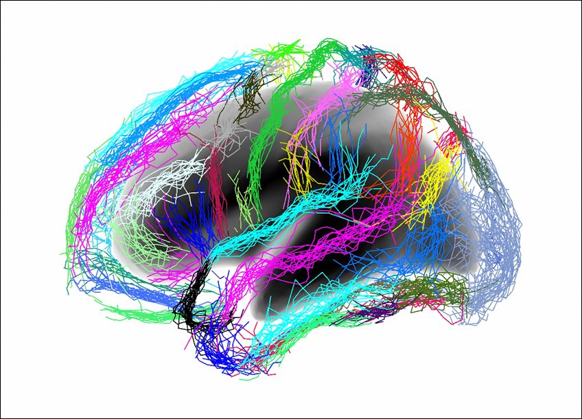
From this, the researchers were able to identify similarities between overly intensive physical training and excessive mental work. This excessive physical activity leads to reduced activity of the lateral prefrontal cortex (a key region for cognitive control), similar to that observed during mental effort. A reduction in brain activity expressed by impulsive decisions, in which short-term gratification is prioritized over long-term goals. In the case of top athletes, such impulsiveness can, for example, lead to the decision to stop right in the middle of a sporting performance or abandon a race in order to end the pain felt during physical exertion.

Beyond these top athletes, the researchers consider that, clinically, fatigue and reduced cognitive control could constitute the first stage in the development of burnout syndrome, which affects many people across various professional domains.

The next step for the researchers is to propose and test interventions in order to avoid the onset of actual burnout – namely the total exhaustion of the individual.

Identified: A Protein Essential for Chikungunya Virus Replication

Fibroblast infected with the chikungunya virus. © Inserm/Thérèse Couderc/Marie-Christine Prévost/Marc Lecuit

Chikungunya is an infectious disease caused by a mosquito-borne virus transmitted to humans. First detected in Africa, the virus has been responsible for recent epidemics in the Americas, Asia and the Indian Ocean – particularly the Reunion Island. Chikungunya is characterized by high fever and intense joint and muscle pain that can last for several months. The mechanisms of infection of human cells with the virus remain very poorly understood. Led by Ali Amara in collaboration with Marc Lecuit researchers from Inserm, Institut Pasteur, CNRS and Université de Paris have identified a protein that is crucial in order for the virus to replicate within its target cells. This research, published in the journal Nature, opens up therapeutic avenues in the fight against chikungunya.

Originally from Africa, chikungunya is aptly named. It derives from a word in the Kimakonde language, meaning “to become contorted”, because the severe muscle and joint pains endured by the patients prevent them from moving normally or performing their daily activities.

While the clinical manifestations of the disease are well understood, the mechanisms by which the virus infects human cells and multiplies remain poorly elucidated. Several studies had already identified certain host cell factors implicated in the replication of the virus. However, none had succeeded in explaining why the virus preferentially targets the muscle and joint cells, causing these clinical signs.

Researchers from Inserm, CNRS and Université de Paris led by Dr. Ali Amara at the AP-HP Saint-Louis Hospital Research Institute in Paris, in collaboration with Marc Lecuit’s team from Institut Pasteur, Inserm and Université de Paris, have identified that the FHL1 protein is a key cellular factor for the replication and pathogenesis of chikungunya. FHL1 is a molecule present mainly in the muscle cells and fibroblasts, the preferred targets of the virus. Usually, FHL1 contributes to healthy muscle physiology and it is now thought to be diverted from that function by the virus to ensure its replication in the target cells.

To conduct this study, Amara’s team used the CRISPR-Cas9 technology to systematically screen the genome of human cells in order to identify the host factors necessary for viral replication. In doing so, it isolated the gene coding for the FHL1 protein. The team then conducted a series of experiments showing the incapacity of the virus to infect cells whose FHL1 expression had been abolished.

In addition, the researchers have shown that the virus was incapable of multiplying within cells derived from patients suffering Emery-Dreifuss muscular dystrophy – a rare genetic disease. This muscle disease is the result of mutations of the FHL1 gene responsible for the breakdown of the FHL1 protein. The researchers have shown that the cells of these patients are resistant to the virus.

Finally, the researchers performed in vivo experiments in mice whose Fhl1 gene was invalidated. They have shown that these animals are totally resistant to infection and do not develop the disease, whereas the virus multiplies and causes major muscle lesions in mice expressing a functional FHL1 protein. These observations demonstrate that the FHL1 protein plays a key role in chikungunya virus replication and pathogenesis.

The precise role played by FHL1 in the viral infection is not fully understood. The researchers have discovered that FHL1 interacts with a viral protein known as nsP3. It is when binding to this that FHL1 participates in the replication of the virus.

“We now want to understand this interaction in molecular detail. The next step is to define why FHL1 is so specific to the chikungunya virus, and to decipher its mechanism of action at the molecular level. Elucidating the molecular structure of the FHL1-nsP3 complex could represent a major step forward in the development of antivirals that block the replication of the virus“, emphasize Ali Amara and Laurent Meertens, the Inserm researchers in charge of the study.

At present, only symptomatic treatments are available for patients infected with chikungunya.

Disarming a probiotic to improve its benefits

Escherichia coli © Inserm/Cloup, Emilie/Nougayrede, Jean-Philippe

For more than a century, the Nissle 1917 strain of the bacterium Escherichia coli has been used as a probiotic to treat gastrointestinal disorders. However, this bacterium also produces a toxin, colibactin, which has deleterious effects on host DNA and might cause colon cancer. It is therefore crucial to understand the mechanisms at play in the strain if we wish to limit undesirable side effects. In a recent study, researchers at INRA, INSERM, the University of Toulouse III – Paul Sabatier, and ENVT managed to decipher certain pathways by which Escherichia coli Nissle 1917 produces both beneficial and toxic compounds. They then successfully created a modified strain with the same probiotic properties but an unactivated version of the toxin. This work was published on September 23, 2019, in PLOS Pathogens.

A German doctor discovered the bacterial strain that became known as Escherichia coli Nissle 1917 during World War 1. There had been an outbreak of dysentery in a group of soldiers; only one remained unafflicted. The doctor, Alfred Nissle, isolated the strain from this soldier and used it to treat other soldiers. Since then, E. coli Nissle 1917 has been used as a probiotic to treat various gastrointestinal disorders.

In 2006, researchers at INRA, INSERM, the University of Toulouse III – Paul Sabatier, and ENVT discovered that the genome of E. coli Nissle 1917 contained a group of genes that jointly encode a toxin: colibactin. In certain E. coli strains, colibactin acts as a virulence factor and can promote colon cancer. Until now, researchers had been unable to decouple the strain’s probiotic properties from its toxic side effects. It was therefore necessary to delve into the mechanisms underpinning both the strain’s synthesis of beneficial compounds, which make it a probiotic, and its synthesis of harmful compounds, notably colibactin, which can have negative impacts.

In a new study, this same team of researchers uncovered the important role played by the protein ClbP. It is an enzyme that has a key function in the strain’s probiotic action but that also activates colibactin. 

Using this discovery, the scientists were able to create a strain in which a tiny relevant portion of the enzyme was modified. This strain had the same probiotic properties as the original but could not activate colibactin, negating its toxicity. In vivo assays in mice confirmed the functionality of the new strain. Mice infected with Salmonella typhimurium (one of the bacteria that can cause dysentery) developed less severe symptoms of salmonellosis when treated with the modified E. coli Nissle 1917 strain.

Comparable results were obtained with the original E. coli Nissle 1917 strain.

This research underscores the importance of taking a closer look at the bacteria that we use as probiotics. Indeed, like traditional medications, they can have side effects.

This study may have further implications because genomic analyses have revealed that other E. coli strains naturally produce modified versions of this enzyme. Future work should assess whether such bacteria could also serve as probiotics.


Illustration of the experimental approach. © INRA, INRA, Eric Oswald


Illustration of the experimental approach © INRA, INRA, Eric Oswald

A Single Dose of Yellow Fever Vaccine Does Not Offer Lasting Protection to all Children

WHO©C. Onuekwe

The World Health Organization (WHO) recommends a single dose of the yellow fever vaccine for individuals aged 9 months or older living in or traveling to areas at risk of disease transmission, but there is a lack of data on its long-term efficacy when administered to infants. José Enrique Mejía, Inserm researcher at Unit 1043 Center for Pathophysiology of Toulouse Purpan and Cristina Domingo from the Robert Koch Institute in Berlin have recently shown that around half of children initially protected by the vaccination at 9 months of age lose that protection within the next 2 to 5 years, due to disappearance of the neutralizing antibodies. This research has been published in The Lancet Infectious Diseases.

Yellow fever is a viral infection spread by various species of mosquito and is rife in 34 countries in Africa and 13 in Latin America. Infection may be asymptomatic and go unnoticed or, on the contrary, it may progress rapidly to severe illness with fever, headache, muscle pain, nausea, vomiting and fatigue. The virus attacks the liver cells, often causing jaundice from which the disease gets its name. Severe bleeding occurs in 25 to 50 % of cases, with high levels of mortality observed 7 to 10 days after the onset of symptoms.

Since 2013, WHO recommends a single dose of the vaccine for life-long protection. This recommendation is based on proof of long-term efficacy, in vitro and in vivo, established in adults and children over 2 years of age.

But data on the long-term efficacy of primary vaccination in infants are absent, despite 9-12-month-olds being the main targets of routine vaccination in countries in which yellow fever is endemic. In this respect, WHO recommended research into the long-term persistence of the immunity conferred by vaccination in this age group. This research was performed by José Enrique Mejía from Unit 1043 Center for Pathophysiology of Toulouse Purpan in partnership with Cristina Domingo from Robert Koch Institute in Berlin, and researchers from the USA, Ghana and Mali, with support from the Wellcome Trust.

Their study verified whether children to whom the vaccine was administered at around 9 months of age were still protected several years later. The team studied two cohorts, one from Mali (587 children) and the other from Ghana (436 children), in whom the levels of specific antibodies to the yellow fever virus had been measured 4 weeks after vaccination. They then repeated the measurement several years later, with findings from previous studies enabling them to estimate that levels above 0.5 IU/ml should protect children from infection.

In the Malian cohort, 4.5 years after vaccination, only half of the children continued to present levels of antibodies above 0.5 IU/ml. And 19.3 % presented detectable antibodies but at levels below this recommended threshold (<0.5 IU/ml). The proportion of children seropositive for these antibodies was therefore 69.7 % as opposed to 96.7 % just after vaccination.

In the Ghanaian cohort, 2.5 years after vaccination, only around 30 % of children continued to be protected against infection and 11.7 % continued to present specific antibodies but in low concentrations (<0.5 IU/ml). All in all, 39.4 % of the children were considered seropositive as opposed to 72.7 % just after vaccination.

Irrespective of the differences in vaccine efficacy between these two groups, which could be explained by ethnic and environmental factors (urban/rural population, seasonality of vaccination, diet, exposure to other infectious agents, etc.), the results in both cases show a substantial fall – practically by half – in the levels of protective antibodies in the years following vaccination, and which predict the absence of protection against infection for large numbers of children.

“Our data suggest that a booster may be necessary when the 1st vaccination is performed in 9-12-month-olds, but we will need more precise knowledge of the decrease in antibodies over time. Maintaining immunity to the virus during childhood and in adulthood is fundamental for obtaining vaccine coverage beyond the threshold of 80 % of the population in order to prevent the risk of epidemic”, concludes Mejía.

Samedi 21 septembre 2019 : 26e Journée Mondiale de la maladie d’Alzheimer

©OMS

Initiée par l’association France Alzheimer, la 26ème édition de la journée mondiale de la maladie d’Alzheimer aura  lieu le 21 septembre prochain.

A cette occasion, le Village Alzheimer® sera installé pour sa 7ème édition à Paris (Place de la Bataille-de-Stalingrad), du vendredi 20 au dimanche 22 septembre. Sous le haut patronage du ministère des Solidarités et de la Santé, ce lieu d’information et de sensibilisation permet de faire le point sur la recherche, les avancées technologiques et les solutions au quotidien.

Aujourd’hui, ce sont 900 000 personnes qui sont touchées par la maladie d’Alzheimer, ou une autre maladie apparentée. En 2020, on prévoit 1,275 million de personnes souffrant de cette maladie[1]. Dans le livre Alzheimer : fatalité ou espoir ?” de la collection Choc Santé, fruit d’un partenariat entre l’Inserm et le Muscadier, Francis Eustache fait un point sur les avancées d’hier à aujourd’hui. Ce dernier explique notamment qu’il existe deux formes distinctes de maladies d’Alzheimer :

  • la forme familiale héréditaire (les premiers symptômes apparaissent au même âge de génération en génération, souvent vers l’âge de 60 ans).
  • la forme sporadique (qui constitue la majorité des cas, et survient la plupart du temps vers l’âge de 85 ans).

A partir de 85 ans, 1 femme sur 4 et 1 homme sur 5 sont touchés. Dès l’âge de 65 ans, le risque de développer la maladie d’Alzheimer double tous les 5 ans[2].

©OMS

Aujourd’hui, de nombreuses unités Inserm travaillent sur le sujet, à la recherche de messages d’espoir à véhiculer aux malades.

David Blum, directeur de recherche dans l’unité 1172 (Centre de recherche Jean Pierre Aubert), travaille sur l’impact que peut avoir l’environnement sur la maladie d’Alzheimer, en s’intéressant de près à la protéine tau. David Blum et son groupe s’intéressent particulièrement aux effets de la caféine. Plus généralement, l’équipe “Alzheimer et Tauopathies” à laquelle il appartient, qui est dirigée par Luc Buée, travaille sur le rôle physiologique de la protéine Tau et sur l’impact de sa dysfonction dans le cerveau. En effet, dans le cas de la maladie d’Alzheimer, cette protéine Tau n’est pas normale, et la propagation de ses formes pathologiques engendre des troubles de la mémoire.  

Les derniers communiqués à lire sur la salle de presse de l’Inserm :
Maladie d’Alzheimer : cinq nouveaux marqueurs génétiques identifiés (communiqué de presse du 08/03/2019)
Alzheimer : identification d’agrégats de protéines cibles potentielles pour soigner la maladie (communiqué de presse du 11/01/2019)
Des mécanismes de compensation intellectuelle chez les malades en début d’Alzheimer (communiqué de presse du 01/03/2018)
Alcoolisme et risque de démences (communiqué de presse du 21/02/2018)


[1] Chiffres de l’association France Alzheimer [2] ““Alzheimer : fatalité ou espoir ?” , collection Choc Santé, Le Muscadier

A novel, more effective method for diagnosing Alzheimer’s disease


Inserm/Auzias, Guillaume/Baillet, Sylvie/Colliot, Olivier

Currently, anatomical analysis of the cerebral cortex by MRI can be used to diagnose 80% of patients with Alzheimer’s disease. But what if analysis of another brain structure could provide better results? This was the finding of a collaborative team of researchers from Inserm, the Université de Paris, and the CEA (French Alternative Energies and Atomic Energy Commission), led by Maxime Bertoux, now an Inserm researcher within Unit 1171, Degenerative and Vascular Cognitive Disorders (Inserm / Université de Lille / Lille University Hospital). According to their study, published in Neurobiology of Aging, morphological analysis of the cortical sulci could make it possible to identify Alzheimer’s disease in 91% of cases. The size of these sulci also appears to be associated with the stage of disease progression and cognitive decline. Their research suggests the potential value of this method in diagnosis and patient follow-up.

Anatomical analysis of the brain using MRI (magnetic resonance imaging) typically consists of measuring the thickness of the cerebral cortex (the tissue also known as “gray matter” that covers the two cerebral hemispheres) and the volume of various areas of the brain such as the hippocampus, the atrophy of which is one of the early signs of Alzheimer’s disease. This method can be used to correctly detect the disease in around 80% of patients. The team led by Inserm researcher Maxime Bertoux has shown that MRI analysis of the cortical sulci is even more effective.

The sulci are the grooves of the brain, which tend to get wider with age. This change in the sulci is accompanied by a decrease in the thickness of the surrounding cortex. The acceleration of this process in Alzheimer’s disease had been shown by the same research group in previous studies. This time, the group wanted to find out whether morphological analysis of the sulci could be used as a diagnostic marker for the disease and its stage of progression.

The researchers performed a brain MRI scan of 51 patients with Alzheimer’s disease, some at an early stage and others at an advanced stage, along with 29 control participants without the disease. Diagnosis was made based on a laboratory work-up, which included both a lumbar puncture to test for the presence of disease biomarkers, and positron emission imaging (a PET scan) to show amyloid deposits, the build-up of protein in the form of plaques characteristic of some neurodegenerative diseases.

The researchers then used the Morphologist software program recently developed at NeuroSpin (the Center for Neuroimaging at the CEA), which allowed them to digitally recreate a “mold” of the brain in negative from an MRI scan. The software then extracted a mean value for the width of each sulcus and the thickness of the surrounding cortex from 18 areas of each cerebral hemisphere. Meanwhile, the researchers took standard measurements of the volume of various areas of the brain and of the cortical thickness, in order to compare the two methods.

An algorithm was then used to correlate the health status of each participant (control or patient) to the measurements found. The researchers found that the width of a group of several sulci, in particular in the frontal and temporal lobes, was associated with Alzheimer’s disease. This measure could be used to determine the health status of participants in 91% of cases, compared to only 80% for the standard anatomical measurements. The morphology of the sulci also appears to change with the stages of the disease: in patients with more advanced cognitive decline, they were wider.

“These measures indicating disease progression appear to be correlated with cognitive performance,” explains Bertoux, “which may be very useful for clinical trials investigating the efficacy of a potential drug. In addition, these measures require only an MRI machine and largely automated analysis, meaning they can be taken in a large number of health institutions. The method still requires validation in larger groups of patients, but it could have great clinical potential,” he concludes. Bertoux is already using this new approach to identify the specific characteristics of other neurodegenerative diseases, including frontotemporal dementia.

Elaboration et validation de l’iBox, le premier outil universel de prédiction du risque de perte de rein greffé basé sur l’intelligence artificielle.


Du nouveau dans l’apprentissage automatique via des systèmes biologiques


Inserm: sport and health, a winning combination

Illustration sport et santé bar Ambre Rolland et Inserm : jeune femme portant des haltères

©Ambre Rolland / Inserm

If there is one area with overwhelming scientific evidence for its health benefits, it is physical activity. Over the last few years, Inserm has become a world leader in the field of sport and health research. Here we provide an overview of recent work at the Institute to mark the Sport Unlimitech festival for sport and innovation, which is being held in Lyon from September 19-21, 2019, in partnership with Inserm.

Physical activity in the treatment of chronic disease

The crucial role played by physical activity in the care pathway and treatment of chronic disease was confirmed this year by an Inserm collective expert review that analyzed over 1800 scientific articles. This showed that appropriate physical activity is beneficial regardless of a person’s disease, age, or health status. The expert review looked at the conditions for obtaining therapeutic benefit, and highlighted the importance of tailoring physical activity to suit the medical needs, abilities, and limitations of each individual


Find out more about this expert review and its recommendations

Performance: is it all in the head?

From the study of emotions, to focus and movement, neuroscience is set to revolutionize sport. How can it improve sports performance? How can the muscles of the brain be built up to overcome its limitations and increase its power ever further? Neuroscientists are looking into the exact conditions most conducive to optimal concentration in elite sport. Inserm researcher Jean-Philippe Lachaux’s work in this area uses results produced in the laboratory alongside the experiences of international athletes, obtained through so-called “explicitation” interviews. These interviews allow for detailed analysis—sometimes over several hours—of an athlete’s mental life at a very specific moment of performance (such as saving a penalty, or during a climb).


Innovation in injury prevention

Sports injuries are a constant concern for millions of people, whether they practice extreme sports or team sports. How can they be better diagnosed? How can technology help athletes to protect themselves from injury? Led by Thierry Pozzo, Inserm researchers are now using artificial intelligence (AI) to improve sports training and rehabilitation after injury. This research group is developing solutions that use AI tools and a number of advances in cognitive science to support motor learning and relearning after injury, and sensory and motor rehabilitation.


Technology doping: an inevitable future?

Will technology transform athletes into superpowered machines? Will enhanced humans break new boundaries in performance? Advances in robotics make it possible to envisage overcoming disabilities, or even improving some functions in able-bodied individuals. But far from the “cyborgs” of popular imagination, the devices that are currently available (such as prosthetics, exoskeletons, and wearable robots) are a long way from making technology doping a reality. At present, they are useful only in very specific situations. Instead, the key issue for research is to advance understanding of human-robot interaction in order to improve the performance of existing devices, or to invent new ones. At Inserm, Agnès Roby-Brami’s research focuses on human motor skills and motor disability, and in particular on assistance and rehabilitation robotic devices for patients with brain or spinal damage.

More information on this project


Beyond the body

From extreme situations to disability and elite performance, scientific research is constantly analyzing the body, its limitations, and its adaptability. It studies athletes who are pushing back the biological limitations of their bodies by using increasingly cutting-edge innovations. How far can they go? These questions lie at the heart of the work led by Samuel Vergès, Inserm researcher and coordinator of the “Expédition 5300” project. In early 2019 his team spent six weeks in the world’s highest settlement, situated in Peru at an altitude of 5,300 m, to study how its inhabitants have adapted physiologically to extreme living conditions, but also to analyze the specific altitude-related health problems they face.

More information on this project

Also check out the Body & Sport exhibition at the Cité des sciences et de l’industrie, which is being held until January 2020 in partnership with Inserm.

Alain Eychène, head of Research and Innovation at the French National Cancer Institute and head of the Aviesan Multi-Organization Institute for Cancer


©Inserm

Inserm Research Director Alain Eychène is taking the helm at the French National Cancer Institute’s Research and Innovation division. In this new role, he will also head the Aviesan Multi-Organization Institute (ITMO) for cancer[1] and will manage Inserm’s Cancer institute.

Inserm Research Director Alain Eychène is the team leader for the “Signaling & Cancer Progression” research team at the Institut Curie. The major advances made by his laboratory include identifying and describing the BRAF gene, which mutates frequently in several types of human cancer, and discovering the significance of this oncogene’s role in melanoma progression.

From 2008 to 2012, Alain Eychène belonged to France’s National Committee for Scientific Research (CoNRS) and, from 2009 to 2012, was a member of the Scientific Commission at the Institut Curie’s research center. As Deputy Director of Science at the CNRS Biological Sciences Institute, he was responsible for oncological research and was a deputy director at ITMO Cancer until 2018. In his new position as head of the research and innovation division at the French National Cancer Institute, he is also taking over the management of AVIESAN’s ITMO Cancer.

“Research is one of the cornerstones in the fight against cancer. As the ten-year anti-cancer strategy to be prepared and implemented by the Institute is emerging, Alain Eychène and his teams will have to confront a number of challenges to further our understanding of the mechanisms of cancer development, particularly in pediatric patients and those with poor prognoses. Pooling the efforts of French National Cancer Institute researchers and Inserm researchers is a better way to rise to these challenges.” Prof. Norbert Ifrah, president of the French National Cancer Institute.

“Inserm is very excited about Alain Eychène’s new position. To fully assess the extent of the complexity of cancer pathologies, and to figure out how to confront the multitude of scientific challenges that arise in the cancer field, it is crucial to maintain a superior level of fundamental scientific research. Alain Eychène will guide ITMO Cancer along this path.” Dr. Gilles Bloch, Chairman and Chief Executive Officer of Inserm. President of the Aviesan alliance.

“The presidents of the French National Cancer Institute and Inserm are placing their trust in a biologist with a background in fundamental research, which is a strong message from both institutions to all researchers involved in the battle against cancer in France. I will attempt to build upon the experience of my predecessors in AVIESAN’s ITMO Cancer to take this research forward and bring its results to patients as quickly as possible.” Alain Eychène, head of Research and Innovation at the French National Cancer Institute and head of the AVIESAN multi-organization institute for cancer.


[1] The purpose of the Multi-Organization Institute for Cancer (ITMO Cancer) at AVIESAN (national alliance for life and health sciences) is to unite the research teams studying cancerous pathologies, thereby bringing scientific advances to people suffering from cancer, ensuring the excellence and competitive nature of research in France, and improving coordination among researchers by encouraging interdisciplinary discussion.

fermer