Menu

Study on the medico-economic implications of the level of insecurity in Inpatient Pediatrics

©gettyimages-1038799988

Teams of the Clinical Research Unit in Health Economics “ECO Île-de-France” * at the Hotel Dieu AP-HP, the Clinical Epidemiology Unit and Pediatric Endocrinology-diabetology Service hospital Robert Debré AP-HP, and mixed research unit INSERM / University of Paris U1123 “clinical Epidemiology and economic evaluation applied to vulnerable populations (ECEVE) conducted a study on the association between job insecurity, duration of hospital stay and hospital costs in pediatrics. More than four million pediatric visits were analyzed and insecurity was measured based on the standard of living of residence. There is an association between insecurity and length of stay, especially when the homogeneous group of patients for encoding and price the stay is not specifically pediatric. The precariousness is associated with the costs of care and the financial balance, particularly impacting institutions receiving many insecure patients. The study suggests that hospital funding formula taking into account the socioeconomic status of patients and their age usefully rectify pricing at this activity.This work, which were the subject of an editorial were published October 18, 2019 in the journal JAMA Network Open.

The precariousness affects between 20 and 25% of the French population. It was partially offset by a hospital for allocation granted under a general interest mission (MIG). Eligible institutions are those that receive at least 13% of unstable patients (or> 7,000 stays unstable patients), defined as recipients of the following services: Medical aid of State (AME) cover supplementary universal health (CMU and CMUC) urgent care and assistance payment of additional health. In 2018, 282 schools have been funded for a total median structure by € 267,488. however the indicators used have limitations: they underestimate the number of unstable patients, social benefits are sometimes unknown users, and create a threshold effect due to their binary nature,

Several previous studies in adults have shown that precarious patients had a mean duration of longer stay and therefore generated higher hospital costs than non-insecure patients, but there is little data for children. The authors therefore conducted a national study from the medicalization program databases of information systems (PMSI) for the years 2012 to 2014 and used an ecological indicator of insecurity, measured at the children’s place of life through the median income in the municipality, the percentage of graduates, the unemployment rate and the percentage of workers. 4121187 pediatric visits were included and divided by quintiles of insecurity from national references.

The results of this study showed that:

> Precarious pediatric patients have significantly longer lengths of stay than patients less precarious, even within a homogeneous group of patients.

> The revenue associated with precarious patient stays do not compensate for hospital costs.

> The percentage of unstable patients in the patient base of an institution is significantly associated to its financial equilibrium.

> The percentage of homogeneous groups of nonspecific ill pediatrics in the case-mix of an institution is associated with the deficit of the institution.

These results have major implications for hospital pricing and call for a reform of the precariousness finance in healthcare facilities. A modulation rate of diagnosis-related groups at the individual level based on the patient’s insecurity would better take into account the impact of insecurity on hospital budgets.

Furthermore, homogenous groups specific to pediatric patients should be encouraged as much as possible so that their prices better reflect the resources consumed by these patients and hospitals caring for children are not disadvantaged. Such measures would improve the allocative efficiency of the health system and equity financing between institutions.

* The clinical research unit in health economics of Île-de-France is a transverse structure of the Delegation of Clinical Research and Innovation (DRCI) AP-HP is responsible for steering the research projects developed by AP-HP and track all research activities taking place within the institution:
> nearly 3,000 ongoing research projects across all promoters;
> Nearly 917 research projects with AP-HP promotes and management;
> More than 24 604 patients included in clinical trials to promote AP-HP.
Pediatric Cancers: Why Some Forms of Leukemia Only Affect Children

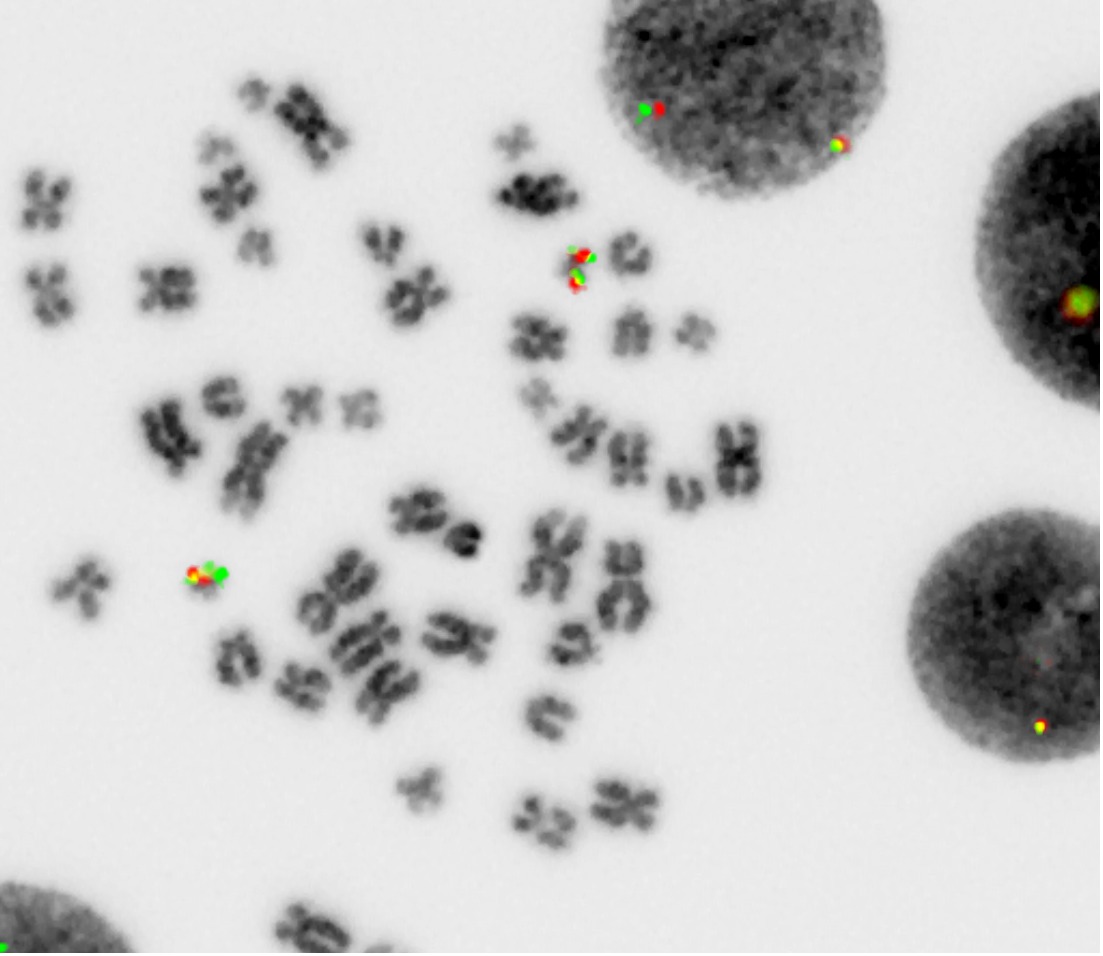
Détection de la fusion ETO2-GLIS2 dans des cellules leucémiques de patients. Le signal rouge correspond au locus ETO2, le signal vert correspond au locus GLIS2 et le signal jaune indique la présence de la fusion ETO2-GLIS2.© Thomas Mercher

Acute myeloid leukemia (AML) mainly affects children, with the prognosis often being poor despite several decades of research into more effective treatments. A new study led by Thomas Mercher (Inserm U1170/Gustave Roussy/Université Paris-Sud-Paris Saclay), and performed in collaboration with Jürg Schwaller (UKBB, Department of Biomedicine, Universität Basel) explains why some forms of leukemia develop in very young children. The study – published in Cancer Discovery, a journal of the American Association for Cancer Research – has also revealed potential new therapeutic targets.

Each year, 2,500 pediatric cancers are diagnosed in France, with one third of cases concerning leukemia – commonly known as blood cancer. Over recent decades, research into pediatric cancer has intensified and treatments have improved, but the prognosis remains particularly unfavorable for these young patients.

Acute myeloid leukemia (AML) accounts for 15% of cases of leukemia diagnosed in children and adolescents. Overall survival at 5 years is around 60%, with relapse being the most common cause of mortality.

Abnormal protein fusion

There are several subtypes of AML. One of the most aggressive, which is linked to treatment resistance and a particularly unfavorable prognosis, is acute megakaryoblastic leukemia (AML-M7). In their new study published in Cancer Discovery, the team focused their efforts specifically on this type of acute myeloid leukemia. Their work is co-financed by the National League Against Cancer.

Through the CONECT-AML collaborative network[1], the scientists obtained samples from young patients with AML-M7. In 2012, their analysis of these samples had already revealed AML-M7 to frequently present genetic alterations that lead to the expression of an abnormal protein resulting from the fusion of the two proteins normally independent in the cell. At that time, although this fusion – known as ETO2-GLIS2 – had been identified in 30% of AML-M7 cases, the researchers could not explain its mechanism of action.

They also wanted to understand why AML-M7 is diagnosed in children who are on average a lot younger (under 2 years of age) than those diagnosed with the other pediatric AML subtypes (on average towards the age of 6).

“One of the objectives of our new study was to look at the mechanism of action of the ETO2-GLIS2 fusion, and to better elucidate its consequences. We wanted to answer two major questions, with the first being why this disease is specific to children – since the fusion is not found in adults, and then what the potential therapeutic avenues could be”, explains Thomas Mercher.

This involved the researchers analyzing the characteristics of human leukemia cells and developing a mouse model to study the consequences of the ETO2-GLIS2 fusion.

Towards new therapeutic avenues

In this model, the researchers showed that this fusion is sufficient in order to rapidly induce aggressive leukemia, if it is activated in fetal hematopoietic cells. However, there is little to link its activation in adult cells with the development of leukemia. Moreover, blocking the ETO2-GLIS2 fusion in the in vivo model brings tumor proliferation to a halt, with the abnormal blood cells once again able to differentiate into normal blood cells.

These findings suggest that some forms of leukemia develop specifically in children because the properties of the fetal cells differ from those of adult cells.

Findings which also make it possible to propose new target mechanisms in fetal cells and pediatric leukemia in order to improve treatments for these patients. “We now want to understand exactly how this fusion works. Targeting it in order to directly inhibit it with molecules that could be used in patients is not something we are able to do at present, so instead we will identify and try to target the surrounding proteins that are important for it to function”, concludes Thomas Mercher.

[1] French network of various teams of researchers, clinical pathologists and pediatric hematologists

The creativity of the human mind rooted in errors ?

©AdobeStock

Why do some of our choices appear to be driven by a desire to explore the unknown? An Inserm team from École Normale Supérieure led by 2017 ERC grant-winner Valentin Wyart has shown that most of these choices are not motivated by curiosity but by errors caused by the brain mechanisms implicated in evaluating our options. These findings have been published in Nature Neuroscience.

Where shall we eat tonight – at our usual restaurant or one we have not tried before? Where shall we spend our next vacation – in the old family cabin or a rental halfway around the world? When we have various options to choose from, our decisions do not always lead us towards the safest one based on our previous experiences. This variability which is characteristic of human decisions is most often described in terms of curiosity: our choices are thought to be the reflection of a compromise between exploiting options that are known and exploring others whose outcomes are more uncertain. Curiosity is even considered to be an attribute of human intelligence – a source of creativity and unexpected discoveries. This interpretation is based on a very strong (albeit rarely explicitly stated) hypothesis, according to which we weigh up our options without ever making any errors.

An Inserm research team from the Laboratory for Cognitive and Computational Neuroscience (LNC2) funded by the European Research Council (ERC) wanted to test this implicit hypothesis upon which many findings are based. The researchers suspected that our capacity to weigh up and review our options is largely overestimated, based on findings published in 2016 in Neuron:

One of our researchers had previously shown that our capacity to make the right choice based on partial indicators is restricted by errors of reasoning when these indicators are combined, and not by hesitancy at the time of making the choice. So we asked ourselves whether these errors of reasoning could be responsible for some of the choices considered as being a matter of curiosity by current theories. “

To back up their suspicions, the researchers studied the behavior of around one hundred subjects using a slot machine-style game that consisted of choosing between two symbols associated with uncertain rewards. They analyzed the behavior of the participants using a new theoretical model developed by Charles Findling, postdoc in the team and co-lead author of the article, which takes into account evaluation errors of the symbols. 

As such, the authors discovered that over half of the choices usually considered as being a matter of curiosity were in reality due to errors of evaluation. “This finding is important, because it implies that many choices in favor of the unknown are made unbeknownst to us, without our being aware of it” explains team leader, Valentin Wyart.

Our participants have the impression of choosing the best symbol and not the most uncertain, but they do it on the basis of wrong information resulting from errors of reasoning. “

The participants played a slot machine-style game which involved choosing between two symbols associated with uncertain rewards. In this example, the participant has to choose between the left-hand symbol which won them money in the previous tests, and the right-hand symbol which has not been tried recently and whose outcome is therefore more uncertain. The current theories describe this type of choice as being the reflection of a compromise between exploiting known options (in this example, selecting the left-hand symbol) and exploring other more uncertain options (selecting the right-hand symbol).

In order to understand where these errors come from, the researchers recorded the brain activity of some of the participants using functional magnetic resonance imaging. They discovered that the activity of the anterior cingulate cortex, a region involved in decision-making, fluctuates with the evaluation errors made by the participants. The greater the activity of this region when evaluating the options, the greater the evaluation errors. For Vasilisa Skvortsova, postdoc and co-lead author of the article, “these errors of evaluation could be regulated via the anterior cingulate cortex by the noradrenaline neuromodulator system, controlling the precision of the mental operations performed by the brain”. In other words, our brain is thought to use its own errors to produce choices in favor of the unknown, without relying on our curiosity. “This is a radically different vision from the current theories that consider these errors as negligible”, insists Wyart.

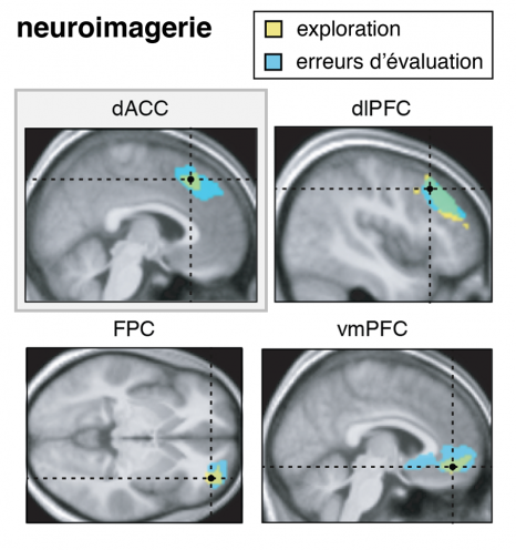
The researchers recorded the participants’ brain activity using functional magnetic resonance imaging. They discovered that the brain regions that activate when the participants explore uncertain symbols (shown in yellow) are the same ones that activate when the participants commit errors of reasoning. The brain regions shown are the dorsal anterior cingulate cortex (dACC), the dorsolateral prefrontal cortex (dlPFC), the frontopolar cortex (FPC) and the ventromedial prefrontal cortex (vmPFC).

Although these findings may appear surprising, are they really? Many major discoveries are the result of errors of reasoning: the discovery of America by Christopher Columbus, who believed he had reached the “East Indies” – a navigation error of 10,000 km, but also the discovery of radioactivity by Henri Becquerel, who initially thought that the radiation emitted by uranium was due to the re-emission of solar energy, or the discovery of the pacemaker by John Hopps when attempting to treat hypothermia with the help of radiofrequency. Going further, “the evolution of the species is also based on random variations of the genome, in other words genetic errors, some of which are retained by natural selection”, reiterates Wyart. So there is in fact nothing surprising about our brain taking advantage of its errors in order to think outside the box.

How People with Autism Might Avoid Socio-Emotional Situations

One hypothesis put forward to explain the repetitive behaviors of people with Autism Spectrum Disorder is a lack of cognitive flexibility. However, this may well not be the case. A recent study by a team of researchers from Inserm and Université de Tours used MRI to track the brain activity of autistic and non-autistic subjects faced with situations similar to those that cause problems in the daily lives of people with the disorder. Their findings, published in Brain and Cognition, suggest that the inflexibility of autistic individuals is actually the result of a strategy used to avoid socio-emotional situations. This research, which suggests now considering the cognitive and socio-emotional domains as closely linked rather than dissociated, opens up new avenues in the understanding and management of autism.

In their daily activities, people with Autism Spectrum Disorder (ASD) experience difficulty adapting their behavior to environmental changes. ASD is characterized by two main diagnostic criteria: the individual experiences persistent difficulties in social communication and is locked into repetitive behavioral patterns, restricted interests and/or activities. But while both criteria need to be present in order to diagnose ASD, little attention has been paid to how they interact.

In a study led by Marie Gomot, Inserm researcher at the Imaging and Brain laboratory (Inserm/Université de Tours), this question was explored by comparing the cognitive management of socio-emotional information and that of non-social information in people with ASD.

While the processes responsible for ASD symptoms have not yet been fully elucidated, one current hypothesis is a lack of cognitive flexibility – in other words, difficulty in alternating between multiple tasks and in analyzing one’s environment in order to adapt to these changes.

To evaluate this flexibility, the researchers used MRI to track the brain activity of ASD and non-ASD participants who underwent a test simulating situations similar to those that cause problems in the daily lives of people with the disorder.

The research team used a modified version of a test traditionally used in neuropsychology in order to test cognitive flexibility while processing non-social or socio-emotional information. Five cards were presented, each illustrated with a different face. The participants were asked to match the central card with one of four surrounding cards, according to one of the following three rules: frame color (non-social information), facial identity (social information) or facial expression (socio-emotional information). In order to evaluate their cognitive flexibility, the participants were asked throughout the test to make different matches (same color, same identity or same facial expression) by changing or maintaining one of the three rules.

The research team saw no significant difference between the ASD and non-ASD participants when it came to the behavioral parameters measuring cognitive flexibility alone – namely the capacity to adopt a new rule. However, the study did reveal the importance of information type for these cognitive flexibility processes in ASD. While the ASD participants needed more attempts than the non-ASD participants in order to adopt the rule linked to socio-emotional information, they had no particular difficulty in adopting those involving the processing of non-emotional information.

In parallel, the MRI revealed a significantly higher level of brain activity in the ASD participants when they were required to demonstrate cognitive flexibility. This brain activity only stabilized when the ASD participants received confirmation that they had found the correct rule to apply, thereby suggesting that people with ASD require a higher level of certainty in order to adapt to a new situation.

“These findings are important because they suggest that the implementation of routines and repetitive behaviors by people with ASD might not be due to a genuine lack of cognitive flexibility but rather to avoid being confronted with certain socio-emotional situations, specifies Gomot. The need for a high level of certainty combined with a poor understanding of the codes that govern socio-emotional interactions would thereby lead to the avoidance of tasks with a socio-emotional component.” And to conclude, “this research confirms the close link between cognitive and emotional dysfunction in ASD and the need for future studies to take them into joint consideration more often.

Rare Diseases: Over 300 Million Patients Affected Worldwide

300 million people are living with a rare disease. Adobe Stock

Rare diseases represent a global problem. Until now, the lack of data made it difficult to estimate their prevalence. Created and coordinated by Inserm, the Orphanet database, which contains the largest amount of epidemiological data on these diseases taken from the scientific literature, has made it possible to obtain a global estimate. Under the coordination of Inserm US14 Director Ana Rath, these data have shown that more than 300 million people worldwide are currently living with a rare disease. The study, published in the European Journal of Human Genetics, is the first to analyze the available data on rare diseases with such precision.

Systemic sclerosis, polycythemia vera and Marfan syndrome… These are obscure conditions, which are still largely unknown by the general public and differ broadly in their clinical expression. They do have one thing in common : they are very rare.

According to the European definition, a disease is considered rare when it affects fewer than 5 in 10,000 people. Few studies have been performed by the scientific community, and there is a lack of health professional expertise and of suitable treatments. This means that the thousands of rare diseases identified over the years cause immense suffering to many patients and their families, throughout the world.

The few epidemiological studies published on the subject so far rarely use general population registries. This made it difficult to establish their exact prevalence.

Yet such figures are needed if we are to identify priorities for health and research policy, understand the societal burden of these diseases, adapt the management of patients and, more generally, promote a real public health policy for rare conditions. “Given that little is known about rare diseases, we could be forgiven for thinking that their sufferers are thin on the ground. But when taken together they represent a large proportion of the population. Although rare diseases are individual and specific, what they have in common is their rarity, and the consequences which result from that”, emphasizes Ana Rath, from Inserm US14 (Information and service platform for rare diseases and orphan drugs).

Under her leadership, study author Stéphanie Nguenguan (Inserm US14), and her colleagues, used the Orphanet database to shed light on the issue.

4% of the world’s population

Created in 1997 by Inserm, Orphanet has progressively transformed into a Consortium of 40 countries, which are principally located in Europe. These partners work together to pool within it the available data on rare diseases taken from the scientific literature, making Orphanet the most comprehensive resource in the field. The large amounts of information it containes can improve the understanding of these conditions.

In their study, Rath’s team examined the data available on the point prevalence of 3,585 rare diseases (namely, the number of people affected at a given time). Rare cancers as well as rare diseases caused by infection or poisoning were excluded from their analysis.

After harmonizing the literature data using a predefined method, following which they added together the point prevalences of the various diseases referenced in the database, they were able to estimate that at any given time, 3.5 to 5.9% of the global population suffers from these conditions. This represents around 300 million people, i.e. 4% of the world’s population.

When taken together, “rare” diseases are not so rare after all, and therefore public health policies at global and national level are needed to address this issue, according to the authors. Such a policy is becoming reality in France, which launched its 3rd National Rare Diseases Plan a year ago. “In all likelihood, our data represent a low estimation of the reality. The majority of rare diseases are not traceable in healthcare systems and in many countries there are no national registries. Making patients visible within their respective healthcare systems by implementing means to record their precise diagnoses would make it possible in the future not only to review our estimations, but more fundamentally to improve the adaptation of support and reimbursement policies”, specifies Rath.

Other observations were made during this study, with the researchers showing for example that out of the more than 6,000 diseases described in Orphanet, 72% are genetic and 70% start in childhood. Furthermore, among the diseases analyzed in the study, 149 alone are responsible for 80% of cases of rare diseases identified worldwide.

Future research must now focus on collecting and analyzing the data on the rare diseases which had been excluded from this study. Cancers and other rare diseases caused by infectious agents or linked to environmental factors will be the subject of new analyses. But the researchers’ priority remains the same: namely, to broaden the field of knowledge on rare diseases in order to offer patients better treatment and ensure that, in the future, no-one is left behind.


Skin graft: a new molecular target for activating stem cells

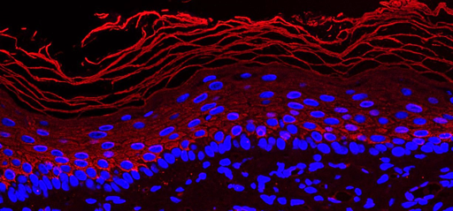
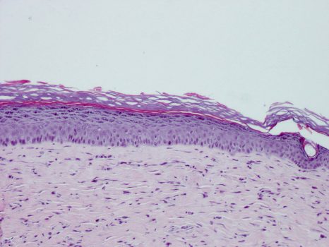
Reconstruction of a pluristratified epidermis using keratinocytes from human embryonic stem cells, hESC. IStem, Génopole d’Evry. Inserm/Baldeschi, Christine

A CEA, INSERM and the University of Paris team, produced in collaboration with I-Stem, the AFM-Téléthon laboratory, and the University of Évry has just published a paper in which it demonstrates the central role of the transcription factor KLF4 in regulating the proliferation of epidermal stem cells and their ability to regenerate this tissue. This study opens perspectives for regenerative skin medicine. It was published on 21 October in Nature Biomedical Engineering.

Human skin completely renews itself every month thanks to the presence of stem cells in the deepest layer, which generate all the upper layers of this tissue. The deciphering of genes that regulate stemness remains an enigma that is only partially resolved, in particular for human skin.

The discoveries of a French research team from the CEA, INSERM and the University of Paris, produced in collaboration with I-Stem, the AFM-Téléthon laboratory, and the University of Évry, opens perspectives for regenerative cutaneous medicine, in particular for the bio-engineering of skin grafts for tissue reconstruction. Massive ex vivo expansion of epidermal cells (called keratinocytes) is needed for the production of grafts. It is performed using a skin sample from the patient that contains adult keratinocytes and a minority population of keratinocyte stem cells. This expansion phase involves a risk: it may be accompanied by a quantitative loss or degradation of stem cells, leading to a loss of regenerative potential.

The results of the paper published in Nature Biomedical Engineering show that reducing the expression of the KLF4 gene during graft preparation promotes rapid expansion of functional stem cells1, without damaging their genomic stability. Keratinocytes expanded under these conditions have an increased long-term regenerative potential in in vitro epidermal reconstruction models and in vivo grafts2. KLF4 is therefore a new molecular target for preserving the functionality of stem cells and making progress in the bio-engineering of skin grafts. These results constitute a proof of concept, which requires additional developments to envisage clinical applications, including the treatment of severe burns and chronic ulcers, and breast reconstruction.

This work has been extended to other types of cells of interest for cutaneous cell therapy. In the future, keratinocytes produced from pluripotent stem cells could be an alternative to adult stem cells in certain reconstructed tissue bio-engineering applications.

One of the difficulties encountered in this area is that fact that the keratinocytes obtained do not have all the functions of adult stem cells. In particular, they are deficient in terms of their proliferation potential. The study has shown that manipulation of KLF4 expression is also suitable for these cells, as reducing its expression in keratinocytes derived from embryonic stem cells (ESC) improves their proliferation capacity and their ability to reconstruct skin.

Skin cells in culture                    Skin graft obtained by bio-engineering

Human skin stem cells expanded in culture can be used for skin regeneration© LGRK, IRCM, CEA-Jacob

1.A single functional stem cell is able to regenerate the skin throughout a person’s life. This is due to its very long-term proliferation capacity, its immature nature and its capacity for three-dimensional organisation.

2. Reconstructed human skin xenograft on an animal model

Some Persistent Organic Pollutants Could Increase Breast Cancer Aggressiveness

Photo d'imagerie en microscopie électronique montrant la transformation des cellules mammaires tumorales dans le cancer du sein

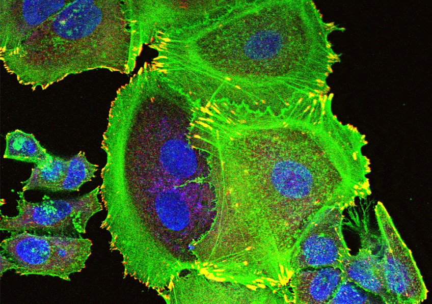
Transformation of tumorous mammary cells in breast cancer ©Xavier Coumoul/Inserm/Université de Paris

Although persistent organic pollutants (POPs) are already suspected to promote breast cancer, there has been little research into how they affect its aggressiveness. A research team from Inserm and Université de Paris in the Environmental Toxicology, Therapeutic Targets, Cell Signaling and Biomarkers laboratory has performed a preliminary study to explore the hypothesis that POPs could promote the development of breast cancer metastases. Its findings suggest a link between the aggressiveness of breast cancer and the levels of some POPs in adipose tissue, particularly in overweight women. While this research published in Environment International opens up new avenues for studying the impact of POPs on breast cancer, its findings must be taken with precaution given the limited number of subjects involved.

Breast cancer is a major public health concern with over 2 million new cases diagnosed and over 600,000 deaths worldwide in 2018. The presence of metastases distant from the original tumor is a marker of its aggressiveness. While the 5-year survival rate for cancer located only in the breast is 99%, this decreases to 85% if it has spread to lymph nodes and falls to 26% if distant metastases are present.

Recent studies have suggested that exposure to persistent organic pollutants or POPs (endocrine-disrupting and/or carcinogenic environmental pollutants that cannot be eliminated by the body) and which accumulate in the food chain1, is a risk factor for breast cancer. However, the influence of these POPs on its level of aggressiveness has received little attention.

A research team led by Xavier Coumoul in the Environmental Toxicology, Therapeutic Targets, Cell Signaling and Biomarkers laboratory (Inserm/Université de Paris) has been the first to test the hypothesis that exposure to POPs could affect the stage of development of metastases in breast cancer.

POPs are highly lipophilic and as such are stored in the adipose tissue. The researchers measured the levels of 49 POPs – including the Seveso dioxin (a waste product from incineration processes) and several PCBs (generated by various industrial processes) – in samples of adipose tissue surrounding the tumors of 91 women with breast cancer2. With excess weight (BMI > 25) known to promote and worsen breast cancer, special attention was paid to the women concerned.

Biological and statistical analysis of these samples enabled the researchers to reveal a link between the presence of distant metastases and dioxin levels in the adipose tissue of overweight women. What is more, in all patients, the concentrations of dioxin and two of the PCBs measured appeared to be linked to the size of the tumor as well as the invasion level and metastatic stage of the lymph nodes. Women with higher concentrations of PCBs were also found to have a higher risk of breast cancer recurrence.

These findings suggest therefore that the higher the concentration of POPs in the adipose tissue, the more aggressive the breast cancer, particularly in overweight women.

To explain this link, the research team has issued several hypotheses based on previous studies. One hypothesis is that dioxin and some PCBs send a signal which promotes tumor cell migration (an essential mechanism in the metastatic process), thereby reinforcing the cancer’s aggressiveness.

According to study leader Xavier Coumoul, “the adipocytes – the cells of the adipose tissue that store fats – play a major role as cells associated with the development of breast cancer. The adipose tissue works like an ʺendocrineʺ gland (secreting hormones into the bloodstream) and we had previously shown that POPs were responsible for the inflammation of this adipose tissue, changing the nature and behavior of the adipocytes. The excessive secretion of inflammatory molecules and the release of the POPs stored by these adipocytes could then promote the formation of metastases.” 

However, the researcher insists on the fact that this study is only preliminary and that its findings must be considered with precaution, given the limitations in its methodology. A limited number of individuals were studied, which encourages statistical bias and means that some sub-categories of the study population cannot be considered to be highly representative. “While it does not enable us to draw firm conclusions on the link between POPs and the aggressiveness of breast cancer, it does open up a novel avenue for research, particularly in overweight patients. An avenue which should be explored by future studies with larger numbers of patients in order to offer more representative statistical findings”, concludes Coumoul.

1 POPs (persistent organic pollutants) have been defined and listed by the Stockholm Convention

2 Men were excluded from the study to limit statistical bias due to sex-specific biological differences.

A new discovery! How our memories stabilise while we sleep

Photo by Kinga Cichewicz on Unsplash

Scientists at the Center for Interdisciplinary Research in Biology (CNRS/Collège de France/INSERM)1 have shown that delta waves emitted while we sleep are not generalized periods of silence during which the cortex rests, as has been described for decades in the scientific literature. Instead, they isolate assemblies of neurons that play an essential role in long-term memory formation. These results were published on 18 October 2019 in Science.

While we sleep, the hippocampus reactivates itself spontaneously by generating activity similar to that while we are awake. It sends information to the cortex, which reacts in turn. This exchange is often followed by a period of silence called a ‘delta wave’ then by rhythmic activity called a ‘sleep spindle’. This is when the cortical circuits reorganize to form stable memories. However, the role of delta waves in the formation of new memories is still a puzzle: why does a period of silence interrupt the sequence of information exchanges between the hippocampus and the cortex, and the functional reorganisation of the cortex?

The authors here looked more closely at what happens during delta waves themselves. They discovered, surprisingly, that the cortex is not entirely silent but that a few neurons remain active and form assemblies, i.e. small, coactive sets that code information. This unexpected observation suggests that the small number of neurons that activate when all the others stay quiet can carry out important calculations while protected from possible disturbances. And the discoveries from this work go even further! Spontaneous reactivations of the hippocampus determine which cortical neurons remain active during the delta waves and reveal transmission of information between the two cerebral structures. In addition, the assemblies activated during the delta waves are formed of neurons that have participated in learning a spatial memory task during the day. Together these elements suggest that these processes are involved in memory consolidation. To demonstrate it, in rats the scientists caused artificial delta waves to isolate either neurons associated with reactivations in the hippocampus, or random neurons.

Result: when the right neurons were isolated, the rats managed to stabilise their memories and succeed at the spatial test the next day.

These results substantially change how we understand the cortex. Delta waves are therefore a means of selectively isolating assemblies of chosen neurons, which send crucial information between the periods of hippocampo-cortical dialog and the reorganisation of cortical circuits, to form long-term memories.

1 An associate member of the Université PSL, since 2009 the Collège de France has had a deliberate policy of welcoming independent teams who can benefit from shared technical and scientific services and an exceptional multidisciplinary environment. Twenty-two teams are currently housed in the Center for Interdisciplinary Research in Biology et de Physique of the Collège de France. Supported in particular by the CNRS, this facility is open to researchers in France and elsewhere. It helps to make Paris an attractive place to conduct research.

Aging with HIV Linked to Increased Risk of Cognitive Impairment

Adobe Stock 

Thanks to antiretroviral therapies, it is possible to grow old with HIV under control. However, this chronic infection may not leave cognitive function unscathed. That is why Alain Makinson (Translational Research on HIV and Infectious Diseases unit, Montpellier University Hospital, Université de Montpellier, Inserm, IRD) and his team were interested in exploring the development of neurocognitive impairment (NCI), such as diminished attention, memory and motor capacity, in patients living with HIV in the ANRS EP58 HAND 55-70 study. In their latest research, published in Clinical Infectious diseases, the scientists describe the results of their observations of 200 people living with HIV enrolled in six French centers. The data collected from these patients were then compared, using the same neurocognitive evaluation methods, with those of a control population of 1,000 people of the same age, sex and education taken from Constances – a cohort recruited from the general population, with over 200,000 volunteers. The researchers reveal that among those living with HIV between the ages of 55 and 70, the risk of developing mild (and in some cases, symptomless) neurocognitive impairment increases by 50%.

Thanks to antiretroviral therapies, people living with the human immunodeficiency virus (PLHIV) can bring it under control. However, while HIV is no longer their major cause of death – having decreased substantially since the advent of these therapies – other risks must be considered. Several recent studies have highlighted the increased prevalence of neurocognitive impairment (NCI) in PLHIV compared with uninfected individuals – an increase that is all the more pronounced in those whose virus is not controlled. However, this NCI could also be caused by cardiovascular factors or the onset of depression, which is more common in this population.

In order to try to determine the nature of the link between living with HIV and developing NCI (such as diminished attention, memory and motor capacity), Alain Makinson and his coworkers studied the data of 200 PLHIV, between 55 and 70 years of age, whose HIV was under control and who were enrolled between January 2016 and October 2017 in the ANRS EP58 HAND (HIV-Associated Neurocognitive Disorder) study. Each patient was compared with five HIV-uninfected individuals of the same age, sex and education, from Constances – a cohort recruited from the general population. The same methods were used for both populations when performing the cognitive tests and collecting the study data. All in all, 1,200 people took part in this study.

Although the impairments observed in the study were either mild or without apparent symptoms (i.e. with zero to low impact on daily activities despite abnormal test results), those living with HIV were more affected by NCI: 35% versus 24% for the control group.

The risk of developing NCI is therefore 50% higher for HIV-infected individuals compared with HIV-uninfected individuals, all other criteria being equal (age, sex and education).

The conclusion remained the same after the authors took into account various confounding factors (alcohol consumption, smoking, physical activity, diabetes, hypertension, etc.), and despite the use of several methods to evaluate the cognitive tests.

Despite these highly robust results, a causal link between living with HIV and developing NCI cannot be established – for which there are several possible hypotheses. One is that HIV infection and its treatments cause recurrent brain inflammation. Another is that the complications associated with the immune deficiency can affect cognition before antiretroviral treatment is initiated, but without the subsequent deterioration occurring more rapidly in comparison with the general population. Finally, seropositivity could be associated with other risk factors (particularly drug use), which are difficult to measure fully in the two populations of this study.

The authors wish to continue to follow the same population over a longer period in order to better define the causes of NCI in this aging population and test the hypothesis of accelerated cognitive aging in PLHIV – particularly given that very few studies with a control group are available. Testing the brain inflammation hypothesis by collecting certain specific blood biomarkers represents another avenue for the team to explore in its quest to elucidate the mechanisms behind NCI.

Science serves gender equality

© Tim Mossholder on Unsplash

In a commentary published in the journal Nature Human Behavior, Inserm researcher Violetta Zujovic and her colleagues from the XX initiative committee at the Brain & Spine Institute (Inserm/CNRS/Sorbonne Université) at the AP-HP Pitié-Salpêtrière Hospital explain how a neuroscientific approach can be used to more effectively combat gender inequality. The actions taken by the researchers have increased the number of female speakers invited to take the floor at the research center from 25 to 44% in the space of a year and a half, and have increased the number of women in senior roles from 25 to 31%. Their neuroscience-based approach now constitutes an important mechanism for changing attitudes and behaviors. Among other things, their future work will focus on using “nudges”—a technique originating in neuroscience that tries to influence our behavior in our own interest—to improve gender equality. An example for others to follow.

Although internationally recognized as a country that supports women’s career progression through suitable infrastructure and specific legislation, France performs poorly when it comes to offering equal opportunities to men and women. This is the paradox highlighted by the XX initiative committee, made up of a dozen scientists, in a commentary published in Nature Human Behavior. If the entire community, including the scientific community, recognizes these inequalities, why do gender-related behaviors persist? The published commentary warns of the impact of cognitive biases, which are beyond our control but deeply rooted in our attitudes. Unconscious prejudices and stereotypes have a powerful influence over almost every decision we make.

Despite the norms of equality, and despite the evidence that teams are more successful when they include both men and women, social prejudices continue to silently pull the strings, for example by creating sexist or racist biases. The neuroscientific community is at the forefront of this issue, not only in raising awareness of such forms of unconscious bias, but also because it has the tools at its disposal to understand their cognitive origins and break societal stereotypes.

The XX initiative committee at the Brain & Spine Institute (ICM), which includes male and female neuroscientists from Inserm, the CNRS, and Sorbonne Université, have proposed a roadmap in several stages. Their recommendations highlight a crucial first stage: realization by each individual of their own implicit assumptions derived from existing prejudices, and the impact these have on their decision-making.

This first stage was put into practice by collecting comprehensive data on the male to female ratio by level of responsibility and job title. The internal ICM data gathered in April 2017 showed that just 26% of women held a management or joint management post within the institute, whose staff is 63% female.

Other figures showed that only 25% of invited speakers at the institute’s weekly scientific seminars were female.

The simple fact of presenting the findings of this investigation triggered a collective reassessment process that has resulted in the number of women heading up a research group increasing from 26 to 31%. The entire community has also been involved in efforts to showcase more female researchers, which has had the practical result of the number of female scientists taking the floor at the institute increasing from 25 to 44%.

“Thanks to the collective drive to combat gender inequality, we were also able to revise various institutional forms to remove all gender-specific information,”explains Violetta Zujovic, the Inserm researcher who led this work.

In this societal and economic context, in which we are trapped by unconscious prejudices and gender stereotypes, one of the key recommendations proposed by the researchers is to inform men and women and train them to overcome these forms of bias. The committee is holding a meeting on “Gender Bias: Science and Practice” on April 3, 2020, which is open to all, along with practical workshops providing individuals with the tools to combat their own prejudices and understand how to assess their own values and skills.

The impact of this committee, and the neuroscientific approach it instigated around eighteen months ago, highlights the importance of combining national policies with practical measures. To that end, the neuroscientists are already working on developing “nudges” that will cleverly encourage rather than impose male/female equality. Overall, these measures are designed to enable individuals to understand and recognize the unconscious gender-related prejudices that we perpetuate and pass on, and to individually strive for cultural change.

In May 2019, Inserm also set up a professional parity and equality mission that will shortly set out practical measures for advancing professional equality. These will be incorporated into a multi-year plan and integrated into the institution’s strategy.

fermer