Menu

Do Some Cognitive Biases Contaminate Even Our Simplest Mental Mechanisms?

Enfants apprenant à écrire à l'école

©woodleywonderworks via Flickr


When we implement complex cognitive processes, for example when making decisions, we are subject to cognitive bias. But what about simpler processes, such as those involved in the most basic learning? In a new study analyzing data from all previous research in the field, researchers from Inserm and ENS-PSL show that not only are positivity and confirmation biases present even in the simplest human and animal cognitive processes, but also that incorporating them into learning algorithms would enhance their performance. This research, published in Trends in Cognitive Sciences, suggests that these biases could initially have been a very old evolutionary advantage.

Cognitive biases, such as positivity and confirmation biases, are known to influence our beliefs and decisions. Until recently, it was assumed that they were specific to so-called “high-level” cognitive processes, namely those that come into play when we reason about complex and uncertain proposals.  For example, it is well known that people overestimate the likelihood of desirable events (France winning the World Cup) and underestimate that of undesirable events (a marriage ending in divorce). 

In a study published in Trends in Cognitive Sciences, Stefano Palminteri, Inserm researcher at the ENS-PSL/Inserm Laboratory for Cognitive and Computational Neuroscience and Maël Lebreton, researcher at the Paris School of Economics, challenge this conception of the involvement of positivity and confirmation biases.

The researchers drew on the various existing data in the scientific literature on “reinforcement learning”. This is a basic cognitive process of learning through rewards and punishments that humans share with many animals. This literature review reveals that very simple reinforcement learning tests can reveal behavioral signatures of positivity and confirmation biases in people who are subjected to them. These biases appear to be much more common than previously thought and are even present in the simplest cognitive processes such as learning to make good decisions through trial and error (reward and punishment).

What is more, these biases do not seem to be exclusive to humans: the behavioral signatures also appear in similar tests conducted in animals. This suggests that these biases may have emerged during evolution in a common ancestor, long before the appearance of Homo sapiens, raising the question of why evolution has selected and maintained what may at first glance be perceived as processes that can generate seemingly irrational behavior.

Palminteri and Lebreton believe that they have identified part of the answer to this question through the findings of studies based on computerized simulations. These studies compared the performance of reinforcement learning algorithms – with some algorithms incorporating positivity and confirmation biases and others not. These simulations show that the presence of a confirmation bias in the algorithm actually enables it to learn more effectively in a wide range of situations. These biases could therefore actually promote survival, which would explain why they have not been corrected during the course of evolution.

The article opens up new avenues of research that would allow us to deepen our understanding of the cognitive biases and processes related to reinforcement learning. In particular, the researchers suggest exploring the role of these biases in the development and maintenance of pathological states, such as addiction and depression. These findings also suggest that including these biases in artificial intelligence algorithms could paradoxically improve their performance.

La Guinée et la France renforcent leur coopération à travers la création d’une plateforme de recherche internationale (PRISME)

InScience 2022: An Inserm Festival

InScience 2022

The scientific culture event is back for its second edition. © Anne Defreville/Inserm


“Cultivate your health with Inserm! ” InScience is back from June 1-15, 2022, with numerous events in which you can meet and talk to our researchers. In cities across France, discover our exhibitions, participate in escape games, conferences, film-debates… Not to mention online videos and podcasts, accessible to everyone, everywhere. The objective of this event? Give citizens the keys to play an active role in their own health and participate in debates in line with Inserm’s motto:  “Science for health.”

InScience is a unique event in France in the field of medical research and human health. This scientific culture event is the fruit of significant mobilization by Inserm researchers who, keen to share high-quality and reliable information, wish to promote scientific discourse and show the general public what their roles involve.

Coming to a city near you

For this second edition, Inserm scientists are delighted to be able to reach out to the general public across France, thanks to this packed program.


Here is a glimpse of what we have lined up:

  • In Toulouse, as part of an original and friendly get-together at Eurêkafé, called “Du charabia au limpide”, Inserm researchers will face the challenge of presenting their research project in 180 seconds[1] using language that can be understood by many people as possible.
  • In Lyon, scientists working on multiple sclerosis will present the advances made by research into this autoimmune disease, in the presence of patient associations[2].
  • In Paris, come and attend a conference led by Inserm scientists on the subject of endocrine disruptors. Where do they come from? What can we do to protect ourselves?[3] These questions will all be addressed in an educational way, helping the public to gain a deeper understanding of this complex subject.
  • In Lille, Bastia, and Rennes, discover the video game ViRal and be transported inside the human body thanks to virtual reality.
  • And then in Strasbourg, Inserm will present its book “Fake news santé” and the efforts made to fight health misinformation thanks to the mobilization of researchers since the start of COVID-19[4].

Part of the program will also be dedicated to emerging viruses. In a novel exhibition, Inserm will revisit the major epidemics of the past, the lives of viruses, the weapons our body uses to fight them, and the various measures taken by scientists to combat the epidemics they cause. This exhibition will be held in Mulhouse[5] until June 30 and is also accessible in digital format.

Other original activities include: an escape game in Nantes[6] in which you are locked inside a laboratory with a team of scientists, one of whom has kidney failure and needs help. Or the sound installation hEARt[7], an evolving musical journey in which each heartbeat resonates as a poetic element. This composition by Christophe Ruetsch, produced in collaboration with artists, technicians, and scientists, will be held in the cities of Toulouse and Corte.

InScience 2022 will also be available in digital format with a comprehensive program of online conferences, podcasts, plus unprecedented videos, such as the “Science dans tous les sens” series, presented by an Inserm researcher in Montpellier, and aimed at discovering the senses. Online visitors can immerse themselves in Inserm’s universe of fundamental and applied research into subjects as varied and topical as women’s health[8] and post-traumatic stress disorder[9].  

Consult the entire program on the Inserm website and dedicated Facebook page.


[1] June 2, from 7 p.m. to 9 p.m. at Eurêkafé in Toulouse

[2] June 8, from 6.30 p.m. to 7.30 p.m. at Lyon City Hall

[3] June 2, at 7 p.m. at the Benoite Groult library in Paris 14th

[4] June 2, at 7 p.m. at the MAIF Etoile Space in Strasbourg and broadcast to all MAIF centers in France

[5] At La Nef des Sciences association in Mulhouse

[6] June 4, at the Librairie Ludique Portails bookstore in Nantes

[7] June 9-12, from 10 a.m. to 2 p.m. at Eurêkafé in Toulouse – June 1-5 at the Corte Citadel

[8] June 14, at 7.30 p.m. on the Inserm YouTube channel

[9] June 15, at 6.00 p.m. on the Inserm YouTube channel

Phage Therapy: A Model to Predict Its Efficacy against Pathogenic Bacteria

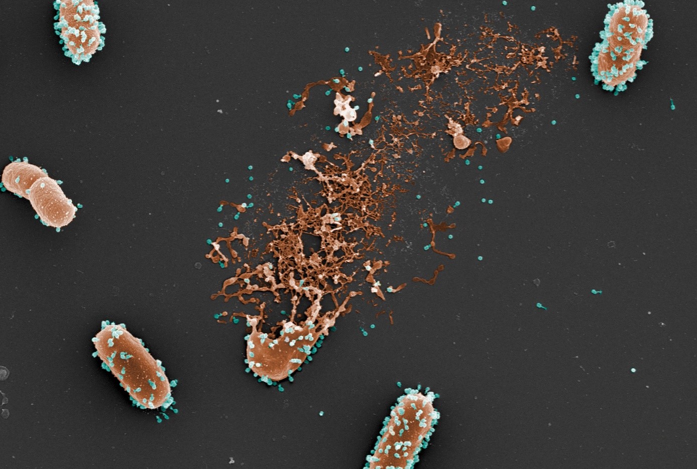
Photo (colorized) of scanning electron microscopy of a bacterium lyzed by the phages (© L. Debarbieux, Institut Pasteur; M. and C. Rohde, Helmholtz Centre for Infection Research).

Antibiotic resistance represents a major public health challenge, associated with a high mortality rate. While bacteriophages – viruses that kill bacteria – could be a solution for fighting antibiotic-resistant pathogens, various obstacles stand in the way of their clinical development. To overcome them, researchers from Inserm, Université Sorbonne Paris Nord and Université Paris-Cité at the IAME Laboratory, in close collaboration with their counterparts at Institut Pasteur and the Paris Public Hospitals Group (AP-HP), have developed a model to better predict the efficacy of phage therapy and possibly develop more robust clinical trials. Their findings have been published in Cell Reports.

The discovery of antibiotics had revolutionized the history of medicine in the 20th century, allowing us to effectively fight bacteria for the first time. However, antibiotic resistance – a phenomenon during which bacteria become resistant following mass, repeated use – has become a major public health issue in recent decades. Each year, these resistant bacteria are estimated to be responsible for 700,000 deaths worldwide. Yet the discovery of new antibacterial agents has been stagnating for several years.

In this context, phage therapy has recently generated renewed interest. This therapeutic approach involves the use of bacteriophages that target and destroy pathogenic bacteria whilst being unable to infect humans. While the concept has been in existence for a long time, its clinical development has been hampered by various limitations. Unlike “conventional” medicines, bacteriophages are complex biologics, whose action in the body, optimal dose, and most effective route of administration are difficult to study and anticipate.

In order to remove some of these obstacles, Jérémie Guedj’s research team at Inserm, in collaboration with Laurent Debarbieux’s team at Institut Pasteur, has developed a new mathematical model to better define the interactions between bacteriophages and pathogenic Escherichia coli bacteria in animals and to identify the key parameters that influence the efficacy of phage therapy.

Supporting clinical development

Various data from in vitro and in vivo experiments were used to construct this model. In particular, the researchers used the bacteriophages’ infection parameters determined in the laboratory (for example, the duration of the infectious cycle of the bacteria, the number of viruses released when a bacterium is destroyed…) and information collected during experiments using a mouse model of lung infection.

Some of the animals were infected with a bioluminescent strain of E. Coli (in order to best monitor it within the body). Among them, some were treated with bacteriophages at different doses and using different routes of administration. The quantities of bacteria and bacteriophages thus measured over time helped to feed the mathematical model and test which were the most important parameters for effective phage therapy. 

Using their model, the scientists show that the route of administration is an important parameter to consider when it comes to improving the animals’ survival: the more rapidly it brings the bacteriophages into contact with the bacteria, the more it is effective. In the animal model, the phage therapy administered intravenously was therefore less effective in comparison with the intratracheal route because fewer bacteriophages were reaching the lungs. On the other hand, when administered by intratracheal route, the model suggests that the dose of the medication given has little effect on the efficacy of the therapy.

Another important point is that this model incorporates data on the animals’ immune response in the context of phage therapy. The model confirms and extends the principle that bacteriophages act in synergy with the immune system of infected animals, enabling more effective elimination of pathogenic bacteria.

“In this study, we propose a new approach to streamline the clinical development of phage therapy, which otherwise continues to have its limitations. Our model could be reused to predict the efficacy of any bacteriophage against the bacteria it targets, once a limited number of in vitro and in vivo data are available on its action. Beyond phage therapy, the model could also be used to test anti-infective therapies based on the association between bacteriophages and antibiotics,” concludes Guedj.

Signature d’un accord cadre entre Santé publique France et l’Inserm

Influenza: A New Avenue for Developing Innovative Treatments

Cellules épithéliales respiratoires humaines

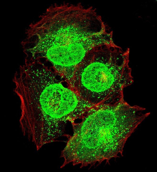
Human respiratory epithelial cells (in red) infected with an influenza virus (in green). © A. Cezard, D. Diakite, A. Guillon, M. Si-Tahar, PST ASB-Microscopy Department.

Seasonal influenza is a major public health issue because it continues to remain associated with considerable mortality, particularly among people who are elderly, immunocompromised, or both. It also has a significant socioeconomic cost. With vaccination and current treatments still being of limited efficacy, research teams are trying to develop new therapeutic approaches. At the Research Center for Respiratory Diseases in Tours, scientists from Inserm, Université de Tours and Tours Regional University Hospital have shown that in a context of influenza infection, a metabolite[1] called succinate, which is naturally present in the body, has an antiviral and anti-inflammatory action. These findings open up new therapeutic prospects based on the use of succinate derivatives. The study has been published in EMBO Journal.

Often considered a mild disease, influenza continues to cause the deaths of 10,000 to 15,000 people each year in France. The socioeconomic cost of the disease is also significant because it is associated with high levels of absenteeism and a major burden on hospitals.

Seasonal influenza vaccination is a central pillar of the preventive strategies deployed to reduce the number of cases and fight the disease. However, its efficacy can vary from year to year, depending on the influenza viruses in circulation and the suitability of the vaccine to them. Drugs that directly target the influenza virus are available for severe cases, but the window of time for effective action with these treatments is very short. What is more, influenza viruses have become resistant to their action.

In this context, the development of innovative therapies is a priority. While current treatments work by targeting certain components of the virus, Inserm Research Director Mustapha Si-Tahar and his colleagues at the Research Center for Respiratory Diseases are trying to better understand the host’s cellular and molecular responses to viral infection, with the long-term aim of developing novel therapeutic strategies aimed at strengthening these responses.

The role of metabolites in immune response

While metabolism1 has long been considered to be a purely energetic mechanism essential for cell function, recent research has shown that some metabolites can also regulate the immune response.

Based on these data, Si-Tahar’s team wondered whether an influenza infection could cause the reprogramming of the metabolism of the target cells of the virus and whether specific metabolites play an especially active role in the immune response.

In mice infected with influenza, the researchers observed that a metabolite called succinate accumulates in the lungs. A phenomenon which was then confirmed in humans by comparing respiratory fluids from intensive care patients with and without influenza pneumonia. The presence of succinate was found to be significantly higher in the influenza patients.

They then exposed cells from the pulmonary epithelium to succinate, thereby demonstrating that the molecule has antiviral activity by blocking multiplication of the virus. Succinate also helps to reduce the strong inflammatory response that is triggered in the lungs following infection with influenza.

The researchers also found that mice exposed to the virus receiving intranasal succinate are better protected against infection and have a higher survival rate than those that do not receive it.

In search of molecular mechanisms

In order to better understand these different phenomena, the scientists sought to decipher the molecular mechanisms behind the antiviral action of succinate.

This involved analyzing the impact of succinate on the various stages of the viral replication cycle and demonstrating that, while there is no influence on the early stages of the cycle (entry, transcription, and translation), there is an influence on a later stage. The findings show that this metabolite prevents a major structural protein of the virus, the “nucleoprotein”, from exiting the nucleus of infected cells, thereby preventing the assembly of the final viral particle and interrupting the multiplication cycle of the virus.

These data all point to the key role of succinate in controlling influenza infection, as well as its therapeutic value.

“Our research has an interesting outlook in that it potentially paves the way for the development of new antiviral treatments derived from succinate,” underlines Si Tahar[2].

Additional studies are needed in order to test the therapeutic potential of succinate and identify other metabolites of interest.


[1] Metabolism refers to all of the chemical reactions that take place inside the body’s cells. A metabolite is an organic substance derived from metabolism.

[2] In keeping with this research, Si-Tahar has since early 2021 coordinated a French National Research Agency (ANR) program entitled “Development of succinate-based formulations and analogues against SARS-CoV-2-induced respiratory infections and influenza viruses.” His team is also the beneficiary of an “ERS-RESPIRE4 Marie Skłodowska-Curie fellowship” to develop a project entitled “Succinate-Producing Probiotics as an Innovative Therapy for Viral Respiratory Infections: a proof-of-concept study”.

Il existerait un lien entre la taille du réseau social et la structure du cerveau

Mère macaque toilettant son jeune singe.

A macaque grooming her offspring. An image that illustrates how macaques form relationships. © Noah Snyder-Mackler

The more social relationships we have, the more certain structures in our brain are developed. This has been the hypothesis of various neuroscience research projects for several years. With previous findings having highlighted the role of our social environment as one of the key factors behind the expansion of the cerebral cortex, researchers from Inserm and Université Lyon Claude Bernard Lyon 1, in collaboration with the University of Pennsylvania, went one step further in elucidating this link. They were more specifically interested in a species of macaques whose brain architecture is comparable to that of humans. By observing these non-human primates in their natural state and by analyzing images of their brains, they discovered that the number of companions they have is predictive of the size of certain brain regions associated with social cognition and empathy. The findings of this study have been published in Science Advances.

The links between social network and brain size have already been the subject of neuroscientific studies. For example, scientists have already looked at variations in the size of the human brain amygdala in relation to the number of Facebook friends that a person has1.

In order to build on this research and attempt to find out more about the organization and functions of the neural networks in humans, research teams have worked with an animal species whose brain characteristics are similar to those of humans: the Rhesus macaque.

In a new study, researchers from Inserm and Université Claude Bernard Lyon 1 at the Stem Cell and Brain Research Institute, in collaboration with the University of Pennsylvania, studied a group of these non-human primates in their natural state for several months before taking images of their brains. The fact that the animals were free ranging enabled the scientists to understand the social group in its full complexity. They were therefore able to measure the intensity2 of the interactions with other individuals and identify an animal’s position within the group social hierarchy.

For example, some of the observations focused on grooming partners, which represent direct, important relationships for macaques.

Alongside these behavioral observations, the scientists analyzed brain scans of individuals from the group, which consisted of 103 rhesus macaques, including 68 adults and 21 young macaques under 6 years of age.

In the adults, they found that the greater the number of social partners, the greater the size of some regions of the brain’s temporal lobe – the ventral dysgranular insula and the mid-superior temporal sulcus3 – regions considered essential for understanding emotions and the perception of how others behave.

In order to better understand how this phenomenon occurs, the scientists also collected brain scans from 21 newborn macaques. The research showed that they are not born with these differences in brain structure size but that they are established during their development.

According to the researchers’ observations, there is therefore no correlation between social network size and brain volume at birth. These findings suggest that exposure to the social environment over the course of life contributes to the maturation of brain networks.

“Which is interesting because if we had seen the same correlation in young macaques, this could have meant that having a very popular mother (with many interactions with the group) could have predisposed the newborn to also become popular. But in fact our data suggest that the differences we see in adults are strongly determined by our social environments, perhaps more than by our innate predisposition,” explains Jérôme Sallet, Inserm Research Director.

Following this study, the researchers now wish to look at anatomical changes at cell level, in order to reveal the mechanisms behind the increased size of the brain regions that were identified using brain imaging.


1 Kanai R., Bahrami B., Roylance R. and Rees G. 2012. Online social network size is reflected in human brain structure, Proc. R. Soc. B.

2 The researchers measured the number of interactions between the animals, their duration and whether these interactions were cooperative or aggressive.

3 The mid-superior temporal sulcus is involved in social cognition and perception.

fermer