Menu

Research shows that treatment with growth hormone in children who have recovered from cancer does not increase the risk of a second tumor.

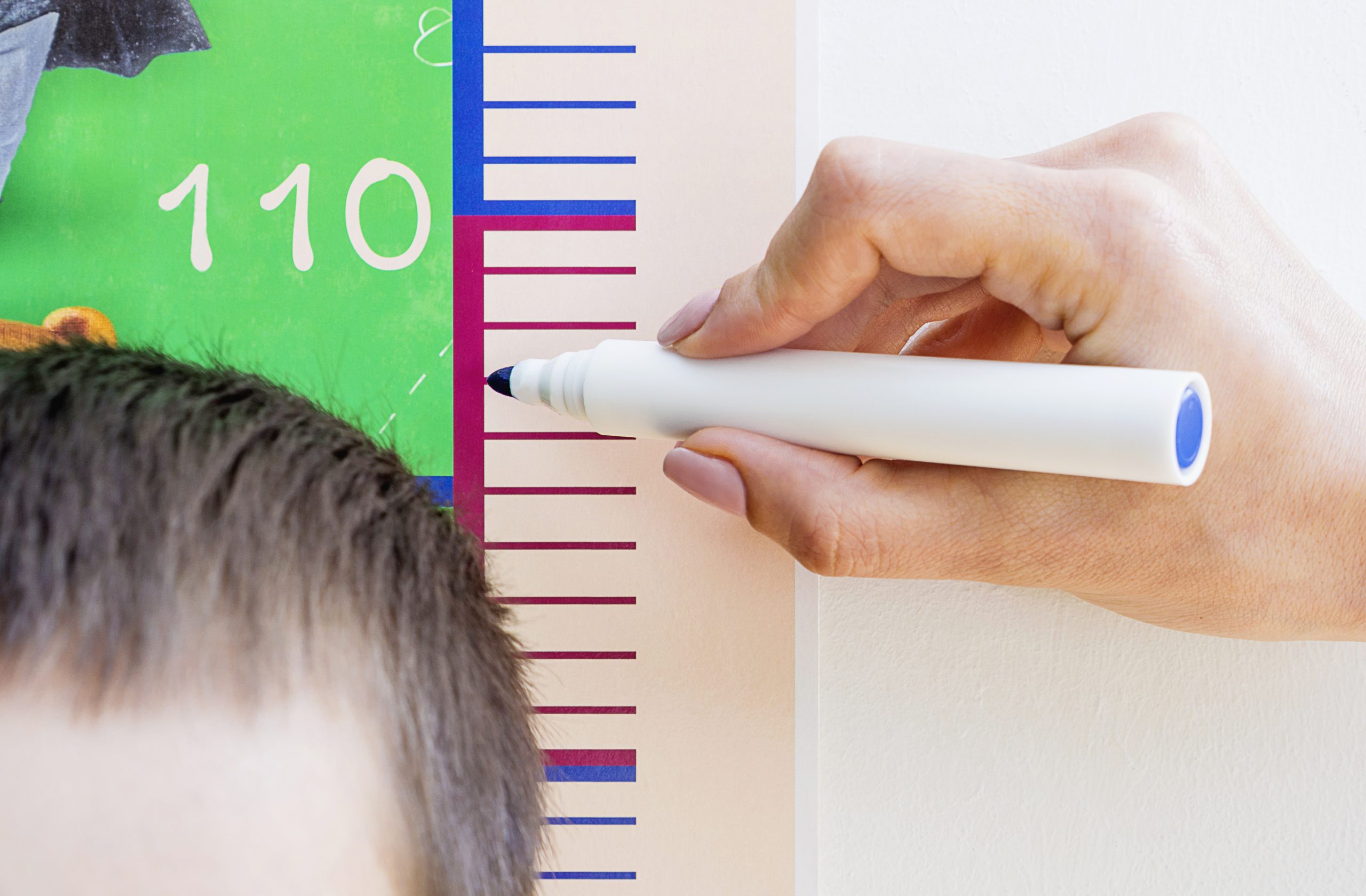
Growth hormone deficiency is a common complication of radiotherapy. © Adobe Stock


Teams from Bicêtre AP-HP hospital, Inserm, Gustave Roussy and the University of Paris-Saclay studied the influence of growth hormone treatment on the risk of a second tumor. in 2,852 adults recovered from childhood cancer. The data confirm that treatment with growth hormone in these children with growth hormone deficiency does not increase the risk of developing a second cancer. This study therefore provides reassuring data on the long-term fate of these children cured of cancer and treated with growth hormone to enable them to reach normal adult height. The results of this study were published in the European Journal of Endocrinology in September 2020.

Growth hormone deficiency is a common complication of brain radiation therapy. Children treated with radiation therapy need growth hormone treatment to reach normal adult height, but there have been concerns about a possible increased risk of another tumor developing in adulthood caused by it. growth hormone treatment.

Researchers from unit 1018 of the “Center for Research in Epidemiology and Population Health (CESP)” (Inserm / Université Paris-Saclay / Gustave Roussy) and from Bicêtre AP-HP hospital, analyzed data from a French cohort, Euro2k, which brings together 2,852 survivors of pediatric cancer diagnosed before the age of 18 before 1986. Among them, 196 had been treated in childhood with growth hormone.

The research team studied the influence of growth hormone treatment on the occurrence of second tumors with a follow-up of 26 years, taking into account the doses of radiation received by all the organs of the body. These were obtained by reconstituting the radiotherapy received for each child. 

In this cohort, 374 survivors developed a second tumor, 40 of whom received growth hormone treatment in childhood. Analysis of the data shows that treatment with growth hormone is not associated with an increased risk of second tumors. However, these researchers found in survivors who received growth hormone treatment for more than 4 years, a slight increase (x2) risk of meningioma, a benign tumor of the meninges favored by high doses of radiotherapy. This slight excess risk of meningioma in survivors who received more than 4 years of treatment with growth hormone is not significant, however, and there is no evidence that the treatment with growth hormone either. responsible.

“This study provides information on the long-term fate of the children whom we treat with growth hormone for a growth hormone deficiency secondary to the treatment of their cancer. These new data allow us to approach the treatment with growth hormone calmly. in these children cured of cancer, when necessary and to reassure families about the absence of an increased risk of second tumors during this treatment. concludes Dr Cécile Thomas-Teinturier, pediatrician-endocrinologist at the Bicêtre AP-HP hospital and first author of the study.

Eliminating Senescent Cells Is Not the Answer to a Long and Healthy Life

Senescent cells. © Inserm/Lemaitre, Jean Marc/U661

Until now considered highly promising in the fight against aging, the therapeutic strategy of eliminating the senescent cells that accumulate in the body is being challenged by the team of Dmitry Bulavin, Inserm researcher at the Institute for Research on Cancer and Aging of Nice (Inserm/CNRS/Université Côte d’Azur). Their research using mice has shown that in the liver the first senescent cells appear in a population of hepatic cells that play a major role in detoxifying the body. When they studied the elimination of these senescent cells, the researchers saw that the effect on liver function deterioration was worse than that of aging. Their findings,published in Cell Metabolism, provide new avenues for ensuring longer life expectancy in good health.

Aging is associated with the deterioration of many functions of the body and the onset of age-related diseases. Eyesight, hearing, muscle, heart and kidney function decline and the risks of cancer, cardiovascular disease and dementia steadily increase.

Also observed is the accumulation of so-called “senescent” cells in the tissues. While unable to divide due to having lost their function, these cells can induce inflammation and the production of oxidized residues that are toxic to the body. Removing these senescent cells from the body to reduce inflammation and restore tissue and organ function is seen as an interesting therapeutic strategy. Medicines whose mode of action is based on eliminating the survival factors of senescent cells (thus leading to their death) are even in development.

Senescent cells in the liver appear in green on the image. ©Dmitry Bulavin

However, before continuing with the development of these therapeutic strategies, Dmitry Bulavin, Inserm researcher at the Institute for Research on Cancer and Aging of Nice (IRCAN, Inserm/CNRS/Université Côte d’Azur) and his team consider that it is first necessary to gain a deeper understanding of the emergence of these cells, their impacts on the body, and to study whether eliminating them would not have any unexpected harmful effects.

To do this, the researchers developed genetically-modified mouse models in order to track in vivo the appearance and localization of senescent cells over time, by monitoring the expression of gene p16 – a senescence marker common to all cell types.

Some of the models studied were also able to spontaneously eliminate their cells that strongly express p16.

Fibrosis instead of senescence

The researchers observed that the first senescent cells appeared primarily and in large quantities in the liver and more particularly in the sinusoidal endothelial cells found on its surface. These cells play a major role in detoxifying the body, enabling the passage of molecular “waste” from the blood to the liver where it is broken down and then eliminated. “To begin with, senescence has no impact on the filtering activity, which continues to function correctly. But over time, this function decreases and toxic residues inducing oxidative stress start to build up in the body. This early-onset mechanism could be a trigger for aging and the onset of age-related diseases. So we focused on this tissue to study the impact of the spontaneous elimination of senescent cells in our animal model”, explains Bulavin.

The team observed that the mice that underwent elimination of their senescent liver cells generally did not do as well as the others.

Not only did they present a blood platelet problem predictive of early mortality, but also hepatic fibrosis (scar tissue in the liver) that appeared with the destruction of the senescent cells. “This repair mechanism is harmful to tissues with the effect on their deterioration being more rapid than the gradual appearance of senescent cells”, explains Bulavin, who considers that the solution therefore does not lie in eliminating all of the senescent cells. “Instead, we must find ways of delaying the effect of senescence. Previous research has shown that senescence is characterized by epigenetic marks, chemical modifications that alter the functioning of DNA, but not its sequence. They prevent the expression of numerous genes. With my team, we will now explore a promising avenue that involves reprogramming the senescent cells to make them lose their senescence and make them functional again”, he concludes.

A Mushroom to the Rescue of Patients with Rare Genetic Diseases

The Lepista inversa fungus has restorative properties to correct certain genetic mutations. © Christine Bailly

An ordinary edible mushroom could be a game-changer when it comes to the treatment of rare genetic diseases. These affect hundreds of millions of people worldwide who often find themselves powerless in the absence of effective therapy. A team led by Fabrice Lejeune, Inserm researcher at the CANcer Heterogeneity, Plasticity and Resistance to THERapies laboratory (Inserm/ CNRS/ Université de Lille/Institut Pasteur de Lille/Lille University Hospital), in collaboration with a team from the French National Museum of Natural History, has shown that an active ingredient contained in the Lepista inversa mushroom has repair properties, making it possible to correct certain genetic mutations, known as “nonsense” mutations. Their findings have been published in Nature Communications[1].

Rare diseases constitute a major public health problem, affecting 300 million people worldwide. Of genetic origin in 80% of cases, there is currently no curative treatment for these hereditary conditions which include cystic fibrosis, hemophilia, and Duchenne muscular dystrophy. However, it has now been established that specific genetic mutations, known as “nonsense” mutations, are implicated in almost 10% of cases of rare genetic diseases.

DNA is made up of nucleotides, organic compounds that code the amino acids implicated in the synthesis of the proteins needed for the body to function correctly. In practice, “nonsense” mutations introduce a “stop codon” in the mutated gene – a sequence of nucleotides that brings the synthesis of the corresponding protein to a premature halt. From that point, the protein is no longer available as such, leading to the onset of the clinical symptoms of the disease.

Identifying how to correct these mutations therefore represents a major challenge for rare genetic disease researchers such as Fabrice Lejeune and his team at the CANcer Heterogeneity, Plasticity and Resistance to THERapies laboratory (Inserm/ CNRS/ Université de Lille/Institut Pasteur de Lille/Lille University Hospital), working in collaboration with the Chemical library / Extract library and JRU 7245 CNRS Communication Molecules and Adaptation of Micro-organisms of the National Museum of Natural History in Paris.

In 2017, the latter had already made a surprising discovery by showing that extracts from the ordinary edible mushroom Lepista inversa could repair nonsense mutations in three cell lines isolated from patients with cystic fibrosis.

Repair properties

In their new study, published in Nature Communications, the research teams have for the first time identified the active substance in the mushroom that is capable of correcting the nonsense mutations associated with the UGA stop codon, the most common of the three stop codons of the human genetic code.

By fractionating extracts of the Lepista inversa mushroom, the researchers were able to identify one of its active substances – compound DAP (2,6 diaminopurine). They showed that this compound repairs nonsense mutations in human cell lines, and also in animal models. It is also of very low toxicity.

This discovery opens up interesting therapeutic avenues for patients with rare genetic diseases. “The idea is to be able to correct the clinical aspects by repairing the nonsense mutations linked to the UGA codon and by restoring the function of the mutated gene. It must be noted that it is not about giving the mushroom to patients to consume directly, given that it contains other compounds whose effects we cannot control, but rather about developing treatments based on the active substance identified here”, emphasizes Lejeune.

The next step for the researchers will involve testing this active substance in other animal models in order to then be able to rapidly launch clinical trials should the results remain promising.


[1] A patent was filed for the discovery via SATT Lutech: Purine derivative for use in the treatment or prevention of diseases caused by a nonsense mutation – October 2017 – S. Rebuffat, S. Amand, C. Maulay-Bailly and F. Lejeune -PCT/EP2017/076846; WO2018073413A1

Cancer: the immune system attacks tumors remotely

Video showing T lymphocytes (green) attacking a tumor (blue and orange). In vivo real-time experiments show how T lymphocytes act both locally and remotely within the tumor. © Ronan Thibaut and Philippe Bousso, Institut Pasteur / Inserm

How does the immune system act to limit tumor development? Using in vivo imaging tools, scientists from the Institut Pasteur and Inserm described the spatiotemporal activity of tumor-infiltrating T lymphocytes, both locally and remotely. Their research was published in the journal Nature Cancer on March 9, 2020.

Some cells in the immune system, like T lymphocytes, are capable of attacking cancer cells. Promising new therapies known as immunotherapies, recognized by the 2018 Nobel Prize in Medicine, attempt to boost the immune system’s response to cancer.

But how exactly do T lymphocytes act in tumors? T lymphocytes are killer cells that are capable of infiltrating a tumor and destroying cancer cells, one by one, through direct contact. This destruction of cancer cells is a highly local phenomenon that only occurs in the immediate vicinity of killer cells. But during these contacts, T lymphocytes also produce soluble molecules known as cytokines. Scientists from the Institut Pasteur and Inserm set out to understand the effect of one of these cytokines, known as interferon-gamma (IFN-γ), on the tumor microenvironment.

They used highly powerful imaging techniques to visualize, in real time and in vivo in mice, both the behavior of T lymphocytes and also the effect of IFN-γ within the tumor. The scientists observed that rather than acting locally, the cytokines spread rapidly within the tumor and affect cancer cells that may be distant from the T cells.

“This remote action within the tumor is very interesting because it enables T lymphocytes to act on a large number of cancer cells, especially those that may have developed mechanisms to escape the immune system,” explains lead author Philippe Bousso, an Inserm researcher and Head of the Dynamics of Immune Responses Unit at the Institut Pasteur.

In their research, the scientists also demonstrated that the number of T lymphocytes that successfully infiltrate the tumor is correlated with the quantity of cytokine produced and determines the extent of tumor cell response. A study of melanoma patient cells supports this model of remote action by immune cells. Stimulating this collective response could therefore represent a key target for future immunotherapy approaches.

A New Blood Component Revealed

Functional extracellular mitochondria revealed in the blood circulation. ©Alain R. Thierry/Inserm

Does the blood we thought to know so well contain elements that had been undetectable until now? The answer is yes, according to a team of researchers from Inserm, Université de Montpellier and the Montpellier Cancer Institute (ICM) working at the Montpellier Cancer Research Institute (IRCM), which has revealed the presence of whole functional mitochondria in the blood circulation. These organelles that are responsible for cellular respiration had hitherto only been found outside cells in very specific cases. The team’s findings, published in The FASEB Journal, will deepen our knowledge of physiology and open up new avenues for treatment.

Mitochondria are organelles that are found in the eukaryotic cells. A place of cellular respiration, they are the cells’ “batteries” and play a major role in energy metabolism and intercellular communication. Their particularity is to possess their own genome, transmitted solely by the mother and separate from the DNA contained in the nucleus. The mitochondria can sometimes be observed outside the cells in the form of fragments encapsulated within microvesicles. Under certain very specific conditions the platelets are also capable of releasing intact mitochondria into the extracellular space.

The work of a team led by Inserm researcher Alain R. Thierry at the Montpellier Cancer Research Institute (Inserm/Université de Montpellier/Montpellier Cancer Institute) has now revolutionized knowledge of this organelle by revealing that whole functioning extracellular mitochondria are in fact found in the bloodstream!

The researchers used previous findings which showed that the plasma of a healthy individual contains up to 50,000 times more mitochondrial DNA than nuclear DNA. They hypothesized that for it to be detectable and quantifiable in the blood in this manner, the mitochondrial DNA had to be protected by a structure of sufficient stability. In order to identify such a structure, plasma samples from around 100 individuals were analyzed.

This analysis revealed the presence in the blood circulation of highly stable structures containing whole mitochondrial genomes. Following examination of their size and density, as well as the integrity of their mitochondrial DNA, these structures observed using electron microscopy (up to 3.7 million per ml of plasma) were revealed to be intact and functional mitochondria.

Throughout the seven-year research period, the scientists used as many technical and methodological approaches as possible to validate this presence of circulating extracellular mitochondria in the blood.

“When we consider the sheer number of extracellular mitochondria found in the blood, we have to ask why such a discovery had not been made before, notes Thierry. Our team has built up expertise in the specific and sensitive detection of DNA in the blood, by working on the fragmentation of extracellular DNA derived from the mitochondria in particular”, he adds.

But what is the role of these extracellular mitochondria? The answer to that could be linked to the structure of the mitochondrial DNA, similar to that of bacterial DNA, which gives it the ability to induce immune and inflammatory responses. Based on this observation, the researchers hypothesize that these circulating mitochondria could be implicated in many physiological and/or pathological processes requiring communication between the cells (such as the mechanisms of inflammation). Indeed, recent studies have demonstrated the ability of certain cells to transfer mitochondria between themselves, such as the stem cells with damaged cells. “The extracellular mitochondria could perform various tasks as messenger for the entire body”, specifies Thierry.

In addition to its importance to our knowledge of physiology, this discovery could lead to improvements in the diagnosis, monitoring and treatment of certain diseases. In fact, the research team is now devoting its attention to evaluating the extracellular mitochondria as biomarkers in non-invasive prenatal diagnosis and cancer.

This research is supported by the Montpellier Integrated Cancer Research Site (SIRIC) (Inserm/CNRS/Université de Montpellier/Montpellier Cancer Institute/Montpellier University Hospital/Université Paul Valéry), funded by Inserm, the National Cancer Institute (INCa) and the Directorate General of Health Care Provision (DGOS).

Meningeal Lymphatic Network: A New Avenue in the Treatment of Brain Tumors

Glioblastomas are the most common tumors of the central nervous system Image taken using a Zeiss Axioimager Z1 widefield epifluorescence microscope. Inserm/Guichet, Pierre-Olivier

Glioblastomas are the most common type of brain tumor, and their prognosis is often highly unfavorable. A collaborative study by Jean-Léon Thomas, Inserm researcher at the Brain & Spine Institute (Inserm/CNRS/Sorbonne Université) and Pitié-Salpêtrière Hospital AP-HP, and Akiko Iwasaki (Department of Immunology, Yale University School of Medicine, USA), has revealed the beneficial role played by the meningeal lymphatic vascular network in treating these tumors – in the short and longer term. Their findings have been published in Nature.

Glioblastomas are not just the most commonly occurring type of brain tumor, they are also the most severe. With an estimated prevalence of 1/100,000, they mainly affect patients between 45 and 70 years of age. Treatment currently involves surgery combined with radiation therapy and chemotherapy. Therapeutic benefit, in terms of survival, remains modest (currently around 18 months), inciting researchers to continue to explore new avenues of potential treatment.

Eric Song (Yale University), first author of this study, Jean-Léon Thomas, Akiko Iwasaki and their colleagues studied the meningeal lymphatic network to see whether it regulates the immune system in response to the presence of a brain tumor. A veritable pipework of lymphatic vessels in the meninges surrounding the brain, the meningeal lymphatic network has been generating particular interest since the publication of studies over the previous five years showing its connection to the lymph nodes of the neck (where immune cells proliferate and differentiate), and its role of draining immune cells into the latter.

In their latest study, published in Nature, the researchers worked with animal models of glioblastoma. They showed that the tumor would disappear following prior enlargement of the meningeal lymphatics – achieved by injecting the meninges with lymphatic growth factor VEGF-C. The growth of the meningeal lymphatic network induced by VEGF-C (as can be seen in the photo) was correlated with the mass entry of immune T cells (CD4 and CD8), which under normal conditions are absent, into the tumor environment.

This short-term response destroys the tumor and is accompanied by the persistence of “memory cells” specifically directed against the tumor cells, which makes it possible to reject the same tumor in the longer term.

Nevertheless, the researchers’ experiments show that it is in combination with an immunotherapy already used in neuro-oncology that the transient VEGF-C treatment is the most effective, enabling complete eradication of the existing glioblastoma. “Our study highlights the fact that reinforcing the network of meningeal lymphatic vessels increases tumor antigen traffic from the meninges to the lymph nodes”, explains Thomas.

With his colleagues, he concludes that the major role of this network is to transport, from the meninges, an immune alert message triggering activation of the lymphocytes directed against the tumor.

The findings of this study therefore open up new avenues in the treatment of brain tumors by targeting the meningeal lymphatic vessels and their associated lymph nodes.

The researchers wish to continue their work by studying the role of the meningeal lymphatic network in other diseases. “We are currently exploring the functional mechanisms and therapeutic potential of this vascular network with novel experimental models, and in other nervous system diseases – neurodegenerative, neurovascular and infectious“, concludes Thomas.

B Cells: New Allies in Sarcoma Immunotherapy?

Tertiary lymphoid structures are cellular aggregates that contain many B-cells (in purple) located near tumors. This is the area where the antitumor immune response starts. ©Antoine Bougouin/Centre de recherche des Cordelier/Inserm, Sorbonne Université, Université de Paris

How can we improve and better personalize the treatment of soft tissue sarcomas, these particularly resistant and aggressive forms of cancer? An international team led by Wolf Hervé Fridman with researchers from Inserm, Sorbonne Université and Université de Paris at the Cordeliers Research Center, in collaboration with the French League against cancer and Institut Bergonié, has shown that B cells also play a major role in predicting of patient’s response to immunotherapy.  It was previously thought only T cells could be used in this way. Their findings, to be published in Nature, pave the way for the personalization of treatments for patients with soft tissue sarcomas.

Soft tissue sarcomas are a heterogenous group of aggressive, chemotherapy-resistant cancers that affect the soft tissues of the body (fat, muscles, fibrous tissue, blood and lymphatic vessels, nerves, etc.). In the current clinical trials, only 15% of patients respond to immunotherapy, which raises the question of the needless exposure of the other patients to the toxicity of these treatments. Identifying markers that predict their response to immunotherapy is therefore crucial. A strategy that until now has been essentially focused on the T cells – immune cells capable of recognizing cells that are infected, cancerous or foreign to the body.

Through research published in Nature, a group led by Wolf Hervé Fridman with members from Inserm, Sorbonne Université and Université de Paris at the Cordeliers Research Center, in collaboration with the “Tumor identity card” team from the French League against cancer, Institut Bergonié, and teams from the USA and Taiwan, studied the question of identifying other potential markers.

They analyzed 608 tumors, classifying them into three groups according to the composition of their microenvironment[1]: immunologically poor tumors (low in immune cells and poorly vascularized), highly vascularized tumors, and immunologically rich tumors. The latter present aggregates of various cell types with high levels of B cells, the immune cells responsible for the production of antibodies. These aggregates are called tertiary lymphoid structures. The researchers observed that an anti-tumor immune response initiates within them, thereby showing that the B cells could play an anti-tumor role.

What is more, in a phase 2 clinical trial, the patients with immunologically rich tumors showed a high response rate (50%) to one immunotherapy: pembrolizumab. These patients also had a higher survival rate than those with immunologically poor or highly vascularized tumors.

A second study by a US team, co-signed by Wolf Hervé Fridman’s team at Cordeliers Research Center (Inserm/Sorbonne Université/Université de Paris), and published in parallel in Nature, extended these observations to include melanoma and kidney cancer.

The results of these studies show that in addition to the T cells that are usually researched, the B cells play an essential role in the response to immunotherapy for certain cancers. These cells bring new hope for the treatment of soft tissue sarcomas, which are particularly resistant to standard therapies. In addition, from a personalized medicine standpoint, these findings could help orient clinical decisions and patient treatment by means of a simple test to identify those whose tumors are immunologically rich.

On the basis of these results, an initial French clinical trial coordinated by Antoine Italiano (Institut Bergonié, Université de Bordeaux), co-author of the first article, and which includes patients with such tumors, is currently ongoing within the French Sarcoma Group.

[1] The tumor microenvironment corresponds to the biological elements that surround the tumor (blood vessels, immune cells, various types of cells, signaling molecules, extracellular matrix, etc.) and with which it interacts.

Metastasis: When Cancer Cells Hitch a Ride Through the Bloodstream

Imagerie de billes fluorescentes filmées à très haute vitesse

Des billes fluorescentes sont filmées à très haute vitesse, et analysées pour suivre leur trajectoire, pour comprendre l’influence des forces fluidiques sur l’arrêt d’une cellule tumorale sur un tapis de cellules endothéliales. ©Harlepp S. /Goetz J

Circulating tumor cells use the blood and lymphatic networks to disseminate within the body, forming metastases distant from the primary tumor. Inserm researcher Jacky Goetz and his Tumor Biomechanics team at the Molecular ImmunoRheumatology laboratory (Inserm/Université de Strasbourg) have helped to show that the flow properties of these biological fluids have a huge influence on the risk of developing metastases. They observed that the slowdown of blood flow where the arteries branch out gives cancer cells the opportunity to attach to and pass through the vessel wall, and then colonize the tissues. The team describes its work in a review article published in Nature Reviews Cancer.

Cancer cells largely exploit the biological fluids in order to disseminate within the body and form metastases in locations remote from the primary tumor. While some access the bloodstream directly, others do so first by escaping from the tumor via the interstitial fluid and lymphatic network to then colonize the lymph nodes before joining the bloodstream.

Jacky Goetz, Inserm Research Director of the Tumor Biomechanics team at the Molecular ImmunoRheumatology laboratory (Inserm/Université de Strasbourg), has been studying these mechanisms for a number of years. His research has been showcased in Nature Reviews Cancer.

Goetz and his team developed a zebrafish embryo model to study the links between blood flow properties, the formation of circumstances favorable to metastases, and their development in the body. The transparency of this animal made it possible to observe in vivo and in real time using a microscope the displacement of cells or tumor extracellular vesicles that had been rendered fluorescent. This enabled the scientists to calculate with precision the flow characteristics — the speed and pressure exerted on the cells — and to correlate these data with the development of metastases.

The researchers were able to observe that the cancer cells circulate rapidly in the large arteries without the ability to stop. However, when the arterial diameter narrows and the blood network branches out, the blood flow slows markedly, giving the tumor cells the opportunity to attach to the vessel wall, pass through it — a phenomenon known as extravasation —, and then colonize the tissues. The team identified a certain number of “hotspots” for this extravasation, corresponding exactly to the most common metastatic sites in humans, there where the blood network is made up of many small capillaries: the brain, lungs and liver. This research was confirmed in collaboration with teams from Germany, in a cohort of 100 patients with brain metastases. Goetz’ team also discovered that it is possible in the zebrafish to change the location of the hotspots, and therefore that of the metastases, by changing the speed of the blood flow.

Also with the help of the zebrafish, they observed that metastatic formation was preceded by the release of tumor‑derived extracellular vesicles. These contain proteins, DNA, RNA, and appear to act as veritable scouts for the tumor cells, possibly to prepare their implantation. The research team showed that the behavior of these vesicles in the blood is similar to that of the tumor cells, and also depends on the force of the blood flow.

Finally, the researchers correlated the force of the flow with the action of two proteins located on the tumor cell surface, which can only act when the blood flow is slowed down. The first, CD44, acts like a brake by attaching to the vessel wall. The second, α5ß1 integrin, enables the cell to stop and to pass through the vessel wall and out of the blood circulation. In zebrafish and mice, the absence of α5ß1 integrin strongly slows metastatic growth.

“Overall, this research shows that if we are to prevent the development of metastases, we cannot just focus on the inherent properties of the tumor, its microenvironment or that of the metastases, we must also consider the role played by the biological fluids. Preventing the circulating cells from stopping or from attaching to the vessel wall could, for example, reduce this risk”, concludes Goetz.

Schéma montrant la circulation, adhérence et extraction des cellules et vésicules tumorales pour former une métastase en fonction de la variation de la vitesse du flux sanguin

Circulation, adherence and extravasation of tumor cells and vesicles to form a metastasis according to changes in blood flow speed. ©Jacky Goetz / Nature Reviews Cancer, 2019


French

English

Cellule cancéreuse

Cancer cell

Globule rouge

Red blood cell

Vésicule extracellulaire issue de la tumeur

Tumor‑derived extracellular vesicle

Remodelage de la paroi endothéliale

Endothelial wall remodeling

Cellule endothéliale

Endothelial cell

Cellules immunitaires

Immune cells

Métastases en croissance

Growing metastasis

Flux sanguin

Blood flow

Extraction hors du vaisseau

Extravasation

Occlusion

Occlusion

Accumulation de cellules cancéreuses

Cancer cell accumulation

Adhésion

Adhesion

Protéines d’adhérence : CD44 et intégrine α5β1

Adherence proteins: CD44 and α5β1 integrin

Fragmentation cellulaire par les cellules immunitaires

Cell fragmentation by the immune cells

Towards a Drug to Combat a Severe Intestinal Disease in Children, Immunocompromised Patients

3D structure of the enzyme with the molecule AN3661 shown against the background of the intestine of an immunocompromised mouse infected with Cryptosporidium. ©Fabrice Laurent and Christopher Swale.

Researchers from Inserm and INRA working in the teams of Mohamed-Ali Hakimi (Institute for Advanced Biosciences – Inserm U 1209 / CNRS JRU 5309 / UGA) and Fabrice Laurent (INRA) have recently discovered a new candidate drug to control cryptosporidiosis, a severe intestinal disease in children, immunocompromised patients, and young ruminants. Beyond this disease, their research represents an opportunity to discover new therapeutic avenues for related infections, such as toxoplasmosis and malaria. Their findings have been published in Science Translational Medicine.

Cryptosporidiosis is a diarrheal disease caused by Cryptosporidium, a microscopic parasite that develops in the intestine of numerous mammals – notably humans. This intestinal parasite is mainly spread through contaminated drinking or pool water, where it can survive for several days in the presence of chlorine, or through contact with infected animals. Over the past 20 years, infection with Cryptosporidium has been recognized as a common cause of waterborne disease in humans. According to a recent study by the US Centers for Disease Control and Prevention (CDC), the number of Cryptosporidium epidemics is even on the increase. In humans, it causes acute and sometimes fatal diarrhea in the most vulnerable populations, including young malnourished children and immunocompromised patients (for example, those infected with HIV). The therapeutic arsenal is currently very limited and in some cases ineffective in eliminating this parasite.

This study conducted by the researchers from Inserm and INRA reveals the discovery of a candidate drug called AN3661, which drastically reduces not just Cryptosporidium infection but also that of Toxoplasma, the parasite responsible for toxoplasmosis.

The study teams have revealed the mechanism of action of this molecule by elucidating the three-dimensional structure of its target, called CPSF3, in Cryptosporidium. AN3661 binds to the heart of the enzyme CPSF3, thereby preventing the maturation of the messenger RNA, a process essential to the parasite’s survival. Preclinical tests using an animal model show remarkable efficacy in vivo with single-dose treatments of the infection in immunocompromised or baby mice.

This major discovery paves the way for new therapeutic strategies and innovations to fight not just cryptosporidiosis but also other related infections, such as toxoplasmosis and malaria.

A Vaccine to Overcome Immunotherapy Resistance

©AdobeStock

For patients with metastatic cancers such as those of the lung or bladder, or melanoma, immunotherapy represents a genuine therapeutic revolution. Unfortunately, it is only effective in 10 to 25% of those eligible to receive it. Researchers from the Cancer Research Center of Lyon (CRCL – Inserm / CNRS / Université Claude Bernard Lyon 1 / Léon Bérard cancer center) and the Léon Bérard and Gustave Roussy cancer centers have shown that a commercially-available vaccine can overcome resistance to immunotherapy. Their study, published in Science Translational Medicine, shows that not only can gastroenteritis vaccines induce the immunogenic death of cancer cells in vitro, but also that combining them with immunotherapy triggers a potent anti-tumor immune response in vivo – where immunotherapy alone had failed.

How can we overcome resistance to immunotherapies, so that as many patients as possible can benefit from these innovations? A team of researchers led by Aurélien Marabelle (Gustave Roussy and Léon Bérard cancer centers), Christophe Caux (Inserm U1052) and Sandrine Valsesia-Wittmann (Léon Bérard cancer center – Inserm UA8) has studied this question. Their idea was to use vaccines to render immunotherapy effective in those cancers in which it has been unsuccessful so far. Then, the aim was to increase the number of patients who could benefit from such therapy in cancers where it has already been shown to be effective.

In this study, our research team looked at pediatric tumors such as neuroblastomas, which are aggressive cancers that do not respond to existing immunotherapies such as anti-PD(L)1 and anti-CTLA4. In our aim to transform how they respond to immunotherapy, we used various vaccines as sources of pro-inflammatory elements because pathogens such as viruses are able to directly stimulate innate immune receptors” explains Marabelle.

Gastroenteritis vaccines

To start with, the researchers tested 14 commercially available vaccines (BCG, Cervarix, TicoVac, etc.) in vitro for their ability to stimulate these innate immune receptors.

Out of the vaccines tested, those used against rotavirus (Rotarix, Rotateq), the virus responsible for gastroenteritis, were identified as having strong pro-inflammatory properties. Unexpectedly, they observed that these vaccines have an oncolytic action – namely the ability to preferentially infect and kill cancer cells in relation to normal cells and induce what is known as immunogenic death.

Vaccine plus immunotherapy – a potent combination

The researchers also conducted in vivo testing of the most pro-inflammatory vaccines using models of neuroblastoma in which the anti-PD(L)1 and anti-CTLA4 immunotherapies are ineffective in humans. This involved injecting these vaccines either systemically or intratumorally (directly into the tumors).

They observed that when the rotavirus vaccines were injected intratumorally, some of the tumors disappeared. When they then administered the vaccine in combination with anti-PD(L)1 or CTLA4 immunotherapies, all of the tumors disappeared.

While the tumors usually do not respond well to either of these treatments used alone, combining them generates a strong systemic antitumor immune response capable of eradicating both injected and non-injected tumors. “Our findings show that the rotavirus strains contained in gastroenteritis vaccines can make usually naturally-resistant tumors sensitive to immunotherapy“, highlights Caux.

The researchers also sought to explain how the rotaviruses exert a stimulant effect on the immune system. They showed that the activation of an innate immune receptor known as RIG-I (retinoic acid induced gene I) was essential for the synergistic effect of the intratumoral rotaviruses with the immunotherapies.

The findings of this study provide a strong scientific rationale in favor of the development of intratumor immunization strategies for immunotherapy-refractory cancers, particularly in pediatric oncology but also in adults,” concludes Valsesia-Wittmann.

This research was supported by the Pediatric Hematology and Oncology Institute (iHOPe), the French League Against Cancer, Inca, the French National Research Agency (ANR) and the patient associations Les Torocinelles, ALBEC and 111 des Arts.

Pediatric Cancers: Why Some Forms of Leukemia Only Affect Children

Détection de la fusion ETO2-GLIS2 dans des cellules leucémiques de patients. Le signal rouge correspond au locus ETO2, le signal vert correspond au locus GLIS2 et le signal jaune indique la présence de la fusion ETO2-GLIS2.© Thomas Mercher

Acute myeloid leukemia (AML) mainly affects children, with the prognosis often being poor despite several decades of research into more effective treatments. A new study led by Thomas Mercher (Inserm U1170/Gustave Roussy/Université Paris-Sud-Paris Saclay), and performed in collaboration with Jürg Schwaller (UKBB, Department of Biomedicine, Universität Basel) explains why some forms of leukemia develop in very young children. The study – published in Cancer Discovery, a journal of the American Association for Cancer Research – has also revealed potential new therapeutic targets.

Each year, 2,500 pediatric cancers are diagnosed in France, with one third of cases concerning leukemia – commonly known as blood cancer. Over recent decades, research into pediatric cancer has intensified and treatments have improved, but the prognosis remains particularly unfavorable for these young patients.

Acute myeloid leukemia (AML) accounts for 15% of cases of leukemia diagnosed in children and adolescents. Overall survival at 5 years is around 60%, with relapse being the most common cause of mortality.

Abnormal protein fusion

There are several subtypes of AML. One of the most aggressive, which is linked to treatment resistance and a particularly unfavorable prognosis, is acute megakaryoblastic leukemia (AML-M7). In their new study published in Cancer Discovery, the team focused their efforts specifically on this type of acute myeloid leukemia. Their work is co-financed by the National League Against Cancer.

Through the CONECT-AML collaborative network[1], the scientists obtained samples from young patients with AML-M7. In 2012, their analysis of these samples had already revealed AML-M7 to frequently present genetic alterations that lead to the expression of an abnormal protein resulting from the fusion of the two proteins normally independent in the cell. At that time, although this fusion – known as ETO2-GLIS2 – had been identified in 30% of AML-M7 cases, the researchers could not explain its mechanism of action.

They also wanted to understand why AML-M7 is diagnosed in children who are on average a lot younger (under 2 years of age) than those diagnosed with the other pediatric AML subtypes (on average towards the age of 6).

“One of the objectives of our new study was to look at the mechanism of action of the ETO2-GLIS2 fusion, and to better elucidate its consequences. We wanted to answer two major questions, with the first being why this disease is specific to children – since the fusion is not found in adults, and then what the potential therapeutic avenues could be”, explains Thomas Mercher.

This involved the researchers analyzing the characteristics of human leukemia cells and developing a mouse model to study the consequences of the ETO2-GLIS2 fusion.

Towards new therapeutic avenues

In this model, the researchers showed that this fusion is sufficient in order to rapidly induce aggressive leukemia, if it is activated in fetal hematopoietic cells. However, there is little to link its activation in adult cells with the development of leukemia. Moreover, blocking the ETO2-GLIS2 fusion in the in vivo model brings tumor proliferation to a halt, with the abnormal blood cells once again able to differentiate into normal blood cells.

These findings suggest that some forms of leukemia develop specifically in children because the properties of the fetal cells differ from those of adult cells.

Findings which also make it possible to propose new target mechanisms in fetal cells and pediatric leukemia in order to improve treatments for these patients. “We now want to understand exactly how this fusion works. Targeting it in order to directly inhibit it with molecules that could be used in patients is not something we are able to do at present, so instead we will identify and try to target the surrounding proteins that are important for it to function”, concludes Thomas Mercher.

[1] French network of various teams of researchers, clinical pathologists and pediatric hematologists

fermer