Menu

Some Persistent Organic Pollutants Could Increase Breast Cancer Aggressiveness

Photo d'imagerie en microscopie électronique montrant la transformation des cellules mammaires tumorales dans le cancer du sein

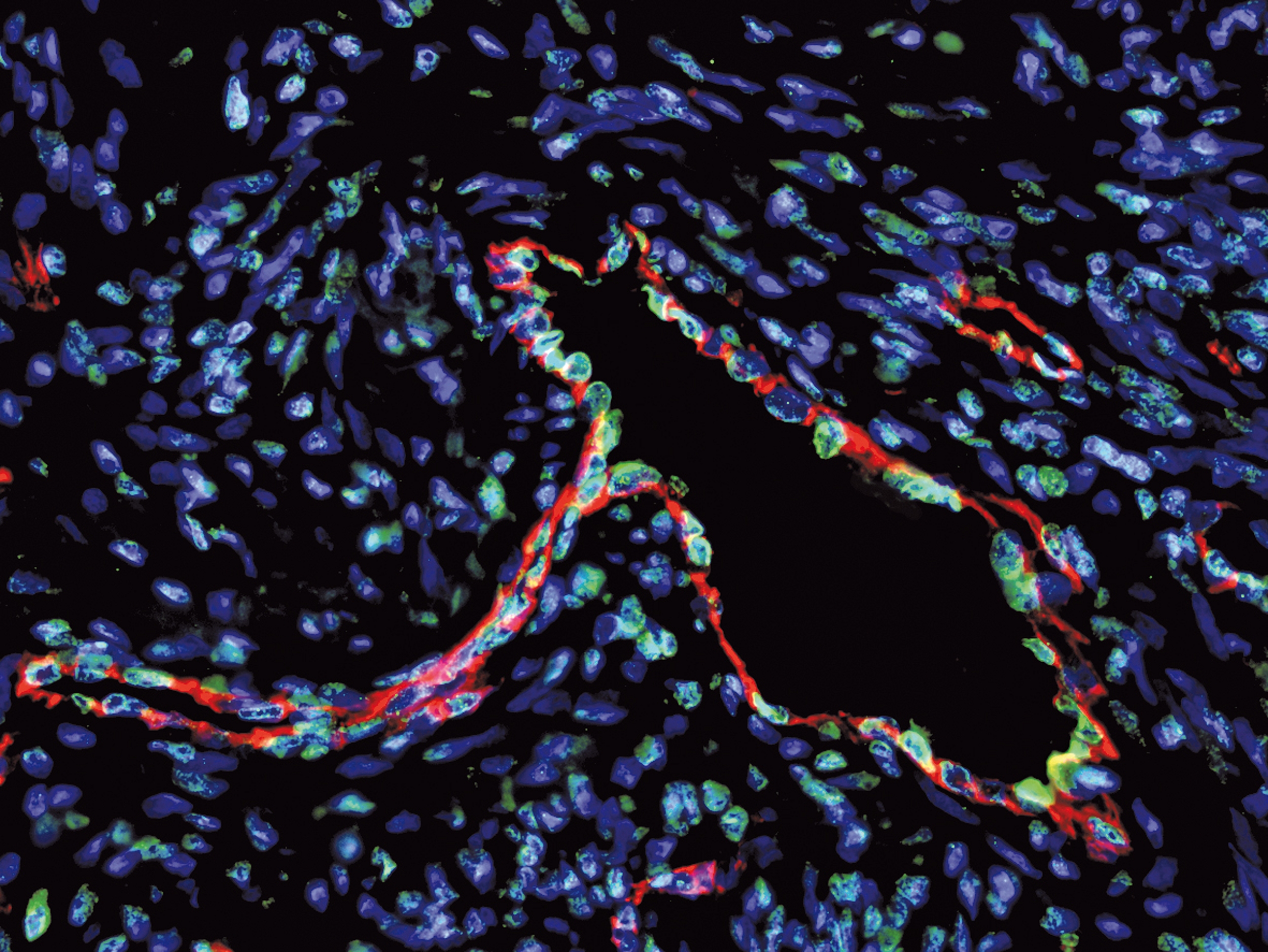
Transformation of tumorous mammary cells in breast cancer ©Xavier Coumoul/Inserm/Université de Paris

Although persistent organic pollutants (POPs) are already suspected to promote breast cancer, there has been little research into how they affect its aggressiveness. A research team from Inserm and Université de Paris in the Environmental Toxicology, Therapeutic Targets, Cell Signaling and Biomarkers laboratory has performed a preliminary study to explore the hypothesis that POPs could promote the development of breast cancer metastases. Its findings suggest a link between the aggressiveness of breast cancer and the levels of some POPs in adipose tissue, particularly in overweight women. While this research published in Environment International opens up new avenues for studying the impact of POPs on breast cancer, its findings must be taken with precaution given the limited number of subjects involved.

Breast cancer is a major public health concern with over 2 million new cases diagnosed and over 600,000 deaths worldwide in 2018. The presence of metastases distant from the original tumor is a marker of its aggressiveness. While the 5-year survival rate for cancer located only in the breast is 99%, this decreases to 85% if it has spread to lymph nodes and falls to 26% if distant metastases are present.

Recent studies have suggested that exposure to persistent organic pollutants or POPs (endocrine-disrupting and/or carcinogenic environmental pollutants that cannot be eliminated by the body) and which accumulate in the food chain1, is a risk factor for breast cancer. However, the influence of these POPs on its level of aggressiveness has received little attention.

A research team led by Xavier Coumoul in the Environmental Toxicology, Therapeutic Targets, Cell Signaling and Biomarkers laboratory (Inserm/Université de Paris) has been the first to test the hypothesis that exposure to POPs could affect the stage of development of metastases in breast cancer.

POPs are highly lipophilic and as such are stored in the adipose tissue. The researchers measured the levels of 49 POPs – including the Seveso dioxin (a waste product from incineration processes) and several PCBs (generated by various industrial processes) – in samples of adipose tissue surrounding the tumors of 91 women with breast cancer2. With excess weight (BMI > 25) known to promote and worsen breast cancer, special attention was paid to the women concerned.

Biological and statistical analysis of these samples enabled the researchers to reveal a link between the presence of distant metastases and dioxin levels in the adipose tissue of overweight women. What is more, in all patients, the concentrations of dioxin and two of the PCBs measured appeared to be linked to the size of the tumor as well as the invasion level and metastatic stage of the lymph nodes. Women with higher concentrations of PCBs were also found to have a higher risk of breast cancer recurrence.

These findings suggest therefore that the higher the concentration of POPs in the adipose tissue, the more aggressive the breast cancer, particularly in overweight women.

To explain this link, the research team has issued several hypotheses based on previous studies. One hypothesis is that dioxin and some PCBs send a signal which promotes tumor cell migration (an essential mechanism in the metastatic process), thereby reinforcing the cancer’s aggressiveness.

According to study leader Xavier Coumoul, “the adipocytes – the cells of the adipose tissue that store fats – play a major role as cells associated with the development of breast cancer. The adipose tissue works like an ʺendocrineʺ gland (secreting hormones into the bloodstream) and we had previously shown that POPs were responsible for the inflammation of this adipose tissue, changing the nature and behavior of the adipocytes. The excessive secretion of inflammatory molecules and the release of the POPs stored by these adipocytes could then promote the formation of metastases.” 

However, the researcher insists on the fact that this study is only preliminary and that its findings must be considered with precaution, given the limitations in its methodology. A limited number of individuals were studied, which encourages statistical bias and means that some sub-categories of the study population cannot be considered to be highly representative. “While it does not enable us to draw firm conclusions on the link between POPs and the aggressiveness of breast cancer, it does open up a novel avenue for research, particularly in overweight patients. An avenue which should be explored by future studies with larger numbers of patients in order to offer more representative statistical findings”, concludes Coumoul.

1 POPs (persistent organic pollutants) have been defined and listed by the Stockholm Convention

2 Men were excluded from the study to limit statistical bias due to sex-specific biological differences.

Hormone therapy has a bigger impact than chemotherapy on women’s quality of life

Cellules cancéreuses. Expression de la protéine PML en rouge et du gène ZNF703 en vert dans des cellules de la lignée de cancer du sein MCF7. ©Inserm/Ginestier, Christophe

Analysis of the CANTO cohort published in the journal Annals of Oncology will upset received wisdom on the effects that hormone therapy and chemotherapy have on the quality of life in women with breast cancer. Contrary to the commonly held view, 2 years after diagnosis, hormone therapy, a highly effective breast cancer treatment worsens quality of life to a greater extent and for a longer time, especially in menopausal patients. The deleterious effects of chemotherapy are more transient. Given that current international guidelines recommend the prescription of hormone therapy for 5 to 10 years, it is important to offer treatment to women who develop severe symptoms due to hormone antagonist medication and to identify those who might benefit from less prolonged or intensive treatment strategies.   

This work was directed by Dr Inès Vaz-Luis, specialist breast cancer oncologist and researcher at Gustave Roussy in the lab “Predictive Biomarkers and Novel Therapeutic Strategies in Oncology” (Inserm/Université Paris-Sud/Gustave Roussy).  

“This analysis of the CANTO cohort shows for the first time that anti-hormonal treatments do not have lesser effects than chemotherapy on women’s quality of life. Quite the contrary, as the diminution in quality of life which is noted at diagnosis is still present two years later, whereas the impact of chemotherapy is more temporary,” explained Dr Vaz-Luis.

In this study, researchers measured quality of life in 4,262 patients with localised breast cancer (stage I to III) at the time of diagnosis and at one and two years thereafter. Primary treatment for these patients was surgical and, for some of them, administration of chemotherapy and/or radiotherapy. About 75-80% of them then took hormone therapy for at least 5 years. Quality of life was measured using a tool which assesses general quality of life in patients with all types of cancer (EORTC QLQ-C30) combined with a tool more specifically designed for use in breast cancer (QLQ-BR23).

For the population studied as a whole there was an overall deterioration in the quality of life at two years from diagnosis. This deterioration was greater in patients who had received hormone therapy, especially after the menopause. By contrast, chemotherapy had a bigger effect on quality of life in non-menopausal patients, especially in terms of worsening of cognitive functions.  

“It is important in the future that we are able to predict which women are going to develop severe symptoms with anti-hormonal treatment so that we can support them,” added Dr Vaz-Luis. While it has been shown that hormone therapy provides a real benefit in reducing the relapse rate of hormone-dependent cancers[1] which represent 75% of all breast cancers, the deterioration in quality of life may also have a negative effect on patient adherence to treatment. It is, therefore, important to offer them symptomatic treatment, in particular for menopausal symptoms, musculoskeletal pain, depression, severe fatigue and cognitive dysfunction; and to combine this with supportive measures such as physical exercise and cognitive behaviour therapy.

“It will also be important in the future to differentiate prior to treatment patients who are at high risk of relapse from those at lower risk in order to tailor hormone treatment. This may be done to avoid escalation of anti-hormonal treatment in certain patients,” concluded Dr Vaz-Luis, emphasising that “hormone therapy is extremely effective in treating breast cancer, resulting in a reduction by approximately 50% in the risk of relapse, and that the finding of adverse effects does not in any way put into question the excellent risk/benefit ratio of this treatment.”

The CANTO cohort (CANcer TOxicities) comprises 12,000 women with breast cancer treated in 26 French centres. It is sponsored by Unicancer and directed by Professor Fabrice André, specialist breast cancer oncologist at Gustave Roussy, Inserm research director and responsible of the lab “Predictive Biomarkers and Novel Therapeutic Strategies in Oncology” (Inserm/Université Paris-Sud/Gustave Roussy). Its objective is to describe adverse effects associated with treatment, to identify the populations at risk of developing them and to adjust therapy accordingly, so as to afford a better quality of life following cancer.

Watch the video on YouTube :

This research was supported by the French National Research Agency (ANR), the Susan G. Komen Association, the Foundation for Cancer Research (ARC), Odyssea and the Gustave Roussy Foundation.


[1] [1] J Clin Oncol. 2019 Feb 10;37(5):423-438 : https://doi.org/10.1200/JCO.18.01160https://doi.org/10.1200/JCO.18.01160

Acting on the immune system even before cancer develops may be feasible

Visualisation en coloration multispectrale d’une sous-population de cellules immunitaires infiltrant une tumeur. Inserm/Jerôme Galon

The immune response is activated from a very early stage in the development of precancerous cells. Unfortunately, this response is simultaneously accompanied by the onset of mechanisms that block it, allowing cancer to develop. This is the first time that the immune response has been described in such detail in precancerous stages, here in lung cancer. This research conducted by the team led by Jérôme Galon, Inserm research director at the centre de recherche des cordeliers (Inserm/Université de Paris/Sorbonne Université) in Paris, shows that using immunotherapies at very early stages could potentially help prevent cancer. This research has been published in Nature.

Our understanding of how the immune system works in the context of cancer is continually improving. Jérôme Galon and his Inserm team have made a significant contribution to such advances. After showing that cancer progression depends among other factors on the presence and functionality of T lymphocytes in the tumor microenvironment and the Immunoscore, the researchers have proven that the immune response and its blocking mechanisms are triggered at very early, precancerous stages.

This means that when a cancer develops, many components of immune surveillance but also the mechanisms designed to evade it have already been implemented.

At the moment, patients with precancerous lung lesions can be monitored and the lesions removed if doctors have the slightest suspicion that they are at risk of developing into cancer. But clinicians may be surprised to discover that it may already be possible at this stage to target the immune system in order to combat progression of these lesions. This is what the research by Jérôme Galon and his team has shown. The researchers had access to 122 lung biopsies from smokers at risk of cancer. They found all stages of precancerous to cancerous lesions in these biopsies. For each biopsy, they studied the immune system in the tumor microenvironment. They performed a genomic analysis of the cells present and looked at them using multispectral fluorescence, an imaging technique that uses specific antibodies to target different types of immune cell. This work enabled them to characterize the nature, quantity, and role of the various immune components in the tumor microenvironment at each precancerous and cancerous stage.

The immune response takes place before cancer

This made it possible for them to compare the development pathways of the cancer and the immune response. At the stage of low-grade dysplasia – an extremely early stage, when only a few morphological abnormalities, DNA repair defects, and greater ability to divide can be seen in the cells – the researchers observed activation of local immune cells and the arrival of naive T lymphocytes, i.e. T cells not trained to specifically destroy abnormal cells. At the stage of high-grade

dysplasia – where there is a higher level of morphological and molecular abnormalities – the researchers then observed mass recruitment of innate and adaptive immune cells, with the presence of B and T lymphocytes specific to abnormal cells and initiation of the immune memory response. But such activation is already accompanied at this stage by the triggering of blocking mechanisms called immune checkpoints and of immune suppressive cytokines, molecules that are also designed to block the immune response. This means that the functioning of the immune system is already altered before the cancer itself develops. This discovery in the context of lung cancer still requires confirmation in other types of cancer. Jérôme Galon is already working on doing so in colon cancer.

The researchers believe that this work will have major implications for the future of patient care. First, they highlight the importance of discovering immune biomarkers that will make it possible to better predict the risk of precancerous lesions developing into cancer.

Second, using immunotherapies designed to inhibit the mechanisms that block the immune system, the much talked about immune checkpoints, could be of benefit to patients in the early stages of cancer prevention.

Inserm Research Director Jérôme Galon Wins European Inventor Award 2019

Inserm: Science for Health

Jérôme Galon©Inserm/Mehrak

Inserm researcher Jérôme Galon has been honored by the European Patent Office (EPO) for his Immunoscore®, a test that predicts relapse risk in patients with certain forms of cancer. An achievement that encapsulates the missions and results of Inserm, a research institute serving good health for all.

The invention of Jérôme Galon – under license from the French National Institute of Health and Medical Research through its private subsidiary Inserm Transfert and marketed under the name Immunoscore® by the company he co-founded – is used to count the immune cells in patient tumor tissue. The more these immune cells are present in the tumor, the better the patient’s chances of survival. In colorectal cancer, the test has a 95% likelihood of predicting patient survival. Immunoscore® gives doctors a more complete picture of the disease, enabling them to offer the most appropriate treatments to their patients.

To perform the test, a specialized scanner produces digital images of tumor samples, from which software will count the number of immune cells. An algorithm then calculates the patient’s Immunoscore® based on the number of T-cells. Clinics throughout the world now use this test to predict relapse risk in patients with colorectal cancer.

“Jérôme Galon’s journey is exemplary in many respects and I offer him my warmest congratulations on winning the European Inventor Award 2019. Through Jérôme, it is the results of our research that are rewarded – when this research succeeds in bringing science and enterprise together. It is also a fine example of our motto at Inserm: Science for health”, declares Inserm Chairman and Chief Executive Officer, Gilles Bloch.

“With 165,000 patent applications filed in Europe every year, I am delighted to receive this award honoring more than 15 years of laboratory work – work that is still ongoing. It is also thanks to the freedom of research afforded to us at Inserm that I was able to embark on this research, over the long term, back when few researchers were interested in cancer immunology. In terms of creating value from this discovery, Inserm Transfert has also played a decisive role by supporting me throughout my journey”, states Galon.

Better air quality: what should the target values be for improving health?

©Photo by Pierre Herman on Unsplash

To produce a significant reduction in mortality due to fine particulates, their average level should be reduced by at least 3 micrograms per m3 as a yearly average, concludes an interdisciplinary study led by researchers from Inserm, the CNRS, INRA, Atmo Auvergne Rhône-Alpes and the Université Grenoble Alpes. This work, published in Environment International, also provides an estimate of the costs of pollution in urban areas: €1200 per inhabitant per year in the Lyon and Grenoble conurbations.

Exposure to fine particulate matter (PM2.5) is recognized to have significant effects on health in urban areas (including cardiovascular and respiratory mortality and disease, and pregnancy and fetal growth problems), but can be reduced. However, public policies designed to reduce atmospheric pollution are often developed without setting the explicit health benefits to be achieved.

So what should the target values be if we want to significantly improve health, decrease health costs, and reduce environmental inequalities? A multidisciplinary team of researchers (epidemiologists from Inserm, biologists and economists from the CNRS and INRA, and air quality specialists from Atmo Auvergne Rhône-Alpes) looked at different hypothetical scenarios for improving air quality in order to identify those that would be most effective.

First, yearly average exposure to PM2.5 was estimated using air quality observation tools and data on the location of housing in the Grenoble and Lyon conurbations (0.4 and 1.4 million inhabitants respectively). Based on previously established dose-response relations, the researchers estimated variations in the number of deaths and cases of lung disease, life expectancy, and associated economic costs for ten different scenarios for reducing PM2.5.

Using the actual situation over the period 2015-2017 as a baseline, with an average concentration of around 14 and 15 µg/m3 in Grenoble and Lyon, and comparing it to a hypothetical situation without human-produced fine particulates (equating to a concentration of 4.9 µg/m3), fine particulate pollution was considered to be responsible for 145 deaths per year in Grenoble (5.6% of deaths, with a 2% margin of error either way) and 16 cases of lung cancer. In Lyon, the respective numbers were 531 and 65. The associated costs, which included tangible costs linked to treatment, but also “intangible” costs linked to the psychological impact on friends and family, came to nearly 500 million euros per year in Grenoble and 1.8 billion/year in Lyon.

10 scenarios tested

The scenarios designed to obtain spatially homogeneous exposure to pollution across the entire study area were the most effective. “Measures that are highly spatially limited – or limited by time, for example, to ‘peak’ pollution periods – would appear to have a much lower impact on both mortality and on reducing health inequalities, ” explains Rémy Slama, Inserm research director.

In relation to mortality, reducing exposure to PM2.5 in line with the WHO (World Health Organization) guideline value for air quality (10 μg/m3) would halve mortality attributable to PM2.5 of human origin, while a reduction of 2.9 μg/m3 (Grenoble) and 3.3 μg/m3 (Lyon) would be required to reduce the mortality attributable to these particulates by a third. It is not simply deaths among vulnerable individuals that would be prevented: life expectancy would also simultaneously increase by around 3 months.

This kind of study can be easily transposed to other conurbations, and may help French cities, many of which have similar levels of pollution to those in Grenoble and Lyon, to focus on scenarios for reducing atmospheric pollution that make it possible to significantly improve health and well-being. The team of researchers is now working to identify the concrete measures that might be taken in relation to the main sources of pollution (heating and road traffic) in order to achieve such a decrease in pollution concentrations.

This study was carried out as part of the QAMECS and MobilAir projects supported by ADEME, Grenoble-Alpes Métropole and IDEX Université Grenoble Alpes.

Human “Jumping Genes” Caught in the Act!

©Photo AdobeStock

Over the course of evolution, the genomes of most living organisms have grown more complex thanks to transposable elements, a.k.a. “jumping genes,” or DNA fragments that can move and copy themselves from one chromosome location to another. Researchers from Inserm, the CNRS, Université Côte d’Azur, and Université de Montpellier were able to capture these “jumping genes” just after they moved. The researchers compared their observations with existing databases. Their work, to be published in Molecular Cell, shows that the integration of “jumping genes” in humans is not random. Instead, it is thought to be influenced by specific genome properties. These results open up new perspectives for interpreting whole genome sequencing data.

Transposable elements, also known as “jumping genes,” are small DNA fragments that can multiply and move in the chromosomes of most living organisms. They have proliferated so intensely in mammals and primates that they make up more than half of our chromosomes! Of course, they don’t jump all at once in all of our cells. Of all the copies present in our DNA, only a small fraction remain active. All the rest are molecular remnants reflecting millions of years of evolution, during which harmful insertions were eliminated and beneficial ones retained.

In humans, the most active jumping genes are L1 retrotransposons. They can alter or destroy genes when they jump, triggering the manifestation of genetic diseases like hemophilia and muscular dystrophy. L1 retrotransposons are also particularly active in some forms of cancer, and could be involved in cellular aging or in some mental illnesses.

Do L1 retrotransposons target specific chromosome regions, or do they choose their positions at random? Teams led by Inserm head researchers Gaël Cristofari and Simona Saccani working at the Nice Institute for Research on Cancer and Aging (IRCAN, Inserm, CNRS, Université Côte d’Azur), along with their colleagues at Université de Montpellier, were able to use a “high-speed” genome sequencing technique to catch actively jumping genes right after they jumped to a new position.

After comparing their observations with genomic and epigenomic databanks, the researchers were able to identify which genome characteristics influenced the integration of the L1 retrotransposons. The most notable characteristic was DNA replication, and natural selection phenomena after integration played a preponderant role.

“We already knew that L1 retrotransposons tend to accumulate in specific areas of our chromosomes, especially heterochromatin. But we didn’t know whether that reflected a particular attraction to those regions, or if they are simply tolerated in those regions and eliminated elsewhere through natural selection. When we know where they jump to and which copies are retained over the course of evolution, we can discover – by deduction – the regions where they can do damage,” explains Cristofari.

Their results make it easier to understand how jumping genes can trigger mutations in humans, and how they contribute to the evolution of our genetic heritage. In the future, this research could be used to interpret whole genome sequencing data, particularly in personalized medicine and vast sequencing programs.

The research was made possible with financial support from the Fondation pour la Recherche Médicale, Cancéropôle PACA, the European Research Council, the French National Research Agency, the Labex Signalife, the Groupement de Recherche sur les Eléments Transposables (CNRS, GDR 3546), the FHU OncoAge, and the European Erasmus Mundus Mobility with Asia program.

Metastatic Lung Cancer: A Targeted Therapy to Improve Treatment Efficacy

©Adobe Stock

When faced with the most aggressive forms of lung cancer, how can the efficacy of chemotherapy be increased? Teams from Inserm, Université Paris Descartes and the Paris public hospitals system AP-HP have maybe hit on a solution. They have developed a targeted therapy which aims to improve the response to platinum salts – the standard chemotherapy used in lung cancer – by neutralizing the activity of a receptor that contributes to its aggressiveness. This research, published in Cancer Letters, shows that in mice this therapy restores the response to chemotherapy and reduces the risk of metastasis by one half to two thirds.

In metastatic lung cancer, life expectancy remains very limited, with death occurring within 5 years in 85 % of patients. The new treatments available for some patient populations present genuine efficacy, but this is of limited duration and successive relapses are common. Increasing the efficacy of existing treatments and finding new ones therefore remains a priority.

A priority that is being addressed by the team of Inserm researcher Patricia Forgez who, in collaboration with teams from the Paris public hospitals system AP-HP (Cochin, Lariboisière and Saint-Antoine hospitals) and Université Paris Descartes is developing a targeted therapy to increase the sensitivity of the most aggressive tumors to platinum salts, an essential chemotherapy in lung cancer.

In previous research, Forgez and her co-workers had shown that lung tumors and especially those at a metastatic stage overexpress the neurotensin receptor. Neurotensin is a small molecule produced in the intestines and brain that is also found to be abnormally overexpressed in tumors where, by binding to its receptor it triggers the continuous cascade of signals stimulating tumor cell growth, survival and migration. This renders them much more aggressive and with little or no sensitivity to platinum salts. By correlating the neurotensin receptor overexpression with the poorer prognosis observed in patients, the researchers had demonstrated that this receptor has a role to play in tumor progression.[1]

In this new study, the research team has developed an antibody to specifically neutralize the form of neurotensin produced by the tumors.[2] 

When tested in multiple experimental mouse models, the researchers observed a 40 to 65 % regression in tumor size and a decrease in aggressiveness. The treated mice presented half as many lymph-node and lung metastases as their untreated counterparts. The researchers also showed that concomitant administration of the antibody with a platinum salt restored or improved treatment efficacy by improving the access of the therapeutic molecule to its target.

The long-term objective is to develop a targeted therapy to block the neurotensin receptor in order to weaken the tumor cells and improve their sensitivity to platinum salts.

“Almost all patients diagnosed with lung cancer receive platinum salts at some point in their treatment, reiterates Jean Trédaniel, study co-author and head of the Paris Saint-Joseph Hospital Group thoracic oncology unit, whether as first-line treatment or following the failure of a targeted therapy or immunotherapy. However, platinum salts are toxic to the body, making it impossible to increase the doses in the event of resistance. Administering this antibody would render the tumor more sensitive to the treatment. What is more, it has been shown in mice to be very well tolerated over the long-term. “

In collaboration with Inserm Transfert, French technology transfer acceleration company SATT Ile-de-France INNOV, and Fair Journey Biologics, the research team is now working on the development of anti-neurotensin antibodies for use in humans, the objective being to embark on a clinical trial. Encouraging results in lung cancer would enable the extension of this therapy to include the other cancers that express neurotensin and its receptor, such as those of the breast, ovary, endometrium, prostate, pancreas, stomach and colon.

[1] This discovery has been protected by a patent filed by Inserm Transfert, the co-owners of which are Inserm and AP-HP. This patent claims that the neurotensin receptor is a marker of tumor aggressiveness and that neurotensin is a potential therapeutic target.

[2] This antibody has been the subject of several patent applications filed by Inserm Transfert on behalf of Inserm and Université Paris Descartes; one of these patents was issued in the USA at the end of 2018.

Early Environmental Exposures and Child Respiratory Health: the Exposome Reveals its Preliminary Results

©Photo by Kelly Sikkema on Unsplash

A team of researchers from Inserm, CNRS, Université Grenoble Alpes and Barcelona Institute for Global Health has shown that prenatal and postnatal exposure to various chemical pollutants is linked to decreased respiratory function in children. These results, based on the concept of the exposome (defined as the totality of an individual’s environmental exposures from conception until old age), were obtained as part of the European HELIX project and have been published in The Lancet Planetary Health.

With the changes in our lifestyles and the development of synthetic chemistry, exposure to environmental contaminants has become multiple and complex. Pregnancy and the early years of life are recognized as being periods of high sensitivity to environmental factors, with potential lifetime health consequences for the child. Researchers from Inserm, CNRS, Université Grenoble Alpes and Barcelona Institute for Global Health have measured a large number of environmental factors to which children are exposed – including through maternal exposure during pregnancy –, and which are defined as the “early life exposome”.

The objective of this approach is to link these exposures to the health of children between 6 and 12 years of age, particularly respiratory function.

The researchers collected data from prenatal and postnatal exposures related to the external environment (pollution of the air with fine particles, noise…), chemical contaminants (endocrine disruptors, metals, persistent organic pollutants…), and lifestyle (diet…) in over 1,000 pregnant women and their children in six European countries. Through 85 prenatal exposures and 125 postnatal exposures, a snapshot of the early-life environment was established for each child. The pregnant women and the children had generally been exposed to dozens of chemical substances at variable levels. Over two-thirds of the chemical exposure biomarkers had levels detectable in at least 9 women or 9 children out of 10.

The analysis suggests that prenatal exposure to perfluorinated compounds (used for their hydrophobic properties in various industrial and consumer products, such as some non-stick kitchen utensils or stain-resistant coatings) and postnatal exposure to ethylparaben (a paraben used as a preservative in cosmetics) and metabolites of phthalates (diethylhexyl phthalate (DEHP), a known endocrine disruptor, and diisononyl phthalate (DINP), used as a plasticizer) could be linked to decreased respiratory function in children.

This study, one of the very first large-scale implementations of the exposome approach, suggests links between pre- and postnatal exposure to chemical substances and deteriorated respiratory function in children. Valérie Siroux, Inserm researcher and joint-coordinator of the study specifies: “Identifying the risk factors of decreased early-life respiratory function is important because lung development is a determinant factor not just of a child’s lifetime respiratory health, but also their general health”.

This exposome approach must be seen as an initial selection step making it possible to identify questionable exposures for which more specific research is needed.

Cancer under pressure: visualizing the activity of the immune system on tumor development

Cancérogenèse : Surexpression de TRF2, marqué en vert, dans les vaisseaux tumoraux, marquage rouge, dans un cancer ovarien. ©Inserm/Wagner, Nicole, 2014

As tumors develop, they evolve genetically. How does the immune system act when faced with tumor cells? How does it exert pressure on the genetic diversity of cancer cells? Scientists from the Institut Pasteur and Inserm used in vivo video techniques and cell-specific staining to visualize the action of immune cells in response to the proliferation of cancer cells. The findings have been published in the journal Science Immunology on November 23, 2018.

Over time, the uncontrolled proliferation of tumor cells results in the accumulation of new mutations and changes to their genome. This gradual process creates significant genetic diversity among the cancer cells in any given patient. And although the cells in the immune system, especially T cells, are potentially able to eliminate these abnormal cells, tumor diversity can have a harmful effect, complicating the action of the immune system and rendering some therapies ineffective. Understanding this frantic race between tumor development and the immune response is key to the success of future immunotherapy techniques.

Scientists in the Dynamics of Immune Responses Unit (Institut Pasteur/Inserm), directed by Philippe Bousso, in collaboration with Ludovic Deriano, Head of the Genome Integrity, Immunity and Cancer Unit (Institut Pasteur), investigated how spontaneous immune responses to tumors influence this tumor heterogeneity. They demonstrated that the immune system can employ mechanisms to significantly reduce tumor diversity, favoring the emergence of more genetically homogeneous tumor cells.

In their study, the scientists marked each cancer cell subclone with a separate color in a mouse model. By monitoring these different colors they were therefore able to characterize the evolution of tumor heterogeneity in time and space. They were also able to observe the contacts between T cells and cancer cells and determine how some tumor cells are destroyed. Their research highlights the drastic impact the immune system can have on tumors by reducing their heterogeneity.


Visualizing the action of stained immune cells.
In this video, the tumor cells are shown in gray. The tumor-specific T-cells, in purple, come into contact with the cancer cells and destroy them. The killed cells are shown in blue. In green, the control cells circulate but do not kill the tumor cells. © Institut Pasteur / Philippe Bousso


Visualizing different clusters of cancer cell clones.
This video illustrates how tumor subclones, each marked by a different color (blue, orange and green), develop in the bone marrow. The vessels are shown in white. © Institut Pasteur / Philippe Bousso

The same impact on the heterogeneity of tumor cells has also been observed in response to immunotherapies that release the brakes on the immune system, an approach which was awarded the Nobel Prize in Physiology or Medicine this year.

This research shows that taking into account the interaction between immunotherapies and tumor heterogeneity could contribute to the development of optimum therapeutic combinations and sequences.

In addition to the organizations mentioned above, this research was funded by the Fondation de France, the French National Cancer Institute (INCa) and the European Research Council (ERC).

A new pathway for modulating anti-tumoral immune response

Cellules cancéreuses d’un mélanome.  Inserm/Valencia, Julio C., 2018

Researchers from Inserm, CNRS, Paris-Sud University, Gustave Roussy, and Institut Curie have identified a new agent in regulating PD-L1 gene expression: the eIF4F complex, which plays a role in controlling protein synthesis.

This complex could become a predictive marker for response to immunotherapy. Furthermore, the researchers demonstrate, for the first time, that inhibition of the eIF4F complex gives rise to an anti-tumoral effect related to decreased PD-L1 expression, which therefore elicits immune system intervention.

The researchers hope that eIF4F inhibitors will be able to be used as anti-cancer agents in the future, alone or, more than likely, in combination with other treatments.

Just a few years ago, the immune system, our defense against disease, seemed unequipped to fight cancer. Advances in immunotherapy are making it possible to correct these shortcomings: the immune system can now learn to recognize and destroy cancer cells. Lymphocytes are thus rediscovering their initial ability to fight rather than protect tumors.

The PD-1 (programmed cell death) molecule, expressed on the surface of T cells, binds to another molecule present on the surface of certain tumor or immune cells, PD-L1. This interaction in a way renders the tumor cell invisible to the immune system, by deactivating (or disarming) T cells.

In recent years, immunotherapy targeting the PD-L1/PD-1 interaction has revolutionized the treatment of melanoma and other types of cancer.

However, many patients do not respond to treatment. These agents are highly effective for several months or years, but only in 10 to 20% of patients, for all types of cancer combined.

The development of biomarkers is therefore a key issue in identifying patients liable to respond to treatment,” explains Professor Caroline Robert, Head of the Dermatology Department at Gustave Roussy.

A high PD-L1 level in tumors is a major indicator since this is often associated with a good response to anti-PD1 agents. However, the mechanisms for regulating PD-L1 expression have not been fully elucidated,” points out Stephan Vagner, Inserm Research Director and Head of the RNA Biology Team at Institut Curie.

In this latest publication, the researchers demonstrate, for the first time, that a complex known as eIF4F, involved in initiating the translation of messenger RNA into proteins, regulates PD-L1 expression and that, by targeting eIF4F in tumor cells, anti-tumoral immunity can be stimulated, thus mimicking the effect of immunotherapy.

In this study, the researchers mainly used melanoma as a model, but also conducted tests with lung cancer, breast cancer, and colon cancer cells.

They will now go on to evaluate the findings of the study on the formation of the eIF4F complex, as a predictive marker for response to immunotherapy.

They are, moreover, developing other melanoma treatment models, based on the use of eIF4F complex inhibitors, in combination with other treatments, to increase therapeutic efficacy and fight resistance.

This study was supported by Inserm, CNRS, Gustave Roussy, and Institut Curie. It was also funded by the Ligue Nationale Contre le Cancer (accredited team), French National Cancer Institute, the ‘Ensemble contre le mélanome’ collective, and the ‘Vaincre le Mélanome’ association, SIRIC Socrate, Fondation Bettencourt Schueller, and Fondation ARC for Cancer Research.

Development of an alternative to bone grafting for edentate patients

©Photo by rawpixel on Unsplash

Decreased jaw bone volume in edentate patients is the main difficulty which arises when fitting dental implants. Bone grafting is currently the most frequent solution for this problem. However, this method has several disadvantages, such as deterioration of the bone graft over time. Between 2010 and 2015, Pierre Layrolle, Inserm researcher, working with Norwegian and German teams as part of the REBORNE European project, tested an innovative technique for bone augmentation in 11 edentate patients, by combining a biomaterial with stem cells. The results, published in the journal Stem Cell Research & Therapy, demonstrate sufficient growth of viable bone in the treated area to allow implants to be fitted, together with the durable preservation of this bone after fitting dental prostheses.

Owing to the lack of mechanical stimulation resulting from mastication among edentate patients, bone wastage affecting the alveolar bone (surrounding and supporting the tooth) can be observed, which represents a third of contraindications for fitting dental implants. This procedure, in fact, requires sufficient bone volume to avoid impinging on the facial nerve, which would result in facial paralysis. Now, without sufficient bone volume in the jawbone to support the soft tissue in the gums, even dentures are proving relatively unsuitable.

While bone augmentation involving biomaterials is successfully used to fill the dental alveoli, bone grafting is currently the most commonly used technique for correcting longstanding multiple tooth loss, with a bone sample taken from the patient’s mandible or skull. In addition to the postoperative risks associated with creating two surgical sites, along with the limited quantity of bone material able to be sourced from a single patient, and postoperative pain, this technique appears relatively unsustainable over time. Transplanted bone tends to resorb very quickly. Owing to the lack of vascularization and continuity with the original underlying bone, it is degraded by immune cells which perceive it as a foreign body.

Pierre Layrolle, Inserm researcher heading up the “Inflammation and cellular communications in bone disorders” team (Unit 1238, Inserm/University of Nantes) worked with Norwegian and German teams as part of the Reborne project from 2010 to 2015, on a long-term solution for alveolar bone augmentation. This European clinical trial followed up 11 edentate (over several years) patients, treated with an innovative mandibular bone augmentation technique based on the “cell therapy” principle.

To replace the bone lamellae (containing the patient’s cells and growth factors) conventionally used in bone augmentation, the research team used a biomaterial, calcium phosphate, to which stem cells taken from patient hip bone marrow samples, cultured for amplification, had been affixed. Although calcium phosphate is unable to regenerate bone, it allows adhesion of stem cells – which have this function. This biomaterial is, moreover, highly resistant to the degrading action of immune cells. The mixture thus obtained was applied to the edentate part of the mandible.

For 5 months, researchers monitored positive bone formation around the graft, then generated a 3D model of the augmented part in order to select the type of implant suited to each patient. The sample taken from the 11 patients when fitting the implants showed that the tissue formed not only generated sufficient quantities of bone enabling dental implants to be fitted, but was also vascularized, unlike conventional bone grafts. The dental implants and prostheses were functional two years after fitting, in the 11 patients.

Further to these positive results, the research team launched the MAXIBONE European project in 2018, once again coordinated by Pierre Layrolle. This clinical trial aims to compare the results of maxillary bone augmentation using cell therapy with conventional bone grafting, on a wider scale on 150 patients, by evaluating the quantity of bone generated by the two types of transplants and their specific cost. This project will also examine the possibility of replacing the patient’s stem cells (autologous cells) with donor stem cells in the context of cell therapy. This could offset the technical difficulties related to the cost and complexity of sampling and storing autologous cells, but also mitigate the variations observed in terms of each individual’s ability to effectively generate bone.

The FP7-Reborne project, a phase 2 clinical trial, received European funding from 2010 to 2015.

The H2020-Maxibone project, a phase 3 clinical trial, has received two European grants and is scheduled to start in early 2019.

Pierre Layrolle is due to attend the Nantes international science fiction festival Les Utopiales from October 31 to November 5, 2018, to demonstrate 3D bioprinting. He will lead the round table “In foreign flesh: accepting a foreign body”, on Wednesday, October 31, at 14:00.

fermer