Menu

Fatty acids, essential for the nervous system… of the gut (as well)

How does a fatty acid deficiency explain Crohn’s disease?

Two Inserm research teams have just shown that failure of the intestine to produce a lipid “messenger” is associated with Crohn’s disease, a common and highly disabling inflammatory bowel disease (IBD). This messenger, derived from an essential fatty acid, regulates the permeability of the intestinal barrier, and could thus become a choice target in the management of IBD. These studies also throw fresh light on the role of the enteric glial cells. Like their counterparts in the central nervous system, for too long considered to play a “secondary role,” the intestinal glial cells are starting to “show their hand,” which is actually indispensable to intestinal homeostasis. Details of this work are published in Gastroenterology.

The enteric nervous system plays a central role in controlling the homeostasis of the digestive functions, such as motor function, and in controlling the functions of the intestinal epithelial barrier. This integrative nervous system, located along the entire length of the digestive tract, is made up of neurons and glial cells. Much remains to be discovered about the role of the enteric glial cells. A growing number of studies shows that they regulate, in a manner analogous to the astrocytes in the brain, the functions of the enteric neurons, as well as those of the intestinal epithelial barrier, such as the proliferation of epithelial cells, their migration and permeability (paracellular and transcellular), and repair processes. Glial cells regulate these functions by liberating different glial mediators, including certain lipid metabolites of n-6 polyunsaturated fatty acids (PUFA).

Otherwise, characterisation of enteric glial cell (EGC) involvement in diseases associated with dysfunctions of the intestinal barrier remains limited to description of the altered expression of glial markers. These dysfunctions in the intestinal barrier (increased permeability, failure in repair) are recognised as playing a possible key role in inflammatory bowel disease (IBD) by helping to trigger recurrences. Reducing these dysfunctions is also a likely mechanism of action of the biotherapies used in the management of IBD.

In this context, Malvyne Rolli Derkinderen and Michel Neunlist, researchers in the Inserm/Nantes University Joint Research Unit “Neuropathies of Enteric Nervous System and Digestive Diseases: Involvement of Enteric Glial Cells,” and their collaborators at the Toulouse Purpan Digestive Health Research Institute (IRSD) initially sought to characterise the production of lipid mediators derived from n-6 fatty acids in animal and human enteric glial cells. They then analysed the impact of the most abundantly produced PUFA metabolites on the functions of the intestinal epithelial barrier, and ultimately demonstrated that production of one of them was defective in patients with Crohn’s disease.

shema neunlist en

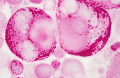
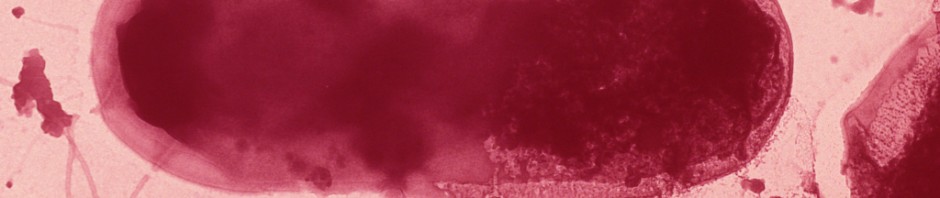
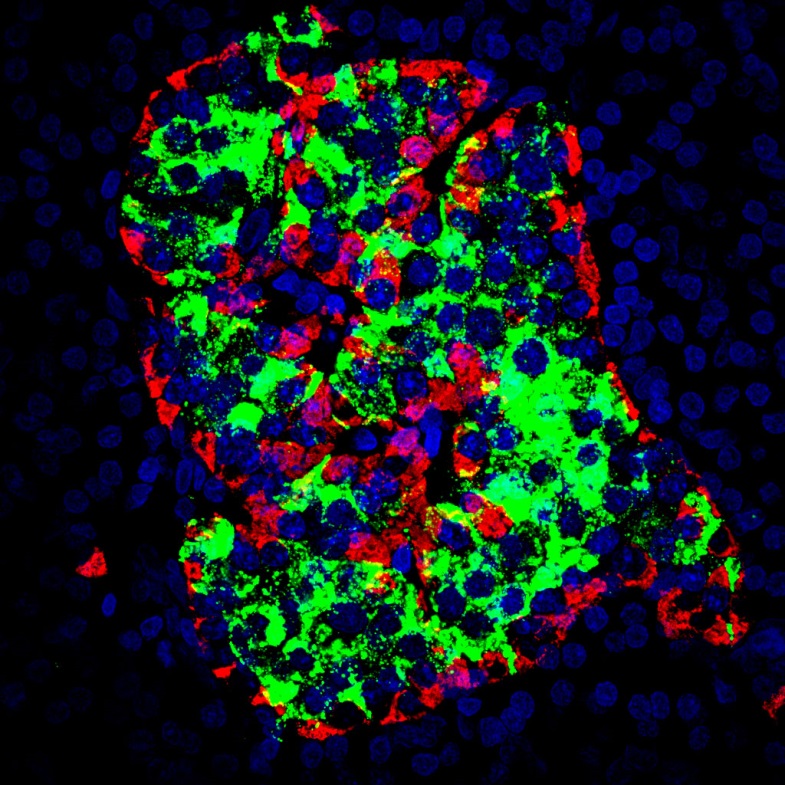
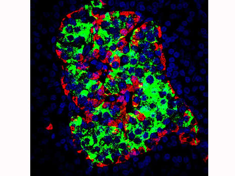
In green: under physiological conditions, the enteric glial cells produce lipid metabolites including 15-HETE, which strengthens the resistance of the intestinal epithelial barrier, and prevents entry of pathogens. In red: during Crohn’s disease, there is a deficiency in glial production of 15-HETE, which leads to an increase in the permeability of the barrier, and facilitates the entry of pathogens. This may contribute to disease recurrence or severity.

Thus the researchers showed that human (as well as rat) EGC are able to produce n-6 PUFA, especially 15-HETE, synthesised by 15-lipoxygenase-2. This 15-HETE strengthens the intestinal epithelial barrier, and reduces paracellular permeability in vivo and in vitro, particularly by increasing the expression of tight-junction molecules including zonula occludens-1. In EGC isolated from patients with Crohn’s disease, the researchers demonstrated defective production of 15-HETE, associated with a loss of ability of the EGC to control the permeability of the intestinal epithelial barrier.

This work therefore identifies n-6 PUFA as a source of metabolites with potentially beneficial effects on the functions of the intestinal epithelial barrier in IBD. For Camille Pochard and Sabrina Coquelorge, the two first authors,

“These results help to underscore the role of the glial cells in particular, and of the enteric nervous system in general, in the physiopathological processes involved in IBD, and to identify new targets of therapeutic interest.”

An anti-diabetic drug corrects dyspraxia associated with neonatal diabetes

Researchers from Inserm and physicians from the Department of Paediatric Endocrinology, Gynaecology and Diabetology at Necker Hospital for Sick Children (AP-HP, Inserm U1016, Paris Descartes University, Imagine Institute) have developed an improved treatment for a rare form of early childhood diabetes associated with cognitive disorders. Their work, conducted in collaboration with the Neurophysiology Department of Pitié-Salpêtrière Hospital, AP-HP, shows that a drug used for decades to treat type 2 diabetes in older subjects acts on the brain of these children. It reduces their hyperactivity and improves their ability to perform precise movements such as writing. The work has recently been the subject of a publication in the journal Diabetes Care.

bébé

©Fotolia

Neonatal diabetes is a disease that develops in the first months of life. There are many causes, but several years ago, researchers discovered that a mutation in the potassium channels that maintain membrane polarisation is often responsible. In the pancreas, this genetic defect affects secretion of insulin, which becomes trapped in the insulin-producing cells. However, the consequences do not end there, since these defective receptors are also present in the muscle and brain cells. They cause hypotonia and dyspraxia in the children.

Although treatment with insulin injections controls the children’s blood sugar levels, it does not correct the other symptoms.

In 2006, this same team of researchers and physicians had shown that use of glibenclamide, a drug used for decades to treat type 2 diabetes in older patients, eliminated the need for insulin injections in its own patients. This drug allows insulin secretion in accordance with the quantity of sugar present in the child’s bloodstream. It therefore enables better control of blood sugar levels without causing hypoglycaemic episodes.


In the study which has just been published*, the team shows that this drug also reduces the hyperactivity observed in the children, problems with fine movement (writing, spatial orientation), and impairment of muscle tone and complex brain functions (task planning) in the children. This enables a clear improvement in their socialisation and family and school life.

The study was carried out in 19 children with this rare form of neonatal diabetes, in whom insulin injections were replaced with oral glibenclamide. Electrophysiological muscle tests, brain imaging and tests for fine and precise psychomotor skills were carried out before and 12 months after introducing the drug. Results made it possible to show that the neuropsychomotor improvement observed was not related to an action of the drug on muscle, but to an effect of the drug on the brain.

“This study is the first to show that an oral diabetes can also act directly on the brain of these children,” explains Dr Jacques Beltrand, coordinator of the study. “It also shows that the effect is better in young children. This drug should therefore be given to these patients as early as possible.”

This work repositions a drug which previously had no indication for paediatric use, and was not available in a suitable dosage form for infants. The team of researchers from Inserm and Necker Hospital for Sick Children, AP-HP, is now involved in the development of a syrup suitable for children, which will allow easy administration and the most accurate dose possible (NEOGLI Study, NCT02375828).

Obesity: surgery corrects diabetes even before weight loss occurs

Each year, obesity kills 2.8 million people worldwide. When confronting morbid overweight—and its direct consequence, type 2 diabetes—surgery is used as a last resort. Strangely, in patients who have had such an operation, the diabetes disappears before and independently of weight loss. Inserm researchers, with the help of physicians from Bichat Hospital, AP-HP, have shown that the surgically reconfigured intestine might be at the root of this improved glycaemic control. This study is published in Gastroenterology.

Overweight, even to a mild degree, is never harmless. As a cause of cardiovascular disease, joint and muscle problems and the development of type 2 diabetes, it can lead to premature death.

When a person is so overweight that his/her health is under threat, there is sometimes only one solution, i.e. bariatric surgery to reduce the patient’s appetite. The most frequently offered procedures are sleeve gastrectomy and gastric bypass. Both of these solutions involve removal of most of the stomach. In sleeve gastrectomy, the stomach is reshaped so that it forms just a tube. In gastric bypass, the food pathway is modified: the surgeon directly connects a small pouch from the top of the stomach to the intestine, forming a “roux limb.” The rest of the stomach, through which food no longer passes, continues to supply the enzymes needed for digestion. These enter the food pathway further down the alimentary tract, at the Y intersection.

Curiously, although patients lose weight rapidly in the months following the operation, physicians have noticed a clear improvement in their type 2 diabetes, practically as soon as they leave hospital. How can this phenomenon be explained? For the researchers at Inserm, the answer is clear: surgery not only affects food intake, but also modifies intestinal metabolism.

In order to confirm their hypothesis, the scientists reproduced both techniques in an obese rat model.

Following bypass, the Roux limb became strikingly enlarged in the rodents, as the cells constituting it proliferated abnormally. Moreover, these cells expressed a type of glucose transporter on their surface that is unusual for the intestine. These observations were also found in patients following surgery at Bichat Hospital.

People with type 2 diabetes secrete insulin, but their organs no longer respond to it. The level of the sugar in the bloodstream is no longer controlled, and remains abnormally high after every meal, resulting in damage to the tissues.

“We labelled glucose in order to follow its metabolic fate. Following surgery, the patient’s newly reconfigured intestine, like that of the rat, begins to consume very large quantities of glucose,” explains Maude Le Gall, Inserm Research Fellow. It diverts dietary and blood glucose for its own consumption. This phenomenon, which develops after barely a few days, might contribute to the rapid improvement in glycaemia and the disappearance of diabetes.”

Gastrectomy, a different mechanism with a similar result

Although gastrectomy does not affect the size of the intestine, it also acts on its cellular function. Absorption of dietary glucose by the intestinal cells becomes reduced. Conversely, the number of cells secreting GLP-1 peptide, which is usually inadequately produced in obese people, increases considerably. GLP-1 acts on the pancreas to increase its production of insulin. As a result, control of glycaemia in response to nutrient intake is more rapid.

“These results prove that sleeve gastrectomy and gastric bypass, while they have very similar effects on diabetes, do not involve the same mechanisms,” adds Maude Le Gall. “At the same time, they indicate the major role played by the intestine in controlling glycaemia.”

Although more studies are needed to understand these processes, this discovery indicates that there is hope of finding new options for treating type 2 diabetes without recourse to surgery.

illustrationobesitefinalsanspicto_en

Ovarian cancer: A promising approach for treating the most aggressive forms using targeted therapy

Thanks to work done at Institute Curie by a team led by Fatima Mechta-Grigoriou, Inserm Research Director, it is now possible to identify, among women with aggressive ovarian cancer, those who could benefit from a promising targeted therapy.inserm_5619

Métastase tumorale dans l’ovaire © Inserm/Evrard, Gilles.

The Stress and Cancer team (an Inserm/Institut Curie team designated by the French National Cancer League), directed by Fatima Mechta-Grigoriou (photo, right), has just taken a major step toward identifying women with an aggressive form of ovarian cancer who are likely to benefit from targeted therapy in the form of a MEK inhibitor.

Finding the Achilles heel of ovarian tumour cells

Moreover, BRAF activates the MEK signalling pathway[1] involved in the development of low-grade ovarian tumours. MEK inhibitors have therefore been proposed as a possible therapeutic solution for these cancers.“75% of ovarian cancers are high grade, i.e. highly aggressive. The mutation profile differs between low- and high-grade tumours,” explains Virginie Mieulet (photo, left), post-doctoral fellow and co-author of this work. “For example, alterations in the KRAS/BRAF oncogenes are present in 70% of less aggressive tumours, but in only 1% of aggressive tumours.”

Fatima Mechta-Grigoriou and her team show that prescription of MEK inhibitors could extend to high-grade tumours. Because the MEK pathway is activated in 50% of high-grade ovarian tumours, even in the absence of KRAS/BRAF mutations. Why? “Because of MAP3K8 accumulation,” explains the researcher. “This protein controls tumour progression by regulating the cell cycle and tumour invasion, thus playing a key role in the development of ovarian cancers.”

“The MAP3K8 protein might serve as a biomarker for identifying patients likely to benefit from a therapy based on MEK inhibition,” adds Fatima Mechta-Grigoriou, Inserm Research Director, “especially since it can be quite simply detected from a tissue section by our physician pathologist colleagues, who, incidentally, have helped us greatly in this work.”

While clinical trials are already underway to assess MEK inhibitors in low-grade ovarian cancers, everything seems to point to the interest of developing a clinical trial in women with high-grade ovarian cancer overexpressing MAP3K8, to evaluate the efficacy of this targeted therapy, in addition to the conventional chemotherapies.

[1] Because of the key role of the MEK signalling pathway in carcinogenesis—activation is observed in 30% of cancers—MEK inhibitors constitute a promising therapeutic approach. Clinical trials are currently underway for several tumour locations (skin melanoma, intestinal tumours, thyroid cancer, etc.).

Ovarian cancer affects nearly 4,600 women in France each year. An ovarian tumour can develop silently for quite a long time, and thus attain a large size before causing symptoms that induce the woman to seek medical advice. This explains why this cancer is often diagnosed at an advanced stage. It is responsible for 3,150 deaths per year. Treatment is based mainly on a combination of surgery and chemotherapy. The choice between the different treatment options depends on the morphology of the tumour cells, their rate of proliferation, disease spread, etc. Analysis of the molecular profile of tumours could soon contribute to this decision and improve management. In most women with ovarian tumours, combination chemotherapies (platinum-taxane chemotherapy) have been shown to be effective. However, once a recurrence is diagnosed, there is no really effective treatment at this time.

ENS@T-HT, launch of a large scale Europe H2020 project coordinated by Inserm for improved diagnosis and treatment of arterial hypertension through an omics-based approach

An international group of scientists from 6 countries bring together their expertise to improve diagnosis and therapeutic care for primary and secondary forms of arterial hypertension. ENS@T-HT, which is coordinated by Maria-Christina Zennaro, Research Director at Inserm (Paris Cardiovascular Research Center), was officially launched this month in Paris and will last 5 years.

Hypertension affects up to 45% of the general population and is responsible for 9.4 million deaths per year worldwide. Even small rises in blood pressure are associated with increased risk of stroke and heart disease. However, despite a large array of available treatments, blood pressure is still not properly controlled in many patients.

Approximately 10% of current hypertension cases could be treated and cured if properly diagnosed. These include disorders of the adrenal gland that increase the production of hormones affecting blood pressure. Correct identification of these disorders is crucial to proper management of the underlying disease and prevention of cardiovascular complications. However, due to the complexity of diagnosis, proper treatment of these conditions can be delayed by several years, exposing patients to increased cardiovascular and metabolic risk and diminished quality of life.

ENS@T-HT is a five-year-long European H2020 research project created to tackle these issues. It received funding of €7.6m and involves 13 academic institutions from France, Germany, Italy, the United Kingdom, the Netherlands and Australia.

The main objective is to develop a programme to improve diagnosis of various adrenal forms of hypertension, enabling curative treatments and preventing complications.

This will be achieved using various cutting-edge ‘omics’ techniques to identify biomarkers in patients’ blood that provide a distinctive signature for their condition. Useful biomarkers will also permit the stratification of patients, so that those most likely to benefit from particular treatments are identified in order to maximise the effectiveness and cost efficiency of treatment.

Maria-Christina Zennaro, coordinator of ENS@T-HT (Inserm Unit 970, Paris Cardiovascular research center) says: « The ambition of this project is twofold: first we want to establish omics-derived biomarkers and validate their accuracy in the diagnosis of patients with adrenal forms of hypertension. Second, and most importantly for patients, we want to use these biomarkers to accelerate and optimize the diagnosis and management of these conditions. We can then stratify those patients who could most benefit from specific targeted treatment ».

More detail about ENS@T-HT, a multiple-step-project with access to unique cohorts of patients in Europe

— In an initial exploratory phase partners will establish omics-derived signatures of patients with PA, PPGL and CS through bioinformatics modelling of large datasets derived from multiple platforms.

— The signatures will be validated as stratification biomarkers by establishing reference values and variability in healthy controls.

— They will subsequently be used in a prospective clinical study to identify endocrine forms of hypertension and to stratify patients with arterial hypertension. The usefulness and cost-effectiveness of this approach will be evaluated in comparison to current standard of care outcomes and costs.

ENS@T-HT is based on the exploitation of unique cohorts of patients with PA, PPGL and CS recruited by reference centers for adrenal disorders organized within the European Network for the Study of Adrenal Tumors ENS@T (www.ensat.org). ENSAT-HT will take advantage of the prospective collaboration of six European Society of Hypertension ESH Centres of Excellence (https://www.eshonline.org/), providing a unique capability for the recruitment and workup of a large cohort of hypertensive patients.

Website : https://www.ensat-ht.eu/

Intestinal microbiota: a key player in regulating our body’s iron

Do the bacteria in our intestine influence the metabolism of iron, an essential component for a healthy body? For the first time, teams from Inra and Inserm, in collaboration with the CNRS (French National Centre for Scientific Research), have demonstrated how bacteria can alter the distribution and storage capabilities of iron in the intestinal cells. Microbiota may be considered a new pathophysiological regulator in intestinal iron absorption. This research is published online in The Faseb Journal on 15 September 2015.bacterie e.coli

Escherichia Coli, enterobacteria, normal host in the digestive tract. ©Inserm

Iron is a vital element that the body cannot do without. The regulation and control of iron in the body are essential to good health. An iron deficiency can have a negative impact, while an iron overload can also be a health risk. Many societal issues are currently being raised, particularly the effectiveness and the need for people to supplement their diets with iron.

The bacteria that make up the microbiota in the intestine and the intestinal cells live in symbiosis and each require iron to survive. Dietary iron’s only gateway to the body is through the intestine. When the body requires iron, its absorption is promoted by the intestinal cells. The cells also reduce absorption capacity when iron intake needs to be decreased. These defined regulatory mechanisms also respond to the hormone hepcidin, which was discovered several years ago by an Inserm research team.

Researchers at Inra and Inserm, in collaboration with the CNRS, are interested in the effect that the microbiota has on the intestinal absorption of iron, regardless of hormonal effects. To do this, they compared animals (rodents) deprived of intestinal microbiota (“axenic” animals) with animals whose microbiota is controlled. Without the microbiota, intestinal cells have very low iron stores and the transport systems to the body are quite scarce. However, as soon as the microbiota are present in the intestine, the intestinal cells acquire a high iron storage capacity (in the form of ferritin) and promote its transport to the body (increased ferroportin). 

Thus, the intestinal cells adapt their ability to store and distribute iron when microbiota bacteria are present.

The identification of this new pathway for iron metabolism control will lead to better control of iron intake and should help better understand iron anomalies in diseases involving microbiota imbalances called “dysbiosis”.

Eating better on a small budget: possible but not so easy

Although it is accepted that the diet of the most disadvantaged populations does not comply closely with nutritional recommendations, the relationship between budget and a dietary balance is not so simple. The journal Nutrition Reviews has just published a literature review conducted by the Joint Research Unit for Nutrition, Obesity and Risk of Thrombosis (INRA – -Inserm – Aix Marseille University), in collaboration with the University of Washington Center for Public Health Nutrition, on the contribution of food prices to social inequality regarding food choices and nutrition.sea bream  and ingredients

© Fotolia

At the end of the years 1980-1990, it was known that the most disadvantaged populations had the dietary behaviours least in line with nutritional recommendations, with a lower intake of fruits and vegetables, and a higher intake of refined cereal products (white bread, pasta, white rice, etc.), and perhaps of sweet products. However, these variations in dietary behaviours with socioeconomic status were mainly attributed to educational differences, and few nutritionists focused on the cost of a balanced diet. Thus the first French National Nutrition and Health Programme (PNNS, launched in 2001) was mainly based on large-scale dissemination of nutrition education messages. Subsequently, the impact of food environment (price and availability of foods, advertising, etc.) on food choices was gradually demonstrated, encouraging the development of nutrition policies centred not only on demand (consumer behaviour), but also on the food supply.

In this literature review, the researchers have focused on the economic determinants of food choices. They confirm that when foods are compared on the INCA scale, with price expressed in €/100 kcal (and not in €/kg), products rich in vitamins, iron, calcium and fibre are more expensive sources of calories than energy-rich, high-fat or high-sugar products. Whether along the production chain or for the consumer, products such as crisps, confectionery and biscuits, as well as pasta and rice, are easy to carry, store and prepare, and are non-perishable, which explains why they are cheaper than fresh produce[1].

Consistent with these observations on foods, a balanced diet generally costs more than an unbalanced diet, making it harder to achieve nutritional balance where budgets are limited.

However, observation and modelling studies show that it is possible to assemble a balanced basket of goods on a modest budget, provided that the latter is equal to or more than €3.5 per day per person.

This is possible on condition that one is prepared to depart more from dietary habits and cultural norms, and in particular to consume foods that are of good nutritional quality for their price, such as canned or frozen vegetables. 

Studies available to date, and analysed in this review, are based on mean or standard prices, but no-one buys food at the mean price. Over time, segmentation of agri-food markets has made it possible to make products available to consumers at all price levels, from entry level price to premium price, with everyone benefiting from good nutritional quality. It is therefore possible that a balanced diet is now within reach of all. What would be concluded from studies, if real prices were considered and not mean prices?

While awaiting this future research, it is important to take budgetary constraints and differences in food prices into consideration when drawing up dietary advice.

[1] The price of fresh foods includes the price of transport and refrigerated storage, and the higher risks of loss and waste.

The small intestine is involved in chronic inflammation in obese people

Obesity is caused by many complex factors, some of which are yet unknown. Researchers from the French National Centre for Scientific Research (CNRS), Inserm, Pierre and Marie Curie University (UPMC) and Paris Descartes University, in collaboration with clinician researchers from the Paris Public Hospitals (AP-HP), have just shown that severe obesity is accompanied by inflammation of the small intestine and a strengthening of the immune defences in that area. This phenomenon reduces enterocyte[1] sensitivity to insulin and increases nutrient absorption, thus exacerbating the disease. This work, carried out at the Cordelier Research Centre (Inserm/UPMC/Paris Descartes University) and the Institute of Cardiometabolism and Nutrition (ICAN – Inserm/UPMC/AP-HP), is published on 18 June in the journal Cell Metabolism.

Study of the mechanisms involved in human obesity is particularly interesting in the jejunum, a portion of the small intestine that plays a major role in the absorption of lipids and carbohydrates. Because of its location in the body, the jejunum is difficult to study, and its contribution to this metabolic disease was not well known. In this study, the researchers were able to obtain jejunum samples from patients during a surgical operation performed to reduce their obesity and associated diseases (gastric bypass). Samples from 185 individuals suffering from severe obesity were compared with jejunum samples from 33 non-obese individuals, who were operated on for other reasons.

The research teams, coordinated by Edith Brot-Laroche and Karine Clément, observed a state of chronic inflammation in the small intestine in these obese individuals, and colonisation of the jejunal epithelium by T lymphocytes, at a density that increased with degree of obesity. These immune system cells produce cytokines[2] that inhibit the insulin sensitivity of the absorbent cells of the intestinal epithelium. Since the action of insulin regulates nutrient absorption and blood sugar level, this immune system phenomenon thus contributes to the exacerbation of the patient’s clinical situation.

obésity

Diagram explaining the mechanisms associated with jejunal inflammation in obese people© Edith Brot-Laroche/Armelle Leturque/Sébastien André/Karine Clément

Additional clinical studies also showed that, in obese people, the increased density of T lymphocytes in the intestine is probably related to complications associated with obesity such as liver disease (NASH) and dyslipidaemia[3].

The results of this study have also shown that the projections of the intestinal mucosa in these patients, the villi, are longer than in non-obese subjects. This means that the small intestine’s surface area for exchange is increased by 250% (an increase in area equivalent to two tennis courts), and that the patients absorb more nutrients. This phenomenon, which is due to a reduction in apoptosis, a mechanism of cell death, also enhances the inflammatory action of the immune system in this region, exacerbating the disease.

Although insulin resistance in the adipose, hepatic, pancreatic and muscle tissue had already been observed in obese people, these studies highlight the existence of similar mechanisms in the small intestine, and offer opportunities for noninvasive therapeutic interventions to help reduce the inflammatory state of the intestine and combat obesity.

This project, which brought together two teams from IHU-ICAN, the teams led by Edith Brot-Laroche and Armelle Leturque at the Cordelier Research Centre and by Karine Clément at Pitié Salpêtrière Hospital, was supported by ANR-ALIA Nutra2sense, the MetaCardis European project, clinical research programes (AP-HP) and Investissements d’Avenir (ANR-IHU).

[1] Intestinal epithelial cells specialised in nutrient absorption.

[2] Molecules used in cellular communication.

[3] Dyslipidaemia is an abnormally high or low concentration of lipids (cholesterol, triglycerides, phospholipids or free fatty acids) in the bloodstream.

A new therapeutic approach for rare and serious kidney diseases

In an article published in the journal JASN on 22 May last, a team at the Paris Cardiovascular Research Centre (PARCC) (Paris Descartes University / Inserm / AP-HP) proposes a new approach for the treatment of serious kidney diseases. Under the leadership of Pierre-Louis Tharaux, this team focused on the ability of kidney cells to react to inflammation.

The kidney is an essential organ of the body. Its function is to filter the blood and eliminate wastes collected by the urine. This filtering function is mainly performed by specialised cells, the podocytes, located in structures known as glomeruli. In some people, an abnormal inflammatory process is triggered and results in lesions in the kidney and in these glomeruli, leading to serious kidney failure, constituting a genuine diagnostic and therapeutic emergency.

To date, the only treatment for these diseases, known as glomerulonephritis, involves targeting the immune system in order to reduce the inflammation causing this condition. Unfortunately, these therapies are only partly effective, and expose the patients being treated to a high risk of infection. For this reason, the team led by Pierre-Louis Tharaux at the Paris Cardiovascular Research Centre (PARCC) chose a different approach: they studied the possibility of stopping renal destruction by influencing the manner in which the kidney cells (the podocytes) react to inflammation.

In people without disease, a receptor known as PPARγ (peroxisome proliferator-activated receptor-gamma) is present in the kidney podocytes. While studying this molecule, the researchers discovered that it partly disappeared from the glomerular cells in a mouse model of the disease. Similarly, this reduction was also observed in the kidneys of affected patients, suggesting an important role for this receptor in the development of glomerulonephritis. To confirm their hypothesis, the scientists completely deleted PPARγ from the podocytes of mice. They then observed a clear aggravation in the severity of the kidney damage in these mice, thereby corroborating the requirement for PPARg to protect the kidney.

For therapeutic purposes, the team then had the idea of administering a PPARγ activator, pioglitazone, to these mice. Clinically developed to treat some kinds of type 2 diabetes, it was known for its beneficial anti-inflammatory effect. Surprisingly, its administration to mice, up to 4 days after onset of the disease, considerably reduced renal inflammation, and enabled the structure and function of the kidney to be maintained. This efficacy was lost when the PPARγ gene was absent from the podocytes, indicating that the core effect of the drug comes from its action on these cells rather than from a general anti-inflammatory effect, as had been thought.

“These results suggest a new indication for this class of drugs activating the PPARg pathway, which could turn out to be of benefit in cases of glomerulonephritis by preventing serious kidney failure, which still affects many patients,” says Pierre-Louis Tharaux.

IRF5, a new player in the occurrence of obesity complications

Metabolic complications of obesity and overweight, such as type 2 diabetes, are an important challenge to public health. Teams led by Nicolas Venteclef, Dominique Langin, Karine Clément and Irina Udalova (Kennedy Institute of Rheumatology, University of Oxford, UK) in collaboration with several teams[1], have succeeded in elucidating part of the mechanisms involved in the development of these metabolic complications associated with obesity. Results of these studies are published online in the journal Nature Medicine.

Currently, over one and a half billion people worldwide suffer from overweight or obesity. We have known for about a decade that a chronic state of inflammation is present in obese patients. This state might play a fundamental role in the development of associated metabolic diseases. This inflammation results from abnormal activity of the immune system observed both systemically (bloodstream) and locally (in metabolic organs such as the liver, muscles, pancreas and especially the adipose tissue).

Following excessive weight gain, the adipose tissue develops in an abnormal manner in the intra-abdominal region (android obesity), and becomes an important source of pro-inflammatory mediators, the “chemical messengers” that activate inflammation, with harmful metabolic consequences. This phenomenon is particularly provoked by the accumulation of pro-inflammatory macrophages in this tissue. Paradoxically, some obese subjects do not develop metabolic alterations. Indeed, when adipose tissue expansion occurs in the more superficial deposits, such as the subcutaneous adipose tissue (gynoid obesity), the risk of developing metabolic complications is reduced.

In an earlier study (Dalmas et al. Diabetes 2014), the team led by Karine Clément (Guerre-Millo and coll., UMR_S 1166, Paris, France), in collaboration with Nicolas Venteclef, had observed the importance of inflammatory and prodiabetogenic cross-talk between macrophages and lymphocytes in the visceral adipose tissue of obese patients.

By characterising these macrophages, they were able to identify transcription factor IRF5 (Interferon Regulatory Factor 5) as the orchestral conductor of macrophage activation in adipose tissue in obesity.

In order to demonstrate the importance of IRF5 in obesity and type 2 diabetes, the authors generated mice lacking this factor, and then subjected them to a high-fat diet that usually induces obesity and type 2 diabetes. Surprisingly, mice deficient in IRF5 did develop obesity, but without metabolic complications, in contrast to wild-type mice expressing IRF5. This beneficial adaptation by IRF5-deficient mice can be explained by preferential storage of fat in the subcutaneous (protective) and not the intra-abdominal (harmful) region. Decoding of molecular and cellular mechanisms made it possible to show a substantial reprogramming of inflammation in the visceral adipose tissue when IRF5 is absent, which helps to limit its expansion. Indeed, in the absence of IRF5, obesity induces an immune response characterised by the presence of anti-inflammatory macrophages and reduced immune response activation. This modification induces tissue remodelling that limits the expansion of intra-abdominal adipose tissue. This allows the redistribution of lipids in the intra-abdominal cavity to the subcutaneous deposits, a less harmful form of storage for the body.Schéma_NVenteclefEN

Influence of IRF5 on the nature of adipose tissue in obese patients and its metabolic consequences ©Inserm/N. Venteclef (viscAT: visceral adipose tissue. scAT: subcutaneous adipose tissue)

Data obtained with mice were confirmed in overweight, obese or massively obese patients, by showing significant correlation between IRF5 expression in the visceral adipose tissue and metabolic dysfunctions associated with obesity.

This pioneering study suggests that the immune system (in this case the macrophages of the adipose tissue) directly influences the accumulation of fatty matter in the visceral region, a likely target in the prevention of type 2 diabetes. For the researchers, “It is therefore crucial to decipher the different aspects of inflammation in order to better understand the multifactorial diseases associated with obesity, such as type 2 diabetes.”

The approach implemented in this study encapsulates translational research, which is aimed at developing effective therapies for patients by establishing a fruitful dialogue between clinicians and researchers, in order to produce robust results that are supported by mouse models while being relevant to humans.

This work received financial support from the French National Research Agency (ANR), Inserm, Pierre and Marie Curie University (UPMC), the French Medical Research Foundation (FRM), ICAN and the Ile de France Region (CORDDIM).

[1] This study was carried out in collaboration with researchers from the French National Centre for Scientific Research (CNRS), the Institute of Cardiometabolism and Nutrition (ICAN), Paul Sabatier University, Toulouse, and Charles University, Prague (Czech Republic).
Obesity© Fotolia

Type 2 diabetes: understanding regulation of sugar levels for better treatment

Individuals with type 2 diabetes, who are resistant to insulin, have an excess blood glucose level, which they are now trying to reduce using a new class of diabetes drugs known as the gliflozins. These new drugs lower the sugar level but also produce a paradoxical effect, leading to the secretion of glucagon, a supplementary source of glucose. Joint research units 1190, “Translational Research for Diabetes,” (University of Lille, Inserm and Lille Regional University Hospital), directed by François Pattou, and 1011 “Nuclear Receptors, Cardiovascular Diseases and Diabetes,” directed by Bart Staels[1], describe a new mechanism that controls glucagon secretion in humans, making it possible to elucidate this phenomenon and suggesting a modification of this new type of treatment.

These results, obtained in Lille at the EGID (European Genomic Institute for Diabetes) Laboratory of Excellence, are published in the journal Nature Medicine on 20 April 2015.

The team directed by François Pattou is developing innovative therapies to control the more severe forms of diabetes, a disorder characterised by a high blood sugar levels, i.e. chronic hyperglycaemia. To treat type 1 diabetes, the laboratory is conducting projects based on the production of human islets, which are transplanted into patients. Islet transplantation restores production of insulin, the hormone that controls the level of sugar by storing it when its level in the blood is too high. Analysis of human islets destined for transplantation makes it possible to evaluate the cells and thus improve transplantation. It was in this context that the research team discovered a new mechanism for controlling glucagon secretion in humans, a mechanism that explains a side-effect of a new class of diabetes drugs used to treat type 2 diabetes associated with obesity and characterised by insulin resistance.Îlot de Langherans humain

Human islet of Langerhans (0.3 mm diameter) with alpha cells stained red and beta cells stained green
© Inserm Valery GmyrAccessible on www.serimedis.inserm.fr as soon as the embargo is lifted

When the cells detect a low sugar level (e.g. during fasting), an increase in blood sugar level is required to provide the energy needed by the body. This involves another hormone, glucagon, the role of which is to stimulate sugar production by the liver in order to restore the blood glucose levels to normal as quickly as possible. This hormone, secreted by the alpha cells in the islets of Langerhans in the pancreas, has been somewhat forgotten compared to insulin, which is produced by the beta cells to stimulate storage of sugar. It is, however, an essential part of the physiopathology of diabetes.

In this study, the researchers discovered that a glucose cotransporter, SGLT2, known to reabsorb glucose in the kidney, is present in the alpha cells, and controls glucagon secretion. By measuring the expression of the gene for this transporter in the islets of diabetic donors (type 2), they observed a reduction in SGLT2 expression and an increase in glucagon expression compared with the islets of healthy subjects. This result was confirmed in mice with type 2 diabetes. As they became increasingly obese, expression of the cotransporter declined.

Unexpectedly, by revealing this mechanism, the researchers have explained the paradoxical increase in glucagon level observed in patients using a new class of diabetes drugs, the gliflozins, marketed in the United States and the United Kingdom. This class of drugs targets the glucose transporter located in the kidney, preventing the reabsorption of excess glucose in diabetics and its partial elimination in the urine.

“The diabetes treatment dapagliflozin, by blocking the SGLT2 receptor, stimulates the alpha cells and increases glucagon secretion,” explains François Pattou.

This unexpected effect might at least partially limit the hypoglycaemic effect of this diabetes treatment, and, for the researchers, justifies the simultaneous administration of other drugs that limit glucagon secretion, such as the sulfonylureas or GLP-1 analogues. Before it is marketed in France, which is expected in the next few months, this discovery might enable patients receiving this treatment for type 2 diabetes to optimise its efficacy. 

[1]Université de Lille, Inserm, CHRU Lille, Institut Pasteur de Lille


fermer