Menu

Long term correction of hyperbilirubinemia in animal models of Crigler-Najjar syndrome after AAV vector-mediated liver gene transfer

Dr. Federico MINGOZZI, Team Leader of the Immunology and Liver Gene Transfer unit at Généthon, (Inserm U951/UPMC), France, has demonstrated the long-term efficacy of an optimized AAV-UGT1A1 vector for the correction of Crigler-Najjar syndrome (CN) in two different animal models of the disease. This work, published in July 20, 2016 in Molecular Therapy Methods and Clinical Development was supported by the AFM-Telethon.


Crigler-Najjar syndrome (CN) is a rare inherited liver disorder characterized by a deficiency of the enzyme UDP-glucuronosyltransferase 1A1 (UGT1A1). This enzyme converts bilirubin, a yellow pigment, into conjugated bilirubin. After conjugation, bilirubin becomes water soluble and is excreted in the bile and further eliminated from the organism. UGT1A1 enzyme deficiency leads to the accumulation of bilirubin in all body tissues, especially in the brain. If not treated, the brain accumulation of bilirubin leads to neurological damage and death.

Currently, treatment of CN is based on the ability of blue light to degrade bilirubin. Based on that, patients are exposed to phototherapy for 10 to 12 hours per day. Although the treatment is relatively efficacious and keeps bilirubin under the toxicity level it presents several disadvantages. In fact the long phototherapy sessions prevent patients from conducting a normal life, resulting in lack of compliance and other issues. Additionally, the time of exposure to phototherapy increases with the age due to the decreased body surface/mass ratio, and the therapy does not prevent potentially lethal bilirubin spikes associated for example with trauma or infections. The only curative treatment for CN is liver transplantation, which also presents significant potential risks and long-term complications.
Hence, gene therapy represents an alternative curative option to correct the genetic defect, restore the enzyme expression and the consequent bilirubin conjugation. Researchers at Genethon have designed an optimized AAV vector expressing the UGT1A1 transgene and tested it in two animal models of Crigler-Najjar syndrome, the Gunn rat and in UGT1A1 deficient mouse.

Following a single administration of the therapeutic vector, the researchers observed safe and efficient correction of the disease, with disappearance of bilirubin from serum consequent to restoration of the UGT1A1 activity in liver. Animals were followed for more than one year after vector injection, demonstrating long-lasting efficacy of the approach.
This work is the result of the coordinated efforts of a European Network of collaborators that includes the laboratory of Dr. Mingozzi at Genethon, the laboratories of Dr. Bosma at the Academic Medical Center in the Netherlands and Dr. Muro at the International Centre for Genetic Engineering and Biotechnology in Italy, the Crigler-Najjar patients associations of France, Italy and the Netherlands, and several clinical centers in Europe.
Based on these results, Genethon researchers are now preparing a phase I/II clinical trial that will open for enrollment in the near future.

Epigenomic alterations contribute to obesity-associated diabetes

Obesity is a risk factor for developing type 2 diabetes, yet not all obese humans develop the disease. In a new study, researchers from the Karolinska Institutet in Sweden and from the Institute of Health and Medical Research (INSERM) in France have identified epigenomic alterations that are associated with inflammation and type 2 diabetes. The findings, which are published in the journal Nature Medicine, help to explain how alterations of the epigenome during the progression of obesity can trigger insulin resistance and diabetes.

 Obese individuals differ in their susceptibility to developing insulin resistance and diabetes. Researchers believe that changes in the epigenome could be an important reason behind this difference. Epigenetic alterations increase or decrease gene expression by coupling different kinds of chemical flags to the DNA and to the histone proteins that constitute the chromatin. In contrast to the genes themselves, epigenetic alterations are dynamic and can be altered by environmental influences and nutritional components.

The research team, led by Eckardt Treuter and Nicolas Venteclef, were interested in a so-called co-repressor complex that can modify chromatin epigenetically. This particular complex contains GPS2 (G-protein pathway suppressor 2). The study reveals a key role of the GPS2 complex in controlling the epigenome in macrophages. Macrophages play a central role in the immune defense, but are also involved in the metabolically-triggered low-grade inflammation associated with metabolic diseases, including obesity and type 2 diabetes.

Initially, the INSERM team had found that GPS2 levels are reduced in the adipose tissue in obese people with diabetes, as compared to non-diabetic people. As it turned out, the identified obesity-associated alterations are conserved between humans and mice. The Karolinska team had generated mice that lack GPS2 in macrophages. When fed with a high-fat diet, these genetically modified mice did not become more obese than normal mice. However, they developed complications such as adipose tissue inflammation, systemic insulin resistance, and fatty liver, more rapidly. The characteristics observed in these mice mirror the human situation of “metabolically healthy” versus “unhealthy obese” people, according to the authors.

”The findings point to a causal and potentially reversible relationship between inappropriate expression and function of the complex, the extent of adipose tissue inflammation, and systemic insulin resistance towards type 2 diabetes”, says Nicolas Venteclef at the Cordeliers Research Centre, INSERM, in Paris.

”The study highlights the value of combining mouse and human studies in identifying molecular mechanisms underlying disease”, says Eckardt Treuter at Karolinska Institutet’s Department of Biosciences and Nutrition in Huddinge, South Stockholm. ”In particular, the mouse experiments provide strong evidence that epigenome alterations that originate in either macrophages or fat cells can be the cause, and not just a consequence, of obesity complications towards diabetes.”

The investigators believe that these alterations could be part of an ’epigenetic memory’ that can speed up an inflammatory response under conditions of metabolic stress linked to obesity and diabetes.

The research was supported by grants from, among others, the Swedish Research Council, the French National Agency of Research, the Swedish Cancer Society, the French Foundation for Medical Research, the Novo Nordisk Foundation and the Swedish Diabetes Foundation and the French and European Diabetes Foundations.

Genetics and the gut microbiota together contribute to IBD

Modifications of the gut microbiota in chronic inflammatory bowel disease (Crohn’s disease and ulcerative colitis) are both the cause and consequence of these internal disorders. This has been shown a team of French researchers from Inserm, INRA[1], UPMC and AP-HP, who describe these mechanisms and propose new therapeutic approaches. Their work is published in Nature Medicine on 9 May 2016.

Harry Sokol

(c) Harry Sokol – Inserm

Transfer of the microbiota from Card9−/− mice is sufficient to induce the increased susceptibility to colitis observed in Card9−/− mice.

A histology section of axenic, genetically normal mice given the microbiota of genetically normal mice (left) or of Card9−/− mice (right), 12 days after induction of colitis. The colitis is much more severe in mice with the microbiota of Card9−/− mice.


Inflammatory bowel disease (IBD) is characterised by inappropriate inflammation of the digestive tract. It is characterised by inflammatory flare-ups of variable duration and frequency, depending on the patient. This group of diseases usually affects young adults, and its incidence is highest in industrialised countries. Researchers have already discovered susceptibility genes like NOD2, ATG16L1 or CARD9, but also suspect environmental factors and modifications of the gut flora, although when and how these factors are involved is not known.

The CARD9 gene encodes a protein involved in the immune system, and especially in the recognition of microorganisms. “The association between this predisposition gene, the immune system and bacteria deserved to be explored, given that all these factors are involved in IBD,” explains Harry Sokol, leader of this work.


To do this, his team used mice lacking this gene. The researchers found increased sensitivity in the gut of these mice when it was inflamed, with defective healing of the mucosa associated with an interleukin (IL) 22 deficiency and disruptions in the bacterial flora. Observations that did not really surprise them, given that “CARD9 protein expressed by cells of the immune system contributes to the production of IL22, involved in healing and protection of the gut mucosa, and in the recognition of microorganisms,” recalls Harry Sokol.

Except that on transplanting the gut flora from these genetically modified animals to other genetically intact mice but lacking a gut flora, the latter in turn became hypersensitive to gut inflammation. In addition, they also showed a defect in IL22 production. “In other words, the genetic defect in itself is not sufficient to induce the observed malfunctions. Alterations in the composition of the gut flora arising from the absence of CARD9 play a major role in gut hypersensitivity, and the functional defect in the IL22 pathway,” explains Harry Sokol.


The researchers therefore wanted to understand how this altered gut flora could confer these abnormalities on the recipient animal. They then observed that the bacteria present could not, or could only poorly convert tryptophan, an amino acid obtained from food, into an indole derivative that binds to lymphocytes and stimulates IL22 production. An observation that led the researchers to conclude that “Mutation of the CARD9 gene causes a modification of the gut flora mediated by a malfunction of the immune system. The flora loses its ability to produce indole derivatives, helping to exacerbate immunological abnormalities, especially in the IL22 pathway, conducive to inflammation.

These results show how all these mechanisms are interlinked—genetic, immune system and microbiota,” summarises Harry Sokol. “Thus, the abnormalities of the microbiota in IBD are both the cause and consequence of inflammation.”

But most importantly, the researchers showed that these mechanisms were reversible. By giving drugs that can mimic indole derivatives to mice lacking the CARD9 gene, they observed a remission in symptoms, and a return of the IL22 pathway to normal. Exciting results, but it remains to be shown that the same is true of humans. The researchers have already analysed the stools of about a hundred patients with IBD, and have observed a general decrease in the production of indole derivatives by the gut bacteria compared with healthy subjects. By combining this work with a genetic analysis to search for variants of susceptibility genes, they observed that this defect was particularly severe in patients showing a mutation in the CARD9 gene. The idea is now to compensate for this defect in patients. “We can already very easily find patients with a defect in the production of indole derivatives using a simple stool analysis. It may therefore be sufficient to supplement these patients with bacteria producing these derivatives, or directly administer the derivative in question to them.” Work already underway in the laboratory.


[1] “Interactions of commensals and probiotics with the host,” team, MICALIS Institute (Inra-AgroParisTech), Jouy en Josas

An invisible system to rescue the heart

Heart failure affects over one million people in France. Although the blood system is the first to have been explored for the purpose of improving heart function, a study by Inserm has revealed the potential of a secondary system that had previously received scant attention. The researchers[1] analysed the heart lymphatic system in an animal model. They showed that this system was highly impaired following a myocardial infarction. Using a biotherapy based on the injection of innovative microparticles, they succeeded in regenerating lymphatic vessels in a targeted manner. This treatment promotes lymphatic drainage, thus limiting post-infarct oedema and inflammation. Heart function is thereby improved.

These results are published in the journal Circulation.

When the heart is no longer able to provide an adequate blood supply to meet the body’s needs, we speak of heart failure (HF). This is due to an abnormality of the heart muscle that may be associated with injuries, a filling defect associated with a lung disease, deformation of the heart valves, etc. Fatigue, breathlessness and oedema are the main symptoms. While the blood system is involved in supplying blood to the organs and providing them with oxygen and nutrients, the lymphatic system (see illustration below) transports fluids together with cells of the immune system, and drains away cellular wastes.

lymphatic_system_englishversion-01

The human lymphatic system © Inserm, Léa Lemierre

The heart lymphatic system is especially well developed (see photo below), but its role in cardiovascular diseases had received very little attention until now.

ImageJ=1.50c

Lymphatic vessels in the rat heart visible in red by immunofluorescence © Inserm, Ebba Brakenhielm

Following a myocardial infarctus, the heart lymphatic system undergoes extensive modification. In this study, the Inserm researchers in Rouen show, in addition to this structural abnormality, a deterioration in the functioning of this system, which leads to the development of oedema and chronic cardiac inflammation. To relieve the oedema, they had the idea of stimulating the creation of new cardiac lymphatic vessels in a targeted manner.


The research team used innovative biodegradable microparticles, containing growth factors, previously developed during work on the creation of blood vessels[2]. The researchers injected rats with a new biotherapy agent, based on the release of an encapsulated growth factor specific for lymphatic vessels (VEGF-C).

“When administered to rats, the treatment accelerates the post-infarct cardiac lymphangiogenic response, and improves the lymphatic drainage of the heart in 3 weeks. As a direct effect, it reduces cardiac oedema, inflammation and fibrosis,” explains Ebba Brakenhielm, Inserm Research Fellow.

“This work, the result of 4 years of research, shows the strong involvement of this system in cardiovascular diseases. Indeed, research on these lymphatic vessels, which were previously invisible, has only been developed in the last 10 years at most, and their role in physiopathology is often ignored,” concludes Vincent Richard, Director of the Inserm laboratory in Rouen. Lymphangiogenesis (the process that guides the formation of lymphatic vessels) thus represents a significant new therapeutic approach to explore in cases of heart failure and myocardial infarctus.


[1] Inserm Unit 1096, “New pharmacological targets for endothelial protection and treatment of heart failure” (Inserm/University of Rouen)

[2] 2011: Des microcapsules innovantes pour prévenir les maladies du cœur (Innovative microparticles to prevent heart disease), developed in collaboration with a CNRS team from Reims

The intestinal microbiota: a new ally for optimum growth

The intestinal microbiota is necessary to ensure optimum postnatal growth and contributes to determining the size of adult individuals, notably in the event of undernutrition. The key element in this relationship is Insulin-like Growth Factor-1 (IGF-1), whose production and activity are in part controlled by the microbiota.  This has recently been demonstrated in mice by scientists at the Institut de Génomique Fonctionnelle de Lyon (CNRS/ENS Lyon/Université Claude Bernard Lyon 1), the Laboratoire CarMeN (INSERM/INRA/Université Claude Bernard Lyon 1/Insa Lyon)[1], and Unit BF2I (INRA/INSA Lyon)[2]. These findings, published on 19 February 2016 in Science, and obtained in collaboration with researchers from the Czech Academy of Sciences, also show that some strains of intestinal bacteria belonging to the Lactobacillus plantarum species may favor the postnatal growth of animals, thus offering a new opportunity to combat the harmful effects of chronic infantile undernutrition.

 PhotoCP web microbiote dénutrition

In the mouse, the intestinal microbiota is necessary for optimum postnatal growth and thus contributes to determining the size of adult individuals. Left: an infant mouse reared with its intestinal microbiota; right: a young adult mouse devoid of intestinal microbiota. Note their difference in size. The bacterial colonization of the mice is illustrated by the presence or absence of colonies in bacterial cultures on agar plates. © Vincent Moncorgé

During the juvenile phase, animal growth is influenced by interactions between nutritional intake and hormone signaling. Acute undernutrition for a few days in the mouse results in marked weight loss, which has been widely documented and attributed—among other factors—to a disturbance of the intestinal microbiota. Chronic undernutrition will result in the onset of growth retardation. The complex mechanisms underlying this retardation involve a state of resistance to the action of growth hormone secreted by the pituitary, an endocrine gland situated beneath the brain, which normally stimulates the production by numerous tissues of growth factors such as Insulin-like Growth Factor 1 (IGF-1). This tissue resistance to growth hormone causes a drop in the production of IGF-1, leading to a delayed development and reduced size of an individual compared with age. Until now, the influence of the microbiota on these mechanisms remained unknown.

Under different nutritional conditions, the scientists compared the development of standard mice with a normal microbiota with that of so-called germ-free mice without intestinal microbiota. They were able to demonstrate, for the first time, the role played by the bacteria in the intestinal flora in controlling growth.

Whether under a normal diet or in a situation of undernutrition, the researchers observed that the germ-free mice had not only gained less weight but were also smaller than their standard counterparts. In germ-free specimens, numerous bone growth parameters such as bone length or thickness were reduced, without bone mineral density (the amount of calcium in the bones) being affected. In addition, the team showed that the germ-free mice displayed lower IGF-1 levels, with less activity, than the other mice. By interfering with the activity of IGF-1 in normal mice, or by injecting IGF-1 into the germ-free mice, the scientists determined that the intestinal microbiota favored growth by influencing the production and activity of this important growth factor.

Previous studies[3] in Drosophila had demonstrated the ability of bacterial strains in the Lactobacillus plantarum species to favor postnatal growth in the event of chronic undernutrition. The researchers therefore analyzed the growth of so-called monocolonized mice (i.e. containing a single bacterial strain as their microbiota). They thus demonstrated that mice monocolonized with a specific Lactobacillus plantarum strain (called LpWJL), and reared under standard nutrition or chronic undernutrition, produced more IGF-1, gained more weight and grew better than germ-free mice or those monocolonized with other strains.  These results thus prove that certain strains of Lactobacillus, including LpWJL, are able to favor postnatal growth in mammals.

[1] Unité Cardiovasculaire, Métabolisme, Diabétologie et Nutrition (CarMeN).

[2] Unité Biologie Fonctionnelle Insectes et Interactions.

[3] Lactobacillus plantarum promotes Drosophila systemic growth by modulating hormonal signals through TOR-dependent nutrient sensing, Gilles Storelli, Arnaud Defaye, Berra Erkosar, Pascal Hols, Julien Royet, François Leulier, Cell Metabolism (2011) 14(3):403-414 and Pathogen Virulence Impedes Mutualist-Mediated Enhancement of Host Juvenile Growth via Inhibition of Protein Digestion. Berra Erkosar, Gilles Storelli, Mélanie Mitchell, Loan Bozonnet, Noémie Bozonnet, François Leulier. Cell Host and Microbe (2015) 18(4):445-55.

A study reveals a new target in the treatment of chronic inflammatory bowel disease: the fungal microbiota

A team of researchers (AP-HP, AVENIR-ATIP–Inserm team[1], INRA[2], UPMC[3]) led by Dr Harry Sokol, from the Department of Gastroenterology and Nutrition, Saint Antoine Hospital, AP-HP, used a high-throughput sequencing method to show an imbalance in the fungal microbiota of patients with inflammatory bowel disease (IBD), with variations according to the type of disease and topography of lesions. The fungal component (made up of moulds and yeasts) of the microbiota has still received only very little attention, despite the existence of considerable evidence implicating it in the occurrence of this type of disease.

This work was published online in the journal Gut on 4 February 2016.

PhotoCP intestin

(c) Fotolia

The intestinal microbiota (or flora) plays a role in the occurrence of inflammatory bowel diseases (IBD), such as Crohn’s disease and ulcerative colitis (chronic inflammatory diseases of the digestive system, characterised by attacks and periods of remission). Previous work had already shown an imbalance in the bacterial composition of the microbiota in IBD patients, with an increase in pro-inflammatory bacteria and a reduction in anti-inflammatory bacteria. This alteration might be influenced by genetic factors, and might play an active role in intestinal inflammation.

Dr Harry Sokol’s team analysed the fungal component (i.e. moulds and yeasts) of the microbiota in IBD patients, and thus observed a higher Basidiomycota / Ascomycota ratio, a higher proportion of Candida albicans and a reduced presence of Saccharomyces cerevisiae compared with healthy subjects. This work also showed a disruption in the network of connections between bacteria and fungi in the patient intestine. Finally, Harry Sokol’s team identified imbalances in the fungal microbiota composition that are specific for certain types of IBD. Thus in a patient with Crohn’s disease, fungal diversity is increased compared with bacterial diversity, something that is not observed for ulcerative colitis.

This study, which provides a large scale description of the fungal microbiota and its alterations during IBD, is the largest to date in terms of number of patients analysed.

The results demonstrate the role of the fungal microbiota in the development and severity of IBD, and suggest that it could be considered as a new therapeutic target.

According to Dr Sokol, “We could consider reducing the load of pro-inflammatory fungi, or conversely, supplement the microbiota with protective fungi. In terms of research, this study provides opportunities for a better understanding of the complex relationships between bacteria and fungi in the intestine, and their roles in physiology as well as in human disease.”

[1] AVENIR-ATIP team, “Role of CArd9 in intestinal homeostasis,” at the French National Institute of Health and Medical Research (Inserm)

[2] French National Institute for Agricultural Research (INRA)

[3] Pierre and Marie Curie University (UPMC)

Alzheimer’s disease: a new immunotherapy approach?

A study conducted on mice by researchers at Inserm and UPMC (Pierre and Marie Curie University) offers a new type of immunotherapy approach for treating Alzheimer’s disease. This involves amplifying a specific population of T lymphocytes that regulate immune and neuroinflammatory mechanisms that develop during the disease.

These results are published in the journal Brain.

PhotoCP web Alzheimer

(c) Fotolia

A new immunotherapy strategy for treating Alzheimer’s disease. This may be validated by the new work carried out by the Inserm team “Immune System, Neuroinflammation and Neurodegenerative Diseases” at UMRS 938 “Saint-Antoine Research Centre” (Inserm/UPMC) in Paris. In recent years, a body of substantive work has enabled the start of gaining further insight into complex immune and neuroinflammatory mechanisms associated with Alzheimer’s disease. The Inserm team at the Saint-Antoine Research Centre offers further proof of concept on the efficacy of innovative immunotherapy strategy in mice that is based on an immunomodulation approach.

Researchers have shown, in earlier work with mice, that a specific population of T lymphocytes, known as T regulators (or Treg), modulated specific Ab peptide T lymphocytes that accumulate in the brains of sick people. “Treg cells may act in different ways to modulate T lymphocyte response in general. However, there are other aspects of neuroinflammatory reactions observed in this type of disease”, states Guillaume Dorothée, Inserm Research Fellow in charge of this study. As such, researchers chose to evaluate the effect of Treg cells on disease progression using a mouse model.

Interleukin-2: Therapeutic Strategy

To do this, they either depleted or amplified Treg cells at the early stage of the disease. They found that a Treg deficiency accelerated the onset of cognitive disorders and was associated with a decrease in the presence of microglial cells in deposits of Ab peptide.

“Additional studies seem to suggest a change in the functional profile of these inflammatory cells that are chronically activated during the disease which would have a rather beneficial role early in the disease process”, according to the researcher.

By contrast, prolonged Treg amplification using low doses of interleukin-2 injected intraperitoneally increases the microglial cell response and delays the onset of memory impairment.

This immunomodulation approach involving the injection of low doses of interleukin-2, already tested in some bone marrow transplant clinical protocols and for type 1 diabetes, now seems to be a new therapeutic strategy for Alzheimer’s disease. Researchers are already planning a pilot clinical trial in humans and are also considering the possibility of modulating some specific sub-populations of T lymphocytes to refine the response.

Erectile dysfunction following treatment for prostate cancer: promising results from the first transplants of stem cells into the penis

12 patients suffering from severe erectile dysfunction following prostate cancer received a transplant of stem cells into the penis. After six months, significant improvements in the quality of sexual intercourse, erection, penile rigidity and quality of orgasm were reported by the patients, according to a study by Inserm. This clinical trial was conducted under the direction of Professor René Yiou at the Department of Urology, Henri Mondor University Hospital, Paris Public Hospitals (AP-HP), Créteil.

These results are published in the journal European Urology.


Erectile dysfunction (sexual impotence) remains a common sequela of surgical removal of prostate cancer (radical prostatectomy). It is likely to severely affect the quality of life and self-image of men. Erectile dysfunction is caused by lesions in the vessels and nerves of the penis, which normally run alongside the prostate before reaching the erectile elements.

A pilot clinical trial was conducted by Inserm to repair the cellular lesions in the penis caused by radical prostatectomy. This new therapeutic approach consisted of injecting the penis with stem cells taken from the patients’ bone marrow. Many studies have shown that the bone marrow contains several types of stem cells that can spontaneously transform into cells of the same type as those damaged in the penis following radical prostatectomy (essentially endothelial, smooth muscle and connective tissue cells). They can also secrete substances that promote the repair of damaged blood vessels and nerves. In 2004, Prof. René Yiou’s team (Inserm Unit 955, “Mondor Institute for Biomedical Research”) at Henri Mondor University Hospital (AP-HP) set out to develop new therapeutic strategies to correct the sequelae of prostate surgery, using the regenerative capabilities of stem cells.

The main objective of this phase I/II clinical trial, supported by Inserm, was to test the feasibility and tolerance of an injection of bone marrow stem cells into the penis to treat erectile dysfunction following radical prostatectomy. The secondary objectives of the study were to evaluate the effects of the cell transplant on the quality of erections, sexual intercourse and the penile vessels. Since this treatment had not yet been tested in humans, it was necessary to conduct a first trial on a small number of patients to test patient tolerance to different doses of cells. For this pilot study, patients enrolled had severe erectile dysfunction considered to be irreversible following radical prostatectomy, i.e. the maximum medical treatment (prostaglandin injections into the penis + maximum doses of Viagra® and the use of a vacuum (erection pump[1])) remained ineffective after an average period of two years. Moreover, penile Doppler ultrasound showed that the erectile dysfunction was caused by severe damage to the penile vessels.


Trial methodology

Twelve patients were enrolled for a total duration of 6 months. Four increasing doses of stem cells were tested, with each patient participating in the clinical trial receiving only one injection. The stem cells were taken from the bone marrow in the hip, and prepared by the French National Blood Service (Dr Hélène Rouard).

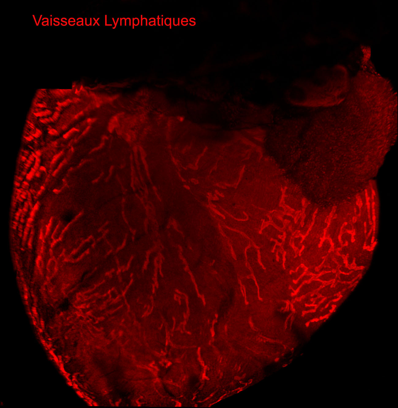
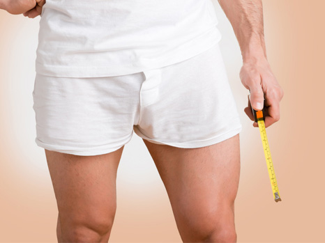
The effects of cell transplantation were assessed by specific self-administered questionnaires that scored the main aspects of sex life, namely satisfaction with intercourse, erectile and orgasm functions, libido, and penile rigidity during intercourse. The penile vessels were studied by Doppler ultrasound before and after cell transplantation. The researchers also assessed changes in the size of the penis after transplantation, since penile shrinkage is a common sequela of radical prostatectomy.

schéma

(c) Inserm / AP-HP

Main results

Tolerance of the treatment was excellent, and the main side-effect reported was transient pain at the puncture site where the bone marrow was taken (buttock region).

Six months after cell transplantation, the researchers noted a significant improvement in the main sexual scores, particularly a 10-point increase in the mean score for erectile function (17.4/30 at 6 months versus 7.3/30 before transplantation) on a scale of 0 to 30 (where 30 corresponded to the best possible erectile function). The other scores that improved significantly concerned overall satisfaction with sexual intercourse: 6.8/10 at 6 months versus 3.9/10 before transplantation (with 10 being the best score); the quality of orgasm: 6.3/10 at 6 months versus 3.5/10 before transplantation; penile rigidity during intercourse: 2.6/4 at 6 months versus 1.3/4 before transplantation (4 indicating maximum rigidity). At least two patients described a return to normal erections similar to the situation prior to radical prostatectomy, without taking drugs. A mean increase of 1 cm in penis length was observed. The clinical benefits were more pronounced at higher doses, and were associated with a normalisation of arterial vascular parameters in the penis measured by Doppler ultrasound. Improvement in sexual scores was maintained 1 year after transplantation, although some patients continued to use a treatment during intercourse.

In light of these good results, the researchers nonetheless wish to recall that: “Because of the small number of patients enrolled in the study, and the absence of a control group that would have received a ‘placebo’ injection, there is a need for caution about the conclusiveness of the efficacy shown for this new therapeutic strategy.” However, it is important to remember that the patients treated all showed severe erectile dysfunction that was unresponsive to the maximum medical treatment, and was associated with substantial damage to the penile vascularisation. The likelihood of spontaneous improvement in erections had been considered close to zero with these criteria. Improvement in sexual scores combined with a normalisation of Doppler ultrasound measurements of the penis argues in favour of a beneficial effect from the stem cell transplant in the medium term.

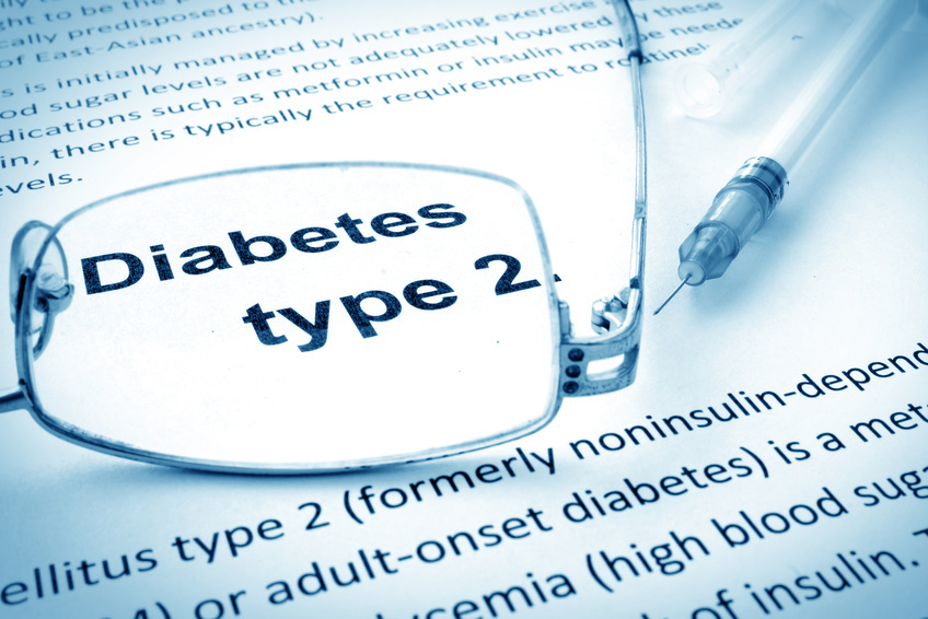
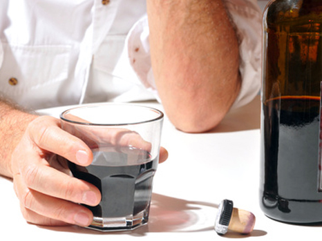
This therapeutic strategy could constitute the first approach to curing erectile dysfunction. For Prof. René Yiou: “If the results of this study are confirmed by other controlled clinical trials, the indications for cell therapy could be extended to other forms of erectile dysfunction that are less severe, or that result from common diseases such as diabetes or other vascular diseases.”

More information at: www.urologie-fonctionnelle.com

[1] The pump creates a vacuum around the penis, causing an influx of blood into the corpora cavernosa, and hence an erection.

Cardiac arrest – Don’t neglect the warning signs!

Over half of the patients who die suddenly have early warning signs, allowing ample time for intervention, according to an Inserm study conducted by Eloi Marijon, a researcher and cardiologist at the Paris Cardiovascular Research Center (Inserm Unit 970 / George Pompidou European Hospital, AP-HP)
This work is published in the journal Annals of Internal Medicine.

PhotoCP web Marijon

(c) Fotolia

Every year, about 50,000 French people die prematurely from cardiac arrest. This means one cardiac arrest every 10 minutes, i.e. ten times more than the number of deaths on the road. After the age of 45, coronary disease is the most frequent cause of myocardial infarction, which, in some cases, will lead to cardiac arrest. In France, only 5-7% of victims of cardiac arrest survive. Until now, these cardiac arrests have been perceived as inevitable, suggesting that nothing could predict the risk of cardiac arrest in the short term.

However, “Over half of the patients who die suddenly have early warning signs, allowing ample time for intervention,” according to a study by Inserm, the Paris public hospitals (AP-HP) and Paris Descartes University published in Annals of Internal Medicine. To obtain this result, Eloi Marijon of the Sudden Death Expertise Centre at the Georges Pompidou European Hospital, AP-HP, in collaboration with the Cedars-Sinai Heart Institute (Los Angeles, California), studied exactly what happened in the 4 weeks preceding the occurrence of a cardiac arrest. For 839 men and women who had died suddenly, events were reconstructed by conducting detailed interviews with witnesses and family members, and by close examination of medical data from the hospitals and private physicians in the region.

Chest pain was the most common symptom. Other warning signs were shortness of breath on exertion and loss of consciousness. Dr Marijon points out that in two out of three cases, the chest pain was absolutely typical of heart trouble, with intense crushing pain. “But it had been intermittent until the occurrence of the cardiac arrest. Conversely, when there was breathing difficulty, it began several days previously, and was usually continuous until cardiac arrest,” says the Inserm researcher and cardiologist.

The team also analysed how these patients “responded” to these symptoms, and how this may have influenced their prognosis (chance of survival). The results are instructive: only 19% of these symptomatic patients called the emergency medical services (equivalent to dialling “15” in France). However, those who made the call showed a 6-fold greater chance of survival (achieving more than 30% survival !!) compared with those who ignored their symptoms.

“The lesson is that if you have these types of symptoms, they must not be ignored. If you are in this situation, go to see your general practitioner as soon as possible. And, most importantly, don’t waste any time,” insists the author of the study.

“These new data should spur the medical community to develop a new strategy for sub-acute prevention,” states Dr Marijon, i.e. to be able to identify subjects at risk of cardiac arrest in the short term.

Until now, prevention of sudden death is essentially based on managing cardiovascular risk factors, implanting defibrillators in patients at greatest risk, etc. But this (long-term) type of prevention has shown its limits. The exponential development of e-health should be an asset for developing this prevention, whether in terms of identifying at-risk subjects, optimising the management of cardiac arrest using geolocation systems, etc.

Individual susceptibility to liver toxicity of alcohol depends on the intestinal microbiota

Despite excessive alcohol consumption, some individuals remain in good health while others develop liver disease. This inequality regarding the toxicity of alcohol for the liver depends on the intestinal microbiota. This has just been demonstrated by scientists and physicians from Paris-Sud University, Inserm, AP-HP (Antoine-Béclère Hospital), INRA (French National Institute for Agricultural Research), AgroParitech and Aix-Marseille University. These results are published online in the journal Gut.

PhotoCP web alcool

(c) Fotolia

Alcoholic liver disease is a major public health problem. The liver is affected to a variable degree, from a simple accumulation of fat to acute alcoholic hepatitis and cirrhosis. Nonetheless, people do not all react in the same way to the toxic effects of alcohol on the liver. For an equivalent intake of alcohol in terms of quantity and duration, only some excessive drinkers will develop liver disease. This unequal risk from the effects of alcohol toxicity indicates that other factors influence the initiation and progression of liver lesions. The intestinal microbiota is the complement of bacteria present in our digestive system. These bacteria perform many metabolic functions. Scientists and physicians from Paris-Sud University, Inserm, AP-HP (Antoine-Béclère Hospital, one of the Paris Public Hospitals), INRA, AgroParitech and Aix-Marseille University have studied the potential role played by the intestinal microbiota in the unequal toxicity of alcohol for the liver.

The scientists found an imbalance in the intestinal microbiota (dysbiosis) of patients with acute alcoholic hepatitis, which was not found in patients who consumed alcohol but did not have serious liver disease.

In order to ascertain whether this dysbiosis plays a causal role in the occurrence of lesions on the liver, the scientists humanised germ-free mice by transferring the microbiota of alcoholic patients to them. One group of mice received the microbiota of alcoholic patients suffering from acute alcoholic hepatitis, and another group of mice received the microbiota of alcoholic patients without serious liver disease. The mice were then fed alcohol. It was observed that the mice from the first group developed inflammation of the liver and adipose tissue, and a greater increase in intestinal permeability than those from the other group. Moreover, some harmful species of bacteria were specifically associated with sensitivity or insensitivity to alcohol. A study of metabolites showed that ursodeoxycholic acid levels were higher in alcohol-resistant mice.

When this type of experiment was repeated with conventional mice, which have the advantage of more closely mimicking the human disease, the work showed that it was possible to reduce the liver lesions in diseased mice by transferring the microbiota from alcoholic patients without liver lesions to them.

This work thus proves that individual susceptibility to alcoholic liver toxicity depends, at least to a substantial extent, on the intestinal microbiota. Moreover, the composition of the intestinal microbiota can be modified by diet, prebiotics, probiotics or by transfer of faecal microbiota. Furthermore, this type of experiment shows that it is possible to transfer alcohol susceptibility from human to mouse via the intestinal microbiota, and makes it possible to anticipate the development of treatments of this nature.

These results thus open up new possibilities, not only for identifying individuals likely to be sensitive to alcohol toxicity, but also for improving the treatment of alcohol-induced liver lesions by modulating the intestinal microbiota

Gut Microbes Signal to the Brain When They’re Full

Press release written by Cell Press

Don’t have room for dessert? The bacteria in your gut may be telling you something. Twenty minutes after a meal, gut microbes produce proteins that can suppress food intake in animals, reports a study published November 24 in Cell Metabolism. The researchers from Inserm and Rouen University also show how these proteins injected into mice and rats act on the brain reducing appetite, suggesting that gut bacteria may help control when and how much we eat.

The new evidence coexists with current models of appetite control, which involve hormones from the gut signalling to brain circuits when we’re hungry or done eating. The bacterial proteins—produced by mutualistic E. coli after they’ve been satiated—were found for the first time to influence the release of gut-brain signals (e.g., GLP-1 and PYY) as well as activate appetite-regulated neurons in the brain.

“There are so many studies now that look at microbiota composition in different pathological conditions but they do not explore the mechanisms behind these associations,” says senior study author Sergueï Fetissov of Rouen University and Inserm’s Nutrition, Gut & Brain Laboratory in France.

“Our study shows that bacterial proteins from E.coli can be involved in the same molecular pathways that are used by the body to signal satiety, and now we need to know how an altered gut microbiome can affect this physiology.”

Mealtime brings an influx of nutrients to the bacteria in your gut. In response, they divide and replace any members lost in the development of stool. The study raises an interesting theory: since gut microbes depend on us for a place to live, it is to their advantage for populations to remain stable. It would make sense, then, if they had a way to communicate to the host when they’re not full, promoting host to ingest nutrients again.

In the laboratory, Fetissov and colleagues found that after 20 minutes of consuming nutrients and expanding numbers, E. coli bacteria from the gut produce different kinds of proteins than they did before their feeding. The 20 minute mark seemed to coincide with the amount of time it takes for a person to begin feeling full or tired after a meal. Excited over this discovery, the researcher began to profile the bacterial proteins pre- and post-feeding.

They saw that injection of small doses of the bacterial proteins produced after feeding reduced food intake in both hungry and free-fed rats and mice. Further analysis revealed that “full” bacterial proteins stimulated the release of peptide YY, a hormone associated with satiety, while “hungry” bacterial hormones did not. The opposite was true for glucagon-like peptide-1 (GLP-1), a hormone known to simulate insulin release.

The investigators next developed an assay that could detect the presence of one of the “full” bacterial proteins, called ClpB in animal blood. Although blood levels of the protein in mice and rats detected 20 minutes after meal consumption did not change, it correlated with ClpB DNA production in the gut, suggesting that it may link gut bacterial composition with the host control of appetite. The researchers also found that ClpB increased firing of neurons that reduce appetite. The role of other E.coli proteins in hunger and satiation, as well as how proteins from other species of bacteria may contribute, is still unknown.

“We now think bacteria physiologically participate in appetite regulation immediately after nutrient provision by multiplying and stimulating the release of satiety hormones from the gut,” Fetisov says. “In addition, we believe gut microbiota produce proteins that can be present in the blood longer term and modulate pathways in the brain.”

fermer