Menu

Antiplatelet agents help reduce the severity of flu

Platelets play a role in severe lung infections associated with flu viruses. This has been highlighted for the first time by a team of researchers from France’s National Institute for Agricultural Research (Inra), National Institute for Health and Medical Research (Inserm) and Claude Bernard (Lyon 1) University in their research published on 1 April 2015 in the American Journal of Respiratory and Critical Care Medicine. Their findings show that antiplatelet drugs are an effective support treatment for severe forms of flu.

Each year, flu epidemics are responsible for 3 to 5 million cases of serious illness worldwide and between 250,000 and 500,000 deaths, primarily among high-risk groups (the very young, the elderly and people with chronic illnesses). In severe flu cases, the lungs suffer excessive, damaging inflammation.

Researchers from Inra, Inserm and Claude Bernard (Lyon 1) University studied the role of platelets[1] during flu infections in mice. Their aim was to understand the mechanisms responsible for the excessive lung inflammation that occurs in the most severe cases. They discovered a mass influx of aggregated, activated platelets, highlighting for the first time the recruitment of platelets during processes linked to the severity of lung infections.

The same team then went on to demonstrate the link between platelet activation in the lungs and overactivation of inflammatory processes. When platelets are overactivated, mortality is higher. Conversely, mice with a platelet function deficiency were protected.

Lastly, the team demonstrated that antiplatelet agents had a beneficial effect on excessive lung inflammation. The researchers tested four different antiplatelet drugs on the mice (two of them already on the market) and three different strains of influenza virus. These were human strains modified to induce severe flu and pneumonia in the mice.

After a median lethal dose of virus was given (level that will kill 50%), antiplatelet agents administered locally or intranasally resulted in almost 100% survival.

 This research therefore suggests that antiplatelet drugs, which already exist in the treatment arsenal, could be used to develop effective anti-inflammatory treatments for severe flu infections. These biological research results could lead on to clinical research to assess the possibility of achieving the same results in human beings.

Image

Lung tissue slices studied under an electron microscope. Mice infected with flu virus were treated intranasally with a solution containing an antiplatelet agent (right) or the solution without the agent (left). The formation of large masses of activated, aggregated platelets, as seen in the lungs of the control mice (left), was prevented in the mice given the antiplatelet agent (right), thus reducing inflammation and promoting survival. © Elisabeth Errazuriz-Cerda, CIQLE

[1] Platelets are blood cells that prevent excessive blood loss through their essential role in clot formation.

Targeting a host receptor instead of the virus: a new experimental approach against hepatitis C virus.

An international collaboration led by Professor Thomas Baumert (Inserm/University of Strasbourg Joint Research Unit 1110, “Institute for Viral and Liver Disease”) has shown that a monoclonal antibody directed specifically against claudin-1, a liver protein essential for infection by the hepatitis C virus (HCV), enables the prevention and treatment of chronic infection by this virus in an animal model. It turns out that this antibody, which was known to inhibit HCV entry and thereby prevent the initiation of infection, can also eliminate infected cells. This discovery, published in a letter in the Nature Biotechnology issue of 23 March 2015, opens the way to developing an approach to hepatitis C that is not only preventive, but therapeutic as well.

Infection with hepatitis C virus (HCV) leads to cirrhosis of the liver and liver cancer, the second leading cause of cancer death in the world. These complications are major indications for liver transplantation, but HCV reinfection of the transplant is a challenge. To date there is no vaccine, and the new treatments developed recently can be accessed by only a minority of patients worldwide because of their high cost. The development of new preventive and therapeutic strategies therefore continues.

The team directed by Prof. Thomas Baumert (Inserm/University of Strasbourg Joint Research Unit 1110, “Institute for Viral and Liver Disease”), in collaboration with international teams, decided to target a liver protein essential for viral infection instead of targeting the virus. They chose claudin-1, a molecule that is important in the initial steps of HCV infection, and involved in cell-cell contacts.

Using mouse models with humanised liver, the researchers show that a monoclonal antibody directed against claudin-1 can prevent HCV infection by blocking the entry of the virus into liver cells. Surprisingly, the researchers also observed that this antibody enables the treatment of chronic HCV infection by inhibiting the activation of intracellular signalling pathways needed by the virus for survival. As a result, the infected cells disappear and are gradually replaced by uninfected cells.

The advantage of this strategy is that it does not need to be combined with an antiviral agent.

 Moreover, by using different viral strains, the researchers show that it is difficult for the virus to escape from this antibody and develop resistance.

“Claudin-1” is a protein that is usually localised in the tight junctions that are the points of contact between adjacent cells. It is interesting to note that tight junction proteins constitute receptors for other pathogens, such as dengue virus and Shigella species. This innovative approach, employing injection of a monoclonal antibody directed against a protein on the host cell, makes it possible to foresee the development of a vaccine strategy and new therapeutic approaches against HCV, and also against other pathogens that use similar infection mechanisms.

This study received support from the European Union (ERC, INTERREG-IV-Upper Rhine [ERDF], FP7), ANRS (French National Agency for Research on AIDS and Viral Hepatitis), the HepSYS and netRNA Laboratories of Excellence of the French National Research Agency (ANR), ARC Foundation for Cancer Research, IHU Strasbourg MIX-Surg, Wilhelm Sander Foundation, Alsace Region, French National Cancer Institute (INCa), French National Institute of Health and Medical Research (Inserm), French National Scientific Research Centre (CNRS), University of Strasbourg, Ghent University (GOA 01G01712), Flanders Research Foundation (FWO) and Cardiex (Nantes).

Manaslu 2015: A scientific expedition at over 5,000 m altitude

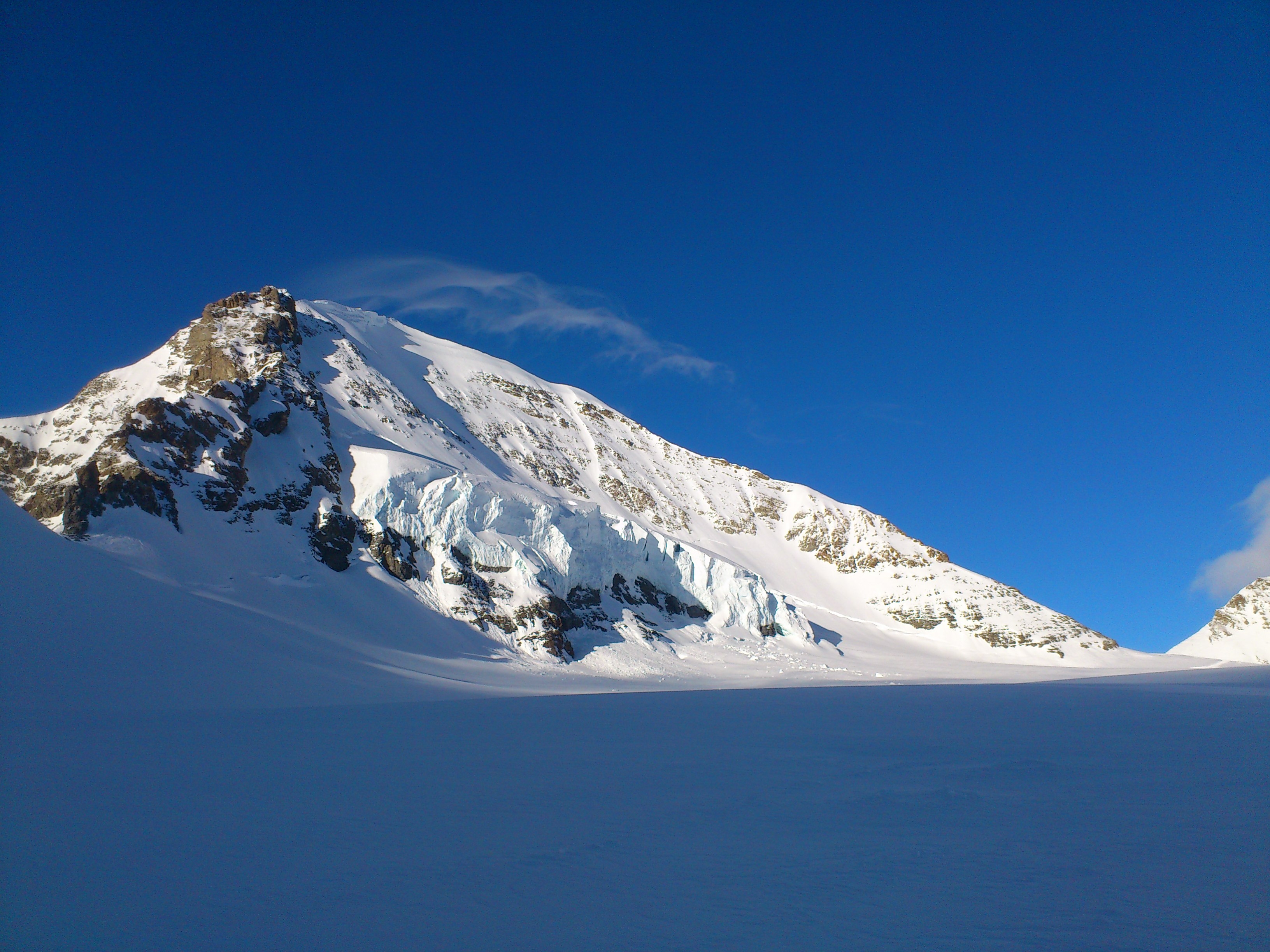
For 5 weeks, 5 international research teams will conduct a scientific expedition in Nepal, in the heart of the Himalayas, close to the summit of Mount Manaslu (8,156 m). The French component of the expedition, coordinated by a researcher from Inserm (Unit 1042, “Hypoxia and Cardiovascular and Respiratory Pathophysiologies,” Inserm/Université Joseph Fourier), will conduct an original project on the impact of altitude on the brain and heart, and on sleep disturbances induced by altitude. The French researchers will also study the benefit of a mask specially designed to improve oxygenation in combating the symptoms of acute mountain sickness. To do this, 50 volunteers will accompany them on this 5,000 m high trek.

Internet users will be able to follow this expedition in real time on the social networks using the hashtag #scienceausommet

Echocardiographie réalisée au sommet du Mont Blanc lors d'une précédente expédition © Samuel Verges

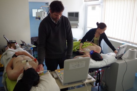
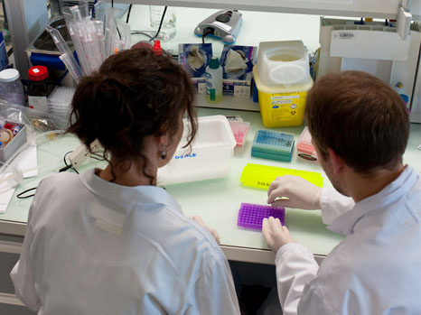
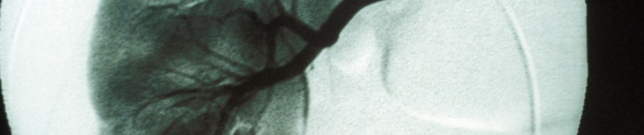
Echocardiography at the summit of Mont Blanc during a previous expedition © Samuel Vergès

The development of recreational activities, and the increasing number of periods spent at medium and high altitude by people who are often inexperienced raises the issue of altitude intolerance. These problems of adaptation are the result of physiopathological mechanisms associated with the progressive reduction in oxygen availability as one rises to higher altitudes.

These illnesses, which are disabling to the point of interrupting one’s period at high altitude, and can be serious or even fatal, are acute mountain sickness (a combination of symptoms such as headache, fatigue, nausea, etc.), high altitude pulmonary oedema, (usually associated with a cough, and breathlessness due to a potentially serious fluid accumulation in the lungs), and high altitude cerebral oedema (fluid leakage in the brain, a particularly serious phenomenon, with marked effects on behaviour). Acute forms occur in people who are poorly acclimatised to altitude, with a rapid onset following exposure (6 h to 4 days). The more devastating forms occur even after acclimatisation, at altitudes above 5,000 metres. Generally speaking, one out of every two people is affected by acute mountain sickness above 4,000 metres, and three out of four people are affected above 5,000 metres.

These conditions may, to a certain extent, be avoided by following simple rules for prevention. However, it also appears that some people are more likely than others to develop these symptoms at high altitudes. Factors predicting altitude tolerance, the mechanisms underlying problems of adaptation, the management of these problems, and the optimum strategies for acclimatisation are still particularly poorly known.

These are the themes that will be studied by the French research team, under the coordination of Samuel Vergès, an Inserm Research Fellow at Unit 1042, “Hypoxia and Cardiovascular and Respiratory Pathophysiologies.” The team is made up of 6 experienced scientists and physicians, all of whom have experience of high mountains and scientific expeditions of this type.

A first research project on the heart and brain

With three main strands, this project is aimed at using innovative and portable techniques (that can be used at high altitude) to define in detail the effects of spending a period at high altitude on the brain and heart, and to assess the effect of a positive expiratory pressure ventilation mask on these cerebral and cardiac responses.

  • Lack of oxygen and the brain

In vitro and in vivo studies show that lack of oxygen impairs neuronal function and may reduce human cognitive performance. A series of studies in the laboratory and at high altitude (VALLOT Observatory, Mont Blanc massif) has revealed significant brain disturbances induced by hypoxia, both in the initial hours of exposure and after several days. The anatomy of the brain, cerebral perfusion and oxygenation, and motor neuron function are particularly affected by hypoxia, a finding that has led to reconsideration of the mechanisms for adapting to altitude, with inclusion of the brain component as a factor that probably plays a major role.

  • Altitude and heart function

High altitude exposure is associated with major cardiac changes. Previous studies have shown that acute hypoxia leads to changes in the relaxation properties of the myocardium. A reduction in the contractile properties of the cardiomycytes might be responsible for this phenomenon, although objective proof has not yet been obtained. Many studies have also shown that exposure to high altitude generates changes in the right ventricle, which may play an important role in the relaxation of the left ventricle, although the mechanical phenomena involved are incompletely known.

In recent times, a new technology based on following the acoustic signatures produced during echocardiography (Speckle Tracking Imaging) has made it possible to measure myocardial speeds and deformations in a comprehensive manner, allowing investigation of the myocardium’s contractility and ability to relax, thus opening up new perspectives in the understanding of the effects of altitude hypoxia on the heart.

  • Development of a mask that minimises the harmful effects of high altitude

Applying positive expiratory pressure (PEP) at mouth level in individuals under high altitude conditions allows an artificial increase in the intra-pulmonary pressure, and has been suggested by some research teams as a possible method for preventing or minimising the harmful effects of high altitude. Researchers at the Hypoxia-Physiopathology Laboratory (HP2) in Grenoble recently demonstrated the utility of this method for increasing oxygenation of the blood and muscles.[1]

Their results suggest that a portable mask type device inducing an increase in expiratory pressure might constitute an original and effective non-pharmacological method for improving altitude acclimatisation and alleviating the symptoms associated with acute mountain sickness. A large-scale field study at a high mountain location is needed to determine whether PEP is likely to become widely used method. The commercialisation of a PEP mask system may be envisaged, given its advantage of being portable, compact, non-medicinal and usable by most people.

A second project on the impact of altitude on sleep

Altitude is known to profoundly disrupt sleep and induce sleep apnoea, known as central sleep apnoea. From an altitude of 2,500 m, a moderate reduction and cyclic oscillations are observed in blood oxygenation. The development of these disorders is also accompanied by a reduction in sleep efficiency, an increase in the time it takes to fall asleep, a shortening in the duration of phases of deep sleep, and a substantial increase in intrasleep awakenings. The quantification of these disorders was for a long time limited to the analysis of oscillations in arterial O2 saturation.

However, at the present time, one question has not had a clear answer: is central apnoea a marker of bad or good adaptation of the body to altitude? This question is important because there are large intersubject variations in the intensity of sleep disturbances induced by altitude exposure. It was recently shown in the laboratory directed by Samuel Vergès that individuals showing severe symptoms of high altitude intolerance (in the course of one night in an altitude simulation laboratory) have less sleep apnoea than comparable individuals who tolerate high altitude well. However, this remains to be established in the field at high altitude.

Study of this question should improve understanding of the mechanisms of adaptation to altitude, and influence the advice and assessments offered to people travelling to high altitude.

The study proposed by the French group within the scientific expedition therefore has the following objectives:

  • To assess the effects of the expiratory resistance mask (ventilation with PEP) on arterial and tissue oxygenation, on pulmonary extravascular leakage and on symptoms of acute mountain sickness during a period at high altitude for a large sample of volunteers.
  • To study, on the basis of new technologies (near-infrared spectroscopy Speckle Tracking echocardiography), the changes to the brain and heart induced by lack of oxygen (hypoxia) at high altitude, and to assess their reversibility through wearing the mask.
  • To compare altitude-induced sleep modifications in trekkers that adapt well to altitude compared to those who show symptoms of acute mountain sickness.


The researchers’ study of the effects of lack of oxygen on the healthy body at high altitude should enable a better understanding of the consequences of lack of oxygen on some patients at normal altitudes, for example those with respiratory diseases. The high altitude is thus a real open-air laboratory, constituting an original study model for the adaptive abilities and limitations of the human body.


An initial phase of study at normal altitude

All participants in the trek and Manaslu climb made themselves available, between 16 and 23 February last, for a battery of scientific and medical assessments at sea level in Wales (Bangor University). All measurements that will be done at high altitude were carried out at sea level, thus enabling reference values to be obtained for every subject, for comparison with measurements taken at high altitude.

test doppler plaine

Measurements at normal altitude © Samuel Vergès

The phase of testing at normal altitude was an opportunity to address technical and logistic issues that needed to be resolved so that the tests can be repeated under the best possible conditions at high altitude at reduced atmospheric pressure (which strongly affects equipment), and in the cold (down to -20°C), and when the bodies of both the researchers and volunteer subjects will be severely affected by the altitude.

A science laboratory installed at 5,000 metres

Five groups of 10 people will leave Kathmandu at intervals of 1 day each to spread the measurements. It will take 10 days to reach the destination. The 5 international research teams will set up their laboratory and all the necessary logistics (tents, production of solar and wind energy, etc.) slightly above the Manaslu base camp, at a little over 5,000 metres altitude. Most of the high altitude measurements will be performed on all those participating in the trek, who will arrive day after day. Other measurements will be performed during the trek and during the Manaslu climb (8,156 m). The entire expedition will last 5 weeks, from 21 March to 26 April.

As with assessments carried out at normal altitude, assessments will involve measuring the pulmonary extravascular fluid by pleuropulmonary ultrasound, cerebral perfusion by transcranial Doppler, cerebral oxygenation by near-red spectroscopy, and cardiac function by Speckle Tracking echocardiography, before and during ventilation under positive expiratory pressure. Symptoms will be assessed by questionnaire. Finally, sleep will be assessed during nights spent in tents at the Manaslu base camp, by placement of various sensors that enable identification of the quality of sleep, and the occurrence of sleep apnoea associated with a drop in blood oxygenation, such as is frequently observed at high altitude. These measurements are identical to the tests known as polygraphy that are carried out in sleep laboratories in hospitals to diagnose sleep apnoea syndrome in patients.

Following 4 days of acclimatisation at 5,000 metres, this battery of tests will be repeated in order to observe the adaptations of the human body to this high altitude. All participants will spend 10 days at base camp, and the team will then complete the Manaslu circuit with a 3-day descent to Beshisahar.

The participants in this trek show a classic profile of populations taking part in treks at high altitude, aged from 22 to 65 years, with relatively varied levels of physical fitness and altitude experience. They have volunteered to complete a trek of several weeks around Manaslu and have also volunteered to participate in scientific experiments in this context, including 5 consecutive days at the camp located at the highest point in the Manaslu circuit, where the laboratories will be installed.


The other 4 international scientific projects conducted as part of the MEDEX project

MEDEX (www.medex.org.uk) was created in Great Britain in 1992 to encourage scientific and medical expeditions. Since then, several large-scale scientific expeditions have been organised at 4-5 year intervals: Everest in 1994, Kanchejunga in 1998, Hongu in 2003 and Hidden Valley in 2008. These expeditions have led to major scientific and medical advances associated with altitude, resulting in scientific publications, news stories for the general public, and training activities for professionals and high mountain climbers.

During this expedition, 4 other scientific projects will be conducted at the same time as the 2 conducted by Samuel Vergès’ team.

  • Coping strategies for those working at altitude – Sue Paddon’s team (Advanced Personnel Management, Australia-UK)
  • Influence of physical and mental preparation on the success of an expedition at high altitude – team led by Sam Olivier and Jamie Macdonald (Bangor University)
  • Disruption of heart rate as an indicator of acute mountain sickness? Jamie Macdonald (Bangor University)
  • Impact of treks on the health of Nepalese staff employed to accompany expeditions – Mary Morrell (Imperial College London)


Following the expedition on social media

To bring a scientific expedition to life for Internet users as if they were there, even though the location (the summit of Manaslu) is particularly inaccessible, Inserm is doing all it can to follow the expedition. Enriched content (photos or videos) will be posted daily on @Insermlive and on the Institute’s Facebook page. From the day-to-day (plan, arrival at location, setting up), to details of scientific experiments (design, goals, measurements, etc.)

A hashtag #scienceausommet

A relay on @inserm_en is also planned for the international community

At the Inserm press room, an instant discussion module will be open to take questions for the researchers before they leave and after they return.

[1] These results have been published in PlosOne

Severe asthma: gallopamil confirmed as a therapeutic approach

A team of Inserm researchers from the Cardio-Thoracic Research Centre of Bordeaux (Inserm/University of Bordeaux and Bordeaux University Hospital) has demonstrated the clinical efficacy of gallopamil in 31 patients with severe asthma. This chronic disease is characterised by remodelling of the bronchi, which exacerbates the obstruction of the airways already seen in “classic” asthma. In contrast to the reference treatment, gallopamil has proved capable of reducing the bronchial smooth muscle mass. This work is published in the 29 January 2015 issue of the American Journal of Respiratory and Critical Care Medicine.

Severe asthma is a chronic condition of the airways that affects between 1 and 3% of the world population to a highly variable extent, depending on the country. It is characterised by persistent breathing difficulty, restricted physical activity, frequent nocturnal attacks, and prolonged asthma attacks that require systemic treatment[1]. These symptoms lead to a considerable number of emergency hospital admissions, have a serious impact on the quality of life for patients, and can even result in death.

In severe asthma, bronchial obstruction causes a strong reduction in respiratory capacity. This bronchial obstruction is due to remodelling of the airways, particularly the thickening of the bronchial smooth muscle (BSM) that surrounds them. This phenomenon is associated with a poor prognosis and resistance to even intensive treatment. Until now, no pharmaceutical drug has succeeded in preventing the excessive proliferation of these muscle cells, including corticosteroids, the reference treatment for severe asthma.

In previous work, Patrick Berger and his colleagues had already demonstrated in vitro and ex vivo that this disproportionate growth was triggered by an abnormal entry of calcium into these bronchial smooth muscle cells[2]. In this same article, the scientists had demonstrated in vitro the anti-proliferative effect of gallopamil, which is normally prescribed for certain heart conditions because of its blocking action on calcium channels.
pelletier

© Inserm, Pelletier L. The left pictures show the characteristic look  of lung inflammation in asthma with thickening of the bronchial wall, a lot of inflammatory cells and an abnormal production of mucus. Mice treated with the calcium channel inhibitor (on the right) are completely protected.

To assess its in vivo efficacy, the researchers then initiated a clinical trial sponsored by Bordeaux University Hospital and supported by the French Ministry and Health and Inserm. For 12 months, they thus measured the effect of the drug on the thickness of the BSM and bronchial wall, and the frequency of asthma attacks in 31 patients.

On analysis, the data showed a significant reduction in BSM in asthmatic patients treated with gallopamil compared with the placebo group. The drug therefore enabled a significant reduction in the thickness of the bronchial wall in patients.

After this phase, both patient groups were monitored for 3 months after stopping treatment. It then became apparent that individuals treated with gallopamil had significantly fewer prolonged attacks than the placebo group.

This pilot study thus provides proof of concept that the calcium channel blocker is able to reduce bronchial remodelling through its action on the smooth muscle cells in individuals suffering from severe asthma.

 Other studies involving larger patient cohorts will have to be put in place to confirm these results. Moreover, although gallopamil seems to affect the occurrence of prolonged asthma attacks, Patrick Berger emphasises the need to test gallopamil over a longer treatment period in order to confirm this observation.

[1] The episodes are also medically referred to as exacerbations.

[2] Berger P. et al., Bronchial smooth muscle remodeling involves calcium-dependent enhanced mitochondrial biogenesis in asthma. J Exp Med 2007; 204:3173-3181


A new MOOC invites the general public to discover a stem cell culture laboratory

University of Nantes, Inserm and Thinkovery have joined forces to produce a new kind of MOOC (Massive Open Online Course). Alternating as it does between theory and practice, “Ouvrez les portes du laboratoire: cellules et cellules souches,” (Open the laboratory doors: cells and stem cells) invites members of the public to discover, by means of enriched video content, the actual work done by researchers at Université de Nantes, Inserm and CNRS.

From 16 March 2015, all Internet users will thus be able to understand how and why researchers grow stem cells in the laboratory, and will be able to attend a course on cell biology.

PhotoCP web

Institut du thorax UMR 915 – Inserm/Latron, Patrice

After more than a year of design and production, the MOOC “Ouvrez les portes du laboratoire: cellules et cellules souches” will begin on 16 March 2015, and will be accessible on the FUN (France Université Numérique/France Digital University) platform. Registration is already open.

To put this novel MOOC together, teams from Université de Nantes, in partnership with Inserm, have joined forces with a new medium devoted to research, Thinkovery.

They have combined their knowledge and ability to produce this six-week online course, and over fifty two- to five-minute videos, mainly with financial support from the Pays de la Loire Region and L’Oréal Research and Innovation. L’Oréal also contributed to preparing the content of this MOOC that covers aspects of somatic hair follicle stem cells.

“We regularly open up our research laboratory to the public, but for one thing it is difficult for us to accommodate everyone, and for another thing, such days do not give the public the opportunity to follow a project from start to finish.

It was this observation that gave us the idea. Based on the same principle as the MOOCs that throw open the doors of lecture theatres, we wanted to throw open the doors of our laboratory via the Internet,” says Patricia Lemarchand, a physician, lecturer and researcher at the Institut du Thorax at Nantes, of the initiative for the project with Loïc Le Gac of the Thinkovery company.

The MOOC alternates between two series:

The “Côté Cours” (Lecture Side) introductory videos on cell biology describe the main concepts needed to understand the “Côté Laboratoire” (Laboratory Side) videos, which immerse the visitor in the daily work of a joint research team.

To register for the MOOC

The MOOC will begin on 16 March 2015

Registration is open! Go to the FUN platform to register for free for the

MOOC “Ouvrez les portes du laboratoire: cellules et cellules souches:”
https://www.france-universite-numerique-mooc.fr/universities/universite-de-nantes/

Stem cell therapy for heart failure: first implant of cardiac cells derived from human embryonic stem cells

On the 21 October 2014, Professor Philippe Menasché and his team from the cardiovascular surgery service of the Georges Pompidou European Hospital, AP-HP, carried out a transplant of cardiac cells derived from human embryonic stem cells*, according to a method developed by the Department of Cell and Tissue Biotherapies of the Saint-Louis hospital, directed by Professor Jérôme Larghero and through research led by this group within Inserm. The surgery, coupled with a coronary bypass*, was carried out on a woman of 68 years suffering from severe heart failure. Ten weeks after the intervention, the patient is feeling well, her condition has improved markedly, with no complications having been observed. This promising advance was presented this Friday, 16 January 2015 at the XXV European Days Conference of the French Society of Cardiology.
inserm_45834

Human embryonic stem cells. Transplantation of undifferentiated human embryonic stem cells into rat heart organotypic cultures. Presence of human cells, in the cardiac parenchyma of the rat two months after injection. The human cells are positive for human nuclear antigen marking (red). Cardiac rat tissue is positive for cardiac troponin 1 marking (green). I-Stem (Institiute for Stem Cell Therapy), Evry Genopole. Inserm/Habeler, Walter

The transplant was carried out as part of a clinical trial developed by the Public Hospitals of Paris (AP-HP) and through the work of the teams from AP-HP, Inserm and the universities of Paris-Descartes and Paris-Diderot. The cardiac cells were prepared according to a technique developed by the Department of Cell and Tissue Biotherapies of the Saint-Louis hospital. The cytogenetics laboratory of the Antoine Béclère Hospital and the French General Agency for Health Products and Equipment also contributed to the preparation of this phase I trial which will enable the verification of the safety and feasibility of the procedure

For 20 years Professor Menasché, currently co-director of an Inserm team within PARCC (Paris Centre for Cardiovascular Research), and his colleagues have been involved in stem cell* therapy for heart failure.

The team first tested the implant of skeletal muscle stem cells in necrosed areas of the heart in the laboratory. These cells were implanted into the heart of a patient with heart failure for the first time in the world on 15 June 2000. Following an initial series of these implants, always coupled with a coronary bypass, the team coordinated a European multi-centre, randomised, placebo-controlled trial whose results have still not been able to establish any significant benefit of these cells on the contractile function of patients’ hearts.
One of the conclusions drawn from this trial was that to be fully efficient, transplanted cells should resemble the cells of the tissue to be repaired as much as possible, in this instance cardiac tissue. It was then decided to venture along the path of embryonic stem cells.  Derived from embryos conceived in in vitro fertilisation, these cells do in fact possess pluripotent properties, that is, they are capable of developing into any type of cell of the body, including of course cardiac cells, as soon as they receive the appropriate signals during the culture cycle in the laboratory.

In 2007, the team then composed of, among others, Michel Pucéat, Director of Research at Inserm, and Philippe Menasché showed that human embryonic stem cells could be differentiated into cardiac cells after being transplanted into the failing hearts of rats.
Since then, many experiments have been carried out on different animal species in order to validate the efficacity of these cells and to optimise conditions which can guarantee maximum safety. At the end of this stage, a bank of pluripotent embryonic stem cells was formed in the conditions which satisfied all regulatory constraints applying to biological products for human use. Then, the Department of Cell and Tissue Biotherapies of the Saint-Louis hospital, still in liaison with the Inserm teams, developed and tested “specialisation” procedures for cells in order to produce “young” cardiac cells from them.
The focus was then on the purification of the cells directed like so in order to ensure that the final product was expunged of any remaining pluripotent cells which could be potentially tumorigenic.

Besides, as initial experience with muscular stem cells showed the limitations of administering cells by multiple injections, their transfer is now performed using a patch that the cells are incorporated into. This patch is then placed on the area of the infarction. To that end, after the purification stage, the cardiac cells are incorporated into a circular fibrin gel which is applied, during the surgical procedure, to the necrosed area with just a few sutures ensuring that it is anchored to the cardiac tissue.

“This type of surgery is aimed at serious heart failure which doesn’t respond to the usual medicinal treatments but is not at the stage of a complete heart transplant. This is a promising advance, which we hope will enrich the therapeutic arsenal available to treat heart failure today” explains Prof. Menasché. “We are continuing the trial, which authorises us to carry out four other transplants. It would seem already that the benefits of the cells are linked mainly to the substances that they secrete.  The direct administration of the substances, without going through a transplant of productive cells, is a path to explore”.

This project has been entirely financed by funds from public intstitutions and societies and was authorised by the French National Agency for the Safety of Medicines and Health Products (ANSM) after agreement with the Agency for Biomedicine for the importation and research on human embryonic cells.

Glossary

Cell therapy: refers to cell transplants aiming to restore the function of tissue or an organ when it has been altered by an accident, illness or ageing. These therapies have benefited from recent scientific advances on stem cells and give millions of patients the hope of regenerative medicine.

Embryonic or pluripotent stem cells: they can renew indefinitely (self-renewal), multiply in a culture and be differentiated into more than 200 types of cell. In the course of development, they are destined to form all types of the body’s tissue.

Coronary bypass: a technique that enables the redirection of the bloodstream towards the cardiac muscle, by using a graft (coming from the saphenous vein or from a thoracic artery….) One end of the graft is connected to the aorta, the main artery supplying the coronary arteries; the other end is connected to the coronary artery, situated just behind the site of the obstruction. This creates a detour enabling the oxygenated blood to circulate towards the heart.

Improved understanding of the diabetic process via the Rfx6 gene

For the first time, the team led by Gérard Gradwohl, an Inserm Research Director at the Institute of Genetics and Molecular and Cellular Biology, Illkirch (IGBMC/Inserm-CNRS-University of Strasbourg), has shown that the Rfx6 gene is essential for the functioning of the cells that produce insulin, the pancreatic beta cells. In adult mice, this gene has proved important not only for allowing the secretion of insulin, but for also playing a major role in beta cell identity. As a logical progression of this work carried out in the rodent, the team led by Raphaël Scharfmann, an Inserm Research Director (Unit 106, “Cochin Institute”/Inserm-Paris Descartes University-CNRS) has confirmed these results in human pancreatic beta cells and in a 6 year old child with neonatal diabetes.
Both of these studies are published in the 11 December 2014 issue of the journal Cell Reports.

ilots langherans

Immunofluorescence, histological section of an islet of Langerhans from an adult mouse pancreas showing expression of Rfx6 transcription factor (red) in the β-cell nuclei (stained green with insulin) ©Julie Piccand 

In 2010, scientists provided evidence that the Rfx6 gene plays a key role in the formation of insulin-producing cells. In mice where this gene was mutated or deleted (absent), severe diabetes appeared at birth, resulting in death of the pups. In human newborns with mutations, neonatal diabetes is diagnosed very early, and then controlled with insulin treatment.

Among the factors involved in neonatal diabetes, Rfx6 is acquiring the status of an essential gene in the control of insulin secretion, and is therefore potentially involved in the diabetic process in adults. Indeed, researchers at IGBMC have created animal models in mice, in which the mature β-cells have been modified so as to inactivate their Rfx6 gene. The result: these mutant mice show glucose intolerance (prediabetes). By what mechanisms? Without Rfx6 expression in β-cells, several critical steps in glucose-induced insulin secretion are disrupted, such as glucose detection, electrical activity of the β-cells and calcium ion flux.

In fact, Rfx6 directly regulates the expression of key genes controlling these processes. This sequence of steps leads to the secretion of insulin according to fluctuations in blood sugar levels, and disruption of the steps causes defective insulin production.

Even more surprisingly, on examining the transcriptome of mutant mice (without Rfx6), Gérard Gradwohl’s team observed that genes that are normally repressed in β-cells become active. These genes, known as “disallowed,” are expressed instead of remaining silent. Their activation then leads to loss of β-cell identity. In a manner of speaking, they undergo dedifferentiation and “forget” their raison d’être and function.

Observations validated in humans

As a logical progression from this work carried out in the rodent, Raphaël Scharfmann’s team has confirmed the results for human β-cells. For 30 years, researchers throughout the world had been trying to reproduce these β-cells in the laboratory in order to study them and understand their dysfunction. In 2011, Raphaël Scharfmann and his academic and industrial collaborators from the EndoCells company were the first in the world to produce functional human β-cell lines in vitro. They therefore possessed the necessary “toolkit” to explore the role of Rfx6 in humans.

Moreover, their genetic analyses of children with neonatal diabetes had shown mutations in the Rfx6 gene, without their knowing what its role might be in the disease.

The researchers therefore sought to crack the Rfx6 mystery simultaneously in normal functional human β-cells, β-cells in which the Rfx6 gene was not expressed, and in a 6 year old child with neonatal diabetes.

Their studies show, like those of the researchers in Strasbourg, that Rfx6 plays a central role in controlling not only insulin production in β-cells, but also the secretion of insulin into the bloodstream.

More specifically, Rfx6 acts on the β-cell membrane by controlling the opening and closing of the Ca2+.(calcium) channels. Normally, glucose enters the pancreatic β-cell by facilitated diffusion. This entry causes a cascade of reactions that lead to the opening of the calcium channels that allow the release of insulin into the bloodstream. Where Rfx6 is mutated and nonfunctional, it prevents the opening of the Ca2+.channels, and blocks subsequent insulin secretion.

For the researchers, this work has three-fold value. It confirms the results obtained in the rodent model. It also makes it possible to scientifically validate the interest of β-cells produced in the laboratory as a genuine tool for exploring the mechanisms of diabetes. Finally, from a therapeutic perspective, it might be very interesting to develop a treatment that modulates the opening and closing of Ca2+ channels.

Avoiding skin graft rejection: it’s possible!

A research team bringing together José Cohen and Philippe Grimbert (Inserm Unit 955/Université Paris Est Créteil [UPEC] and the Centre for Clinical Investigation – Biotherapies 504 [CIC-BT 504]), and their collaborators at Institut Curie and AP-HP (George Pompidou European Hospital) has succeeded in finding a combination of drugs that reduces the risk of rejection following a skin graft. When tested in mice, this treatment seems effective, since no sign of rejection is observed nearly 30 days after transplantation.
These results are published in the American Journal of Transplantation.


peau

©fotolia


For physicians and researchers, a major challenge of transplantation is avoiding rejection of the transplanted organ or tissue by the recipient’s body. During allogeneic transplantation (i.e. of a graft from donor A into a different recipient, B), the immune system of the recipient recognises the graft as a foreign component. It then makes every effort to destroy it in the same way as it would, for example, destroy an infecting virus. To avoid this phenomenon, physicians administer immunosuppressive drugs to make the recipient’s immune system more tolerant. As with every treatment, there are drawbacks: the recipient’s vulnerability to infections is increased, and the treatment may have toxic effects on other organs (kidney). For the last 30 years, there has been little progress in this area of research. The optimisation of these treatments is therefore central to the current efforts.

Inserm researchers under the leadership of José Cohen became interested in a drug with special properties, namely the cytokine interleukin 2 (IL-2). This drug is already used in some treatments for cancer and type 1 diabetes. In cancer, administration of IL-2 in high doses increases antitumour activity by boosting the immune system. Interestingly, Eliane Piaggio, a co-author of this study, had shown that when administered at very low doses in type 1 diabetes, it has the opposite effect. IL-2 thus impedes the action of the immune system, which reacts too strongly against self in this disease.

Given that, in transplantation, the immune response is too strong, the researchers hypothesised that administration of IL-2 might impede the action of the immune system (by analogy with its action in type 1 diabetes), and might therefore reduce graft rejection.

“Our initial experiments proved negative: IL-2 used alone did not give the expected results,” explains José Cohen. “We had to redirect our efforts and our attention to the specific functioning of 2 types of cells from the immune system, namely the regulatory T lymphocytes controlled by IL-2, and the “classical” T lymphocytes.”

The immune system is composed of several categories of cells, each with a specific role in maintaining its balance: it must not be too aggressive or too tolerant. Generally, regulatory T lymphocytes, as their name indicates, act on the other populations of classical T lymphocytes to prevent them from over-reacting. Hence the initial idea of boosting their activity via IL-2. However, this strategy turned out to be inadequate.

The researchers therefore used IL-2 in combination with rapamycin, which has the ability to inhibit the division of classical T lymphocytes. Using this combination, the researchers managed to doubly control the classical T lymphocytes: directly using rapamycin and indirectly using IL-2 (via the regulatory T lymphocytes). Graft rejection was thereby avoided.

“Skin grafting in mice is the most difficult experimental model to control. In our experiment, mice show no sign of rejection 30 days following a skin graft. This is very encouraging when we know that this rejection usually occurs in 10 days: the tissue becomes irreversibly necrotic.”

These results are a first step before clinical evaluation. An advantage of these two drugs is that they have marketing authorisation for use in humans. If the next steps are successful, especially in the liver transplant model, their use in the area of transplantation (any kind of transplantation) might soon begin.

Anorexia / bulimia: a bacterial protein implicated

Eating disorders (ED) such as anorexia nervosa, bulimia, and binge eating disorder affect approximately 5-10% of the general population, but the biological mechanisms involved are unknown. Researchers at Inserm Unit 1073, “Nutrition, inflammation and dysfunction of the gut-brain axis” (Inserm/University of Rouen) have demonstrated the involvement of a protein produced by some intestinal bacteria that may be the source of these disorders. Antibodies produced by the body against this protein also react with the main satiety hormone, which is similar in structure. According to the researchers, it may ultimately be possible to correct this mechanism that causes variations in food intake.

These results are published in the journal Translational Psychiatry, in the online issue of 7 October 2014. See the video of the discovery (English subtitles) :

Anorexia nervosa, bulimia and binge eating disorder are all eating disorders (ED). If the less well defined and atypical forms are included, ED affect 15-20% of the population, particularly adolescents and young adults. Despite various psychiatric, genetic and neurobiological studies, the molecular mechanism responsible for these disorders remains mysterious. The common characteristic of the different forms of ED is dysregulation of food intake, which is decreased or increased, depending on the situation.

Sergueï Fetissov’s team in Inserm Joint Research Unit 1073, “Nutrition, inflammation and dysfunction of the gut-brain axis” (Inserm/University of Rouen), led by Pierre Déchelotte, studies the relationships between the gut and the brain that might explain this dysregulation.

The mimic of the satiety hormone

In this new study, the researchers have identified a protein that happens to be a mimic of the satiety hormone (melanotropin). This protein (ClpB) is produced by certain bacteria, such as Escherichia coli, which are naturally present in the intestinal flora. Where this protein is present, antibodies are produced against it by the body. These will also bind to the satiety hormone because of its structural homology to ClpB, and thereby modify the satietogenic effect of the hormone. The sensation of satiety is reached (anorexia) or not reached (bulimia or overeating). Moreover, the bacterial protein itself seems to have anorexigenic properties.

Variations in food intake in the presence of the bacterial protein

To obtain these results, the researchers modified the composition of the intestinal flora of mice to study their immunological and behavioural response. Food intake and level of antibodies against melanotropin in the 1st group of mice, which were given mutant E. coli bacteria (not producing ClpB) did not change. In contrast, antibody level and food intake did vary in the 2nd group of animals, which received E. coli producing ClpB protein.

The likely involvement of this bacterial protein in disordered eating behaviour in humans was established by analysing data from 60 patients.

The standardised scale “Eating Disorders Inventory-2” was used to diagnose these patients and evaluate of the severity of their disorders, based on a questionnaire regarding their behaviour and emotions (wish to lose weight, bulimia, maturity fears, etc.). Plasma levels of antibodies to ClpB and melanotropin were higher in these patients. Furthermore, their immunological response determined the development of eating disorders in the direction of anorexia or bulimia.

These data thus confirm the involvement of the bacterial protein in the regulation of appetite, and open up new perspectives for the diagnosis and specific treatment of eating disorders.

Correcting the action of the protein mimicking the satiety hormone

“We are presently working to develop a blood test based on detection of the bacterial protein ClpB. If we are successful in this, we will be able to establish specific and individualised treatments for eating disorders,” say Pierre Déchelotte and Sergueï Fetissov, authors of this study.

At the same time, the researchers are using mice to study how to correct the action of the bacterial protein in order to prevent the dysregulation of food intake that it generates. “According to our initial observations, it would indeed be possible to neutralise this bacterial protein using specific antibodies, without affecting the satiety hormone,” they conclude.

This study was carried out in collaboration with other teams and platforms of the Institute for Research and Innovation in Biomedicine (IRIB) Normandy Rouen, and with the University of Tartu (Estonia). It extends other work published by the research team in 2013, which demonstrated molecular mechanisms for increasing the appetite by immunoglobulins that protect the hunger hormone (ghrelin) in obese people.

These works were the object of two demands of patents deposited by Inserm Transfert.

Dimly lit working environments : correcting your body clock is possible!

Researchers at Inserm, led by Claude Gronfier (Inserm Unit 846: Stem Cell and Brain Institute), have, for the first time, conducted a study under real conditions on the body clocks of members of the international polar research station Concordia. The researchers have shown that a particular kind of artificial light is capable of ensuring that their biological rhythms are correctly synchronised despite the absence of sunlight. The full significance of this result can be appreciated with the knowledge that disturbance to this biological clock causes problems with sleep, alertness, cardiovascular problems and even depression.

These results, published in Plos-One, could be rapidly transformed into practical applications for working environments that are dimly to moderately lighted (polar research stations, thermal and nuclear power stations, space missions, offices with no windows, etc.). They could enable the design of lighting strategies intended to maintain the health, productivity and safety of staff.


Horloge biologique, travail et luminosité

Fotolia


The system that allows our body to regulate a certain number of vital functions over a period of about 24 hours is called the body clock (or circadian rhythm). Located deep within the brain, it consists of 20,000 neurons whose pulsatile activity controls the sleep/wake cycle, body temperature, heart rate, the release of hormones, etc. The cycle determined by the internal clock lasts spontaneously between 23.5 to 24.5 hours, depending on the individual. In order to function correctly, it refers to the signals that it receives from the external world and that it interprets as indicators for the purpose of constantly resynchronising itself every 24 hours.
This is why the intake of food, physical exercise and the external temperature, for example, are said to be ‘time setters’. The most important ‘time setter’, however, is light. After inappropriate exposure to light, your entire body clock is thrown out of order with consequences for cognitive functions, sleep, alertness, memory, cardiovascular functions, etc.


For the first time, scientists have been able to study under real conditions how various types of artificial light influence the way the biological clock behaves in situations where the natural light is insufficient. For nine weeks of the polar winter (no sunlight during the day), the staff of the international polar station Concordia were alternately exposed to a standard white light and a white light enriched with blue wavelengths (a particular kind of fluorescent light that is perceived as white by the visual system). For the purposes of the study, the researchers asked the staff not to change their day-to-day habits, particularly the times they got up and went to bed.

Once a week, samples of saliva were taken in order to measure the rates of melatonin (central hormone) secreted by each of the individuals.


The details of the results show that an increase in sleep, better reactions and more motivation were observed during the ‘blue’ weeks. Moreover, while the circadian rhythm tended to shift during the ‘white’ weeks, no disturbance in rhythm was observed during the ‘blue’ weeks. In addition, the effects did not disappear with the passage of time.

On a general level, the study shows that an optimised light spectrum enriched with short wavelengths (blue) can enable the circadian system to synchronise correctly and non-visual functions to be activated in extreme situations where sunlight is not available for long stretches of time.

The effectiveness of such lighting is due to the activation of melanopsin-containing ganglion cells discovered in 2002 in the retina. These photoreceptor cells are basically essential to the transmission of light information to a large number of so-called ‘non-visual’ centres in the brain.

‘Although the benefits of “blue light” for the biological clock have already been demonstrated in the past, all the studies were conducted under conditions that are difficult to reproduce under real conditions’, explained Claude Gronfier, the main author of this work.


These results could quickly lead to practical applications. In working environments where the intensity of the light is not sufficient (polar research stations, thermal and nuclear power stations, space missions, offices with no windows, etc.), they could enable the design of lighting strategies intended to maintain the health, productivity and safety of staff.

‘Beyond a professional context, we envisage this strategy more broadly as a practical approach to the treatment of problems with the circadian rhythms of sleep and non-visual functions in conditions where the lighting is not optimal.’

What should be remembered from this work is as follows:

  • White light enriched with blue is more effective than the standard white light that is found in offices and homes for the purpose of synchronising the biological clock and activating the non-visual functions that are essential to the correct functioning of the body. It is thus not necessary to use blue lights or even LEDs to obtain positive effects.

  • The effectiveness of this light does not require high levels of illumination as is the case in the photic approaches to the treatment of problems with the circadian rhythms of sleep or seasonal affective disorder (5,000 to 10,000 lux are recommended in these approaches.)

  • Due to its effectiveness, this light does not require sessions of exposure to it (between 30 minutes and two hours are recommended in the photic approaches previously mentioned). In this study, the light comes from the lighting of the rooms being used.

  • The effects of this lighting approach do not disappear with the passage of time. This study shows that the effects are the same from the first to the ninth week of observation.

Gronfier 1 (2)Gronfier 1


Composition of standard white light and light enriched with blue

On the left, the spectrum of the white light consists of roughly equal parts of red and green (about 40%), then blue (12%) and infra-red waves (4%). On the right, the proportions are different (42% blue and 14% red). Nevertheless, with the naked eye, a human will perceive a white light in both cases.

Antiphospholipid syndrome : discovery of a promising route to improving patient care

Teams of the Nephrology–Adult Transplant department of the Necker Hospital-Sick Children (AP-HP, department led by Prof Christophe Legendre, Paris Descartes University) and of Dr Fabiola Terzi, Inserm research director of the ‘Therapeutic mechanisms and strategies for chronic renal diseases’ team, have just discovered, in part, the molecular mechanisms underlying the development of vascular lesions in the course of antiphospholipid syndrome. By combining fundamental research and monitoring a single cohort of kidney-transplant patients with antiphospholipid syndrome, the researchers have highlighted a beneficial effect of sirolimus, commonly used as an immunosuppressor in organ transplants, to prevent recurrence of vascular lesions on the transplanted kidney. This study was published in the New England Journal of Medicine on Thursday 24 July.

A rare and still poorly-described disease.

Antiphospholipid syndrome is a rare auto-immune disease with an estimated prevalence between 2% to 5% of the general population. It is characterised by the presence of clots that form repeatedly in the arteries or veins, and in pregnant women is accompanied by repeated episodes of miscarriage. Making the diagnosis requires, in addition to clinical signs, identifying the presence of antiphospholipid antibodies in the blood.

Alongside clinical signs, there is a still poorly-described form of this disease characterised by thickening of the intima[1], the wall of blood vessels, and resulting in a problem of the vessels supplying downstream organs (vessel problem called vasculopathy). These lesions are particularly well described in the kidneys where they lead progressively to terminal kidney failure. The physiopathological mechanisms of this thickening were unknown until now and anticoagulant treatments are ineffective against this phenomenon.

Nephrologists know this syndrome well and find themselves powerless against the rapid advance of the disease and the development of terminal kidney failure requiring dialysis. In an initial study we had shown that, unfortunately, these vascular lesions associated with antiphospholipid syndromes reappeared rapidly on the grafted kidney when these patients were transplanted and very significantly reduced the survival of the grafts. This observation was the basis for our thoughts” explains Dr Guillaume Canaud (Necker Hospital-Sick Children, AP-HP, Inserm Unit 1151, Paris Descartes University)

The origin of this advance: the role of the AKT/mTORC pathway

Work done by the teams Nephrology–Adult Transplant department of the Necker Hospital-Sick Children (AP-HP, department led by Prof Christophe Legendre) and Inserm research unit 1151 shows that the development of vascular lesions during antiphospholipid syndrome is largely associated with activation of the AKT/mTORC pathway in endothelial cells by antiphospholipid antibodies.

When this pathway is activated, whatever the cell type, it induces proliferation and growth of the cell” explains Dr Canaud. “First of all we observed in man that this pathway was highly activated in the vessels of patients with a kidney disorder linked to antiphospholipid syndrome and in vessels of the kidney graft after transplant in patients affected by this syndrome. It is also activated in other vascular regions for an extremely severe form of this syndrome“.

The researchers confirmed in vitro that antiphospholipid antibodies were capable of activating the AKT/mTORC pathway in endothelial cells in cultures. They were later able to test in vitro the impact of different AKT/mTORC pathway inhibitors, including sirolimus, on endothelial cells exposed to antiphospholipid antibodies.

Backed by these laboratory results, the researchers observed the impact of sirolimus treatment in a group of transplant patients with antiphospholipid syndrome.

Among 37 patients with antiphospholipid syndrome, transplanted at the Necker Hospital from 2001 to 2009, 10 received sirolimus as an immunosuppressor. These ten patients, compared with the 27 patients that did not receive sirolimus but another class of immunosuppressor, were spared the recurrence of vascular lesions and saw the survival of their transplant very significantly improved.

For Dr Canaud,For the first time, this study describes the mechanisms that lead to thickening of vessel walls in these patients.

Inhibition of the AKT/mTORC pathway, using sirolimus, makes it possible to prevent the reappearance of vascular lesions after the graft and so improve renal survival (12 years post-transplant).

This observation opens a promising therapeutic route in transplant patients with antiphospholipid syndrome, or even in patients carrying this syndrome but not transplanted”.


[1] The tunica intima is the inner layer of blood vessels, corresponding to the endothelial cells and the underlying connective tissue.

fermer