Menu

Rich or poor in intestinal bacteria: we are not all equal when it comes to obesity-related conditions

Researchers working on the European METACARDIS (Metagenomics in Cardiometabolic Diseases) Project, coordinated by Inserm, have published in the magazine Nature the first results of the investigation into the composition of the intestinal flora of obese patients. They observed that these individuals were deficient in intestinal bacteria (in both quantity and diversity) and were at greater risk of developing cardio-metabolic diseases. At the same time, they managed to improve the composition of the intestinal flora through a specific diet. In future, it will soon be possible to develop a simple test to identify people at risk and offer them a suitable preventive solution.

Centre Biomédical recherche et de valorisation

© Inserm

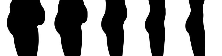
The obesity epidemic affected about 400 million adults in 2005; it will affect 700 million people in 2015 and the numbers will continue to increase. The causes are partially environmental (a more sedentary lifestyle, changes in eating habits, etc.) and partially genetic. Obesity linked to mutations represents a minority of cases. More and more data indicate that the variations in our “other genome”, the microbiome, i.e. the total genome of all the micro-organisms in our body, do more in the development of obesity than variations or polymorphism in the human genome.

Researchers working on the European METACARDIS project participated in two studies whose results have initiated the development and creation of individual treatments for patients suffering from cardio-metabolic disorders, including cardio-vascular diseases and metabolic diseases associated with obesity.

Two types of individuals, depending on the bacterial composition of the digestive tract

The first stage of the METACARDIS project is the study of patient cohorts constituted during previous European or national programmes (the European projects MetaHIT and Micro-Obes supported by the French National Research Agency; > 200 subjects), who provided instant access to much clinical, medical and environmental data as well as biological samples.

An initial study was performed on the MetaHIT cohort, which consisted of 292 Danish adults 123 of them non-obese and 169 obese. The researchers analysed the intestinal bacterial genome of these individuals, using a new technique known as quantitative metagenomics. According to the results, it appears that the two groups of individuals can be differentiated by the richness of the bacteria they carry and the abundance of certain species of intestinal bacteria. One quarter of the people in the cohort were bacteria “poor”, while three-quarters possessed intestinal flora that were “rich” in bacteria. It is the first time that such a distinction has been highlighted in the population. Furthermore, the distinction did not depend on the corpulence of the individuals, because non-obese people were found in both groups, even though the bacteria-deficient group contained more obese people (80%).

Increased risk of complications associated with obesity

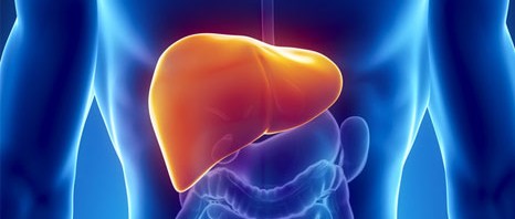
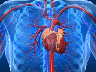
When comparing the two groups, researchers discovered that those who were deficient in intestinal bacteria were at greater risk than those who were bacteria-rich of developing complications linked to obesity such as type II diabetes, lipidic and hepatic problems and increased inflammatory factors in the blood, of the type frequently observed in these conditions.

Species of bacteria limit weight gain

Researchers also observed that obese people in the bacteria-deficient group gained more weight over time than individuals in the other group. In bacteria-poor individuals, eight specific species of bacteria were present in low numbers, or were even absent altogether. These species may have a protective role in preventing weight gain and in the long run their detection will open the way to the development of new probiotics that will help to combat weight gain.

Only six species of bacteria make it possible to differentiate between “poor” and “rich”

The second study, conducted by researchers in the METACARDIS consortium on the MicroObes cohort, focused on a cohort of 49 obese or overweight French adults. This confirmed the results of the first study. There were similarities in the bacteria-poor and rich communities between the French and Danish populations. Furthermore, on the basis of only six species of bacteria that were particularly representative in these communities, it was possible to distinguish between bacteria-rich communities and bacteria-poor communities with 95% accuracy. These results could lead to the devising of a simple method for determining which type of microbial intestinal community lives inside an individual. The purpose of METACARDIS is, on the one hand, to confirm, in a larger European cohort, the importance of these bacterial markers and on the other hand to develop a simple diagnostic test to identify individuals who are at greatest risk of becoming obese, associated with co-morbidity.

A diet that helps to enrich the microbiota

The French study also investigated the impact of a protein- and fibre-rich, low calorie diet on the genetic diversity of the intestinal microbiota. After six weeks, the diet not only produced the expected improvement in the clinical characteristics of the individuals studied, but also increased the richness of intestinal bacteria in the bacteria-poor communities. Researchers were thus able to correlate the increase in bacterial richness with weight loss, a reduction in the mass of fat and in cardio-metabolic risk factors. The way is open not only for the diagnosis of at-risk individuals but also for intervention through targeted nutritional recommendations. The inflammatory level of the blood and fatty tissue was not reduced by the diet as efficaciously in individuals who were species-poor as in those who were species-rich. Other interventions, perhaps involving medication, need to be invented to tackle this problem.

The METACARDIS consortium will pursue its research for another five years in order to better specify the various forms of obesity which vary greatly from one person to another and to detect people at risk from metabolic and cardiovascular complications so as to do more to prevent these complications and treat them in a targeted fashion. Consequently any early changes in the microbiota looks like the right route to pursue since it would be accessible through changes in the diet” explains Prof. Karine Clément, the METACARDIS coordinator. In fact, the ultimate aim of the project is to devise preventive medicines for chronic conditions as an alternative to the curative medicines that are becoming less and less affordable financially for industrialised societies.

The METACARDIS project, coordinated by Inserm and launched last December, represents the first systematic study to correlate intestinal flora and cardio-metabolic conditions in humans. The project is bringing together groups of European doctors and multidisciplinary researchers as well as experts in biotechnology and those from industry can access the latest technologies to promote the development of new preventive and treatment strategies for cardio-metabolic conditions. The teams include the INSERM 872 Unit headed by Karine Clément and Dominique Gauguier at the “Les Cordeliers Research Centre”, the “ICAN” University Hospital Institute headed by Serge Hercberg and INSERM Unit 557 “Nutritional Epidemiology” run by Hervé Blottière, Joël Doré and Dusko Ehrlich of the INRA.

https://www.metacardis.eu

The 12 partners in the METACARDIS project:

Inserm, France

INRA, France

Imperial College London, United Kingdom

University of Copenhagen, Denmark

European Molecular Biology Laboratory, Germany

University of Gothenburg, Sweden

Flemish Institute for Biotechnology, Belgium

Assistance Publique – Hôpitaux de Paris, France

University of Leipzig, Germany

Danone Research, France

Cargill, France

Chalmers University of Technology, Sweden

INSERM Transfert, France

Biobyte Solutions, Germany


Testosterone responsible for worsening iron overload in chronic liver diseases

A research team from Toulouse has just elucidated the mechanisms behind the differences in iron absorption between men and women. The team used mice to demonstrate how the action of testosterone can be “countered” with a drug already used in the treatment of some bronchial cancers.  The results are published this month in the Hepatology review.

artere

The regulation of the iron metabolism differs between men and women. As a consequence, chronic liver diseases associated with a reduced hepcidin production capacity – a hormone that inhibits iron absorption by the duodenum – are often more severe in men than in women.

This is the case for hepatitis C, alcoholic liver disease and hereditary hemochromatosis.

With a view to better understanding the underlying mechanisms of this sexual dimorphism, Marie-Paule Roth and her co-workers at the Centre for Physiopathology in Purpan (Mixed Inserm/CNRS/Université Paul Sabatier Research Unit, Toulouse) have taken advantage of the major differences in hepcidin expression and iron overload observed in male and female mice with iron metabolism disorders.

The researchers showed that testosterone robustly represses hepcidin transcription in these mice since it activates the growth factor receivers in the liver (EGF). Yet, when these EGF receivers are inhibited in male mice by a drug used in the treatment of some bronchial cancers, testosterone-induced repression ceases.   Hepcidin production is then significantly increased. Castrating male mice leads to an increase in hepcidin production, as well as a very strong reduction in iron overload in the heart and pancreas of these mice.

To conclude, Marie-Paule Rothe and her team emphasise: “These results indicate that the testosterone-induced repression of hepcidin may have clinical repercussions for patients who, for different reasons, are only capable of producing hepcidin in limited quantities. Inhibiting the activation of EGF receivers in these patients could help them to produce more hepcidin, thus limiting iron overload that worsens the prognosis of chronic liver disease” they explain.

Boosting Muscle capacity

Type 1 diabetes : regenerate our own insulin cells ?

Patrick Collombat, Inserm Research Director and head of the Avenir team at the Institut de Biologie Valrose in Nice, has published new results concerning Type I diabetes. Researchers show that, in mice, the pancreas contains cells capable of being converted into insulin-producing β cells, something that can be done at any age. They also demonstrate that all pancreatic β cells can be regenerated several times and that chemically-induced diabetes in mice can thus be “treated” repeatedly. The challenge for the researchers is now to show that these procedures can be applied to humans.

This work is published online in the Developmental Cell journal dated 27 June 2013.

Type I diabetes, characterised by the selective loss of pancreatic, insulin-producing β cells, is a condition that affects more than 30 million people worldwide. Despite current treatments, type I diabetic patients have a life expectancy that is reduced by five to eight years. It is in this context that the Avenir “Diabetes Genetics” team have been working to develop new approaches designed to regenerate these cells.

In 2009, researchers at the Valrose Biology Institute (Inserm/University Nice Sophia Antipolis) managed to convert glucagon-producing α cells into β cells in young mice. Today, thanks to the use of transgenic mice, they report the mechanisms resulting in this exchange of cell identity. Specifically, they show that pancreatic ductal cells can be continuously mobilised and literally transformed into α and subsequently into β cells, a process that works at any age. Such transformation is obtained through the forced activation of the Pax4 gene in the α cells of the pancreas. The resulting cascade of events causes the generation of brand-new β cells, thanks to the reactivation of development genes. Throughout this process, α cells are regenerated and gradually adopt the profile of β cells. This means that the pancreas has a virtually inexhaustible source of cells capable of replacing the β cells.

β cell regeneration in the pancreas

Pancreas

© Patrick Collombat / Inserm


Left: pancreas of control mice (non-diabetic)
Right: pancreas of transgenic mice demonstrating massive regeneration of insulin-producing β cells (coloured pink) following chemical induction of diabetes.

By artificially inducing type I diabetes in mice, 

“we also show that all the pancreatic β cells can be regenerated at least three times using this mechanism. Diabetes, induced in this way, in the mouse, can be literally “treated” multiple times thanks to the new stock of functional, insulin-producing β cells”

explains Patrick Collombat, Inserm research director and principal author of the study.

These promising results obtained in the mouse suggest that the pancreas contains cells that can regenerate several times those β cells lost in type I diabetics.

“We are currently working on the possibility of inducing such regeneration by using pharmacological molecules. Thanks to this new data, we shall be concentrating in future years on determining whether these processes can also be made to work in humans, a real challenge in offering better treatments for type I diabetic patients”, he concludes.

Stress: it should never be ignored!

Work pressure, tension at home, financial difficulties … the list of causes of stress grows longer every day. There have been several studies in the past showing that stress can have negative effects on health (cardiovascular diseases, diabetes, high blood pressure and more). The Inserm researchers at unit 1018, “The Epidemiology and Public Health Research Centre”, working in collaboration with researchers from England and Finland have demonstrated that it is essential to be vigilant about this and to take it very seriously when people say that they are stressed, particularly if they believe that stress is affecting their health. According to the study performed by these researchers, with 7268 participants, such people have twice as much risk of a heart attack, compared with others.

These results have been published in European Heart Journal.

Today, stress is recognized as one of the main health problems. When people face a situation that is considered stressful, they may experience several physical, emotional and behavioural symptoms (anxiety, difficulty in concentrating, skin problems, migraines, etc.). Previous studies, particularly the recent studies performed within the Whitehall II cohort[1], composed of several thousand British civil servants, have already shown that the physiological changes associated with stress can have an adverse effect on health.

stess au travail

crédit : ©Fotolia

Herman Nabi, Inserm researcher at Unit 1018 “The Epidemiology and Public Health Research Centre”, and his team went further and studied people who declared themselves to be stressed, in order to look more closely at whether there was a link between their feeling and the occurrence of coronary disease some years later.

Using a questionnaire prepared for the Whitehall II cohort, the participants were invited to answer the following question: “to what extent do you consider the stress or pressure that you have experienced in your life has an effect on your health”, the participants had the following answers to choose from: “not at all”, “a little”, “moderately”, “a lot” or “extremely”.

The participants were also asked about their stress level, as well as about other factors that might affect their health, such as smoking, alcohol consumption, diet and levels of physical activity. Arterial pressure, diabetes, body mass index and socio-demographic data such as marital status, age, sex, ethnicity and socio-economic status were also taken into account.

According to the results, the participants who reported, at the start of the study, that their health was “a lot” or “extremely” affected by stress had more than twice the risk (2.12 times higher) of having or dying from a heart attack, compared with those who had not indicated any effect of stress on their health.

From a clinical point of view, these results suggest that the patient’s perception of the impact of stress on their health may be highly accurate, to the extent that it can predict a health event as serious and common as coronary disease.

In addition, this study also shows that this link is not affected by differences between individuals related to biological, behavioural or psychological factors. However, capacities for dealing with stress do differ massively between individuals depending on the resources available to them, such as support from close friends and family.

According to Hermann Nabi, “the main message is that complaints from patients concerning the effect of stress on their health should not be ignored in a clinical environment, because they may indicate an increased risk of developing and dying of coronary disease. Future studies of stress should include perceptions of patients concerning the effect of stress on their health”.

In the future, as Hermann Nabi emphasizes, “tests will be needed to determine whether the risk of disease can be reduced by increasing the clinical attention given to patients who complain of stress having an effect on their health”.


[1] Created in 1985, the Whitehall II cohort, consisting of British civil servants, is making a major contribution to research in social epidemiology and is considered internationally to be one of the main sources of scientific knowledge concerning social determinant factors for health.

Food contaminants worsen metabolic problems in obese mice

Certain food contaminants are suspected of triggering metabolic disorders, or of worsening them, particularly when they accompany a high-fat diet. In order to get a better understanding of these effects, researchers from the Inserm cardiovascular, metabolism, diabetology and nutrition unit (U1060 « Laboratoire de recherche en cardiovasculaire, métabolisme, diabétologie et nutrition » Inserm/Inra/Université Lyon 1) introduced a “cocktail” of contaminants mixed with low doses of dioxin, PCB, bisphenol A and phtalates into the feeding of mice that had already been rendered obese by a high-fat diet. The results show that metabolic changes occur in these mice, but that the effects differ depending on the gender. Females appeared to be more affected. Their obesity-induced glucose intolerance worsened and their estrogen pathway was altered.

These works have been published in the review Faseb Journal.

assiette-salade

crédit : ©Fotolia

Obesity is a major public health problem because it is a risk factor in the development of metabolic complications (diabetes, cardiovascular diseases, etc.). It is a multi-factorial disorder. In addition to genetic predispositions and a life style that combines overeating with lack of exercise, there is a great volume of proof to suggest that contaminants, particularly in the food we eat, are responsible for the obesity epidemic and the resulting metabolic changes.

Researchers have put forward the hypothesis that contaminants in food could worsen certain metabolic problems already caused by eating an over-rich or a high-fat diet.

In this study, the researchers fed mice a high-fat diet (already a health risk), to which low doses of contaminants had been added. They were given this diet throughout their lives. Their mother had been nourished with this diet prior to their birth and during the gestation and lactation periods. Therefore, they suffered chronic exposure to this diet.

Two environmentally persistent contaminants[1] (dioxin and PCB) and two non-persistent contaminants[2] (phtalate and bisphenol A) were added to the high-fat (obesogenic) diet of the mice. The doses given were low, normally considered not to have any health impacts.  These products were chosen because they are present in human food and because they are known to trigger endocrine disruption. In parallel, a control group of mice was fed with the same obesogenic diet, but without added contaminants.

The researchers then ran glucose tolerance and insulin sensitivity tests. They measured the livers for lipid accumulation and the expression of certain genes that play key roles in the metabolism of the adult mice.

The results show that the effects are highly dependent on the gender of the animal.

Male and female: different effects

In the females fed with a high-fat diet, the addition of contaminants worsened the glucose intolerance and altered the estrogen pathway. In males, it altered the cholesterol and lipid metabolism. There was no change in weight between the exposed mice and the unexposed mice.

The researchers pursued the hypothesis that there was a connection between the observed glucose intolerance and the alteration in the estrogen signaling in exposed females. It is well known that estrogens protect against metabolic disorders. In other words, these works suggest that in obese females, exposure to food contaminants could lower the protection level that estrogens provide against metabolic disorders.

“With this study, we have succeeded in providing proof-of-concept that low doses of contaminants, even at levels normally considered to be without health impacts in humans, do in fact affect humans when subjected to chronic exposure, and when the contaminants are combined with a high-calorie diet” points out Brigitte Le Magueresse Battistoni.

This study was carried out as part of research programmes supported by the Institut Benjamin Delessert (2010), the ANSES (EST-2010/2/2007) and the EFSD (programme 2011) and by Brigitte Le Magueresse-Battistoni and Danielle Naville, 2 researchers who work for team 1 of the CarMeN Unit run by Hubert Vidal.


[1] These products build up throughout the food chain by means of a process known as bioaccumulation, until they end up on our plates.

[2] These products break down more rapidly that the above, but the intensive production of these products, especially in the plastics industry, means that they are omnipresent in our daily lives.

Early exposure to bisphenol A might damage the enamel of teeth.

Are teeth the latest victims of bisphenol A? Yes, according to the conclusions of work carried out by the research team led by Ariane Berdal of the Université Paris-Diderot and Sylvie Babajko, Research Director at Inserm Unit 872 “Centre des Cordeliers”. The researchers have shown that the teeth of rats treated with low daily doses of BPA could be damaged by this. Analysis of the damage shows numerous characteristics that are common with a recently identified pathology of tooth enamel that affects roughly 18% of children between the ages of 6 and 8.

These results have been published in the American Journal of Pathology

Bisphenol A (BPA) is a chemical compound used in the composition of plastics and resins. It is used for example to manufacture food containers such as bottles or babies’ bottles. It is also used for the protective films inside drinks cans and food tins, or as developers on sales receipts. Significant amounts of BPA have also been found in human blood, urine, amniotic liquid and placentas. Recent studies have shown that this industrial compound has adverse effects on the reproduction, development and metabolism of laboratory animals. It is strongly suspected of having the same effects on humans.

As a precautionary measure, the manufacture and commercialisation of babies’ bottles containing bisphenol A were prohibited in Europe in January 2011. The prohibition will be extended to all food containers in France as from July 2015.

So this study shows that teeth are the latest in an already long list of victims of BPA.

The Inserm researchers have shown that the incisors of rats treated with low daily doses of BPA (5 microgrammes/kg/day) could be damaged by this.

This effect has also been observed within a development window of no more than 30 days post-birth in rats, thus demonstrating a range of sensitivity to exposure.

Analysis of these teeth showed numerous characteristics that are common with a tooth enamel pathology known as MIH (Molar Incisor Hypomineralisation) that selectively affects first molars and permanent incisors. This enamel pathology is found in roughly 18% of children between the ages of 6 and 8. Children affected by this pathology present with teeth that are hypersensitive to pain and liable to cavities. It is interesting to note that the period during which these teeth are formed (the first years of life) correspond to the period during which humans are most sensitive to bisphenol A.

Amongst the earliest observations made was the appearance of “white marks” on the incisors of rats treated with endocrine disruptors, one of which was bisphenol A (BPA). The researchers decided to define the characteristics of incisors of rats treated with low doses of BPA and to compare these with the characteristics of teeth in humans suffering from MIH

Macroscopic observation of marks on both series of teeth showed similarities, in particular fragile and brittle enamel.

Microscope observation of the enamel showed a significant reduction of the Ca/P and the Ca/C ratios in affected teeth.  This leads to mineral depletion, making the teeth more fragile and more liable to cavities.

Finally, analysis of the proteins present in the tooth matrix of rats showed an increased quantity of enamelin, a key protein for enamel formation, and a buildup of albumin leading to hypomineralisation. Analysis of the expression of key enamel genes highlighted two BPA target genes: enamelin and kallicrein 4.

According to Sylvie Babajko, the latest author of this article, “Insofar as BPA has the same mechanism of action in rats as in men, it could also be a causal agent of MIH. Therefore, teeth could be used as early markers of exposure to endocrine disruptors acting in the same way as BPA and so could help in early detection of serious pathologies that would otherwise have occurred several years later”.

dents enfants


credit : ©Fotolia

Quinze souris astronautes pour soigner les hommes

Liver disease: understanding it will enable the provision of better treatment

A certain number of patients hospitalised for cirrhosis complications soon develop a syndrome characterised by acute liver failure and/or the failure of other vital organs (ACLF)[1]. This syndrome had no specific diagnostic criteria hitherto. In this prospective study, led by Dr Richard Moreau, INSERM Research Director (Mixed Research Unit 773 “Centre de Recherche biomédicale Bichat-Beaujon”; INSERM/Université Paris Diderot) who is also a practitioner attached to the Hepatology Department of the Beaujon Hospital (AP-HP), researchers studied a cohort of 1343 patients from 12 European countries. The results, published in the learned journal Gastroenterology, describe, for the first time, the specific profile of sufferers from this syndrome that is associated with cirrhosis. This also makes it possible to more clearly define the actual rules of attribution of the organs in those most severely affected, for whom there is a high risk of early death.

Cirrhosis is an irreversible liver disease. It is characterised by chronic inflammation that destroys the liver cells and produces anarchic regeneration in the form of nodules. The disease causes the liver to lose function and is accompanied by multiple complications. When these complications manifest (bleeding in the digestive tract, bacterial infection, accumulation of liquid in the abdomen, etc.), this is known as decompensated cirrhosis and the patients are hospitalised.

A certain number of these patients quickly develop a syndrome characterised by acute liver failure and/or failure of other vital organs1 (ACLF – acute-on-chronic liver failure). The syndrome is associated with a high risk of death at one month and no diagnostic criteria were clearly established hitherto that might make it possible to describe the condition.

Through a consortium[1], the researchers in Mixed Research Unit 773 “Centre de Recherche biomédicale Bichat-Beaujon” (INSERM/Université Paris Diderot), analysed data from 1343 patients hospitalised due to acute cirrhosis complications between February and September 2011 in 29 Hepatology Departments in 12

European countries[2]. This enabled them to define robust diagnostic criteria for ACLF, indicating that one-third of the patients enrolled in the study had developed this syndrome.

The researchers noted that, compared to patients not suffering from ACLF, those who developed acute failure of an organ or organs were younger, were most frequently prey to alcoholism, suffered from a larger number of bacterial infections and had higher levels of white blood cells, as well as other markers of organ inflammation.

Quite unexpectedly, failure was most severe in patients without a previous history of cirrhosis complications. A high number of organ malfunctions (liver, kidneys, brain) were observed in these patients, including white blood cells in the blood and a mortality rate within one month of admission to hospital that was15 times greater than in patients who had a previous history.

“The identification of the criteria to define acute failure of an organ or organs enabled us to show that this is a separate syndrome from cirrhosis complications. In addition to organ failure and the high associated mortality, the development of the disease depends on the patient’s age and medical history”, explains Richard Moreau, INSERM Director of Research, and the Principal Investigator for the study.

We hope to be able to better identify those at risk of early death in order to improve their treatment. Furthermore, these results could lead to improving the current rules for assigning organs for grafting in the severest cases”, he concludes.

Foie

 Crédit photo : ©Fotolia

Liver and cirrhosis

The liver is a vital organ whose main functions are the storage and production of glucose as well as the synthesis and breakdown of other substances (triglycerides, cholesterol, lipoproteins, coagulation factors).
There are about 700,000 cases of cirrhosis in France, of which 30% are at the severe stage, resulting in 10,000 to 15,000 deaths a year. Diagnosis is on average at the age of 50. Not everyone at risk of cirrhosis develops the disease. In fact, it only manifests in 10% to 20% of cases.
The only current treatment for cirrhosis is a liver transplant. The first eligible patients are those whose life expectancy at three months is the lowest. Each year, around 1,000 patients receive liver transplants in France.

Please consult the information on the Inserm.fr 


[1]    Consortium known as CLIF (“Chronic Liver Failure”) including French researchers and their European colleagues; Richard Moreau is the main investigator in the first study conducted under the aegis of this Consortium.

[2] France, Belgium, United Kingdom, Italy, Spain, Germany, Netherlands, Ireland, Switzerland, Austria, Denmark, Czech Republic


[1]    Acute-on-chronic liver failure affecting the kidneys, brain, lungs and/or circulatory system

Treatment with HDL cholesterol to aid thrombolysis

Discovery in the rat reduces bleeding complications by 90% in the only treatment available for embolic stroke

Research performed by the Neurology Department and Stroke Reception and Treatment Unit at the Bichat Hospital (AP-HP/ Paris University Diderot) and the associated INSERM Unit 698 (Prof. Amarenco, Dr Olivier Meilhac) has highlighted the benefits of good cholesterol in reducing bleeding complications in the only treatment available for embolic stroke. The results of this experiment, performed on the rat, have just been published in Stroke .

As the third cause of death in France and throughout the world and the leading cause of disability acquired in adulthood, stroke currently affects 10 million people annually, 150,000 of them in France.

To date, the treatment of reference for stroke caused by occlusion of a cerebral artery that is recognised by the ANSM consists in performing an intravenous injection (in the crook of the elbow) of a medication known as Actilyse® (alteplase) the purpose of which is to dissolve the clot. This makes it possible to cure the patient in 40% of cases, but only if the injection is started within four and a half hours of the first stroke symptoms. An unfortunate complication of the intravenous injection of this medication, however, is a brain haemorrhage with neurological deterioration that leads to death in 6% of cases, though without visible neurological deterioration in 20% of cases.

Researchers have discovered a new treatment that may reduce the risk of the bleeding complication caused by alteplase by as much as 90%. The treatment consists of high-density lipoproteins  (HDL or “good cholesterol”), isolated from human plasma. HDLs are the particles whose job it is to evacuate the bad cholesterol from inside the arteries and send it to the liver, where it is eliminated. They have other favourable actions, in that they are anti-inflammatory, anti-oxidant and anti-protease, preventing the infiltration of white blood cells into the affected area.

The team has produced a hypothesis that, through the protective effect of the blood-brain barrier[1]1, HDL can protect against the bleeding complications caused by alteplase. The team injected rats with alteplase three hours after blocking a brain artery using a filament or a clot. Bleeding occurred in 62% of the rats after the filament was withdrawn and in 46% of rats in which the brain artery was blocked by a clot. In both cases, when alteplase was injected jointly with HDLs, less than 90% fewer bleeding complications were observed. Both these models (occlusion using a filament or the creation of a clot) were used to check the effect of the treatment. The fact that the same type of result was obtained reinforces the evidence of the effect of HDLs.

This discovery, if confirmed in humans through the clinical trial we intend to perform, could revolutionise stroke treatment and offer new opportunities for improving the cure for embolic stroke victims. It is even imaginable that in future, HDL-like particles could be produced through genetic engineering”, states Professor Amarenco, Head of the Neurology Department, Stroke Reception and Treatment Unit at theBichatHospital (AP-HP/University ofParis Diderot) and co-director with Professor Steg of the “Clinical Research into Atherothrombosis” Research Team at the Mixed INSERM-University of Paris Diderot Unit 698.

These studies were partly financed by the INSERM Unit 698 and partly by the Association SOS-Attaque Cerebrale, a stroke charity.

accident vasculaire cérébral

© S Dehaenne/Inserm


[1] the nutrients supplied by the blood need to pass through it to feed the brain.

See Protective effect of high-density lipoprotein-based therapy in a model of embolic stroke.

Obesity and insulin resistance: the lipolysis route

Photo : ©Fotolia

Liver and skeletal muscle resistance to the action of insulin is an early sign of the development of Type 2 Diabetes. The INSERM team at the “Obesity Research Laboratory ” in the Institut des Maladies Métaboliques and Cardiovasculaires (INSERM / Université Toulouse III – Paul Sabatier), headed by Dominique Langin, has shown through results published this week, that there is an association between lipolysis (mobilisation of fat in response to the body’s need for energy) and insulin sensitivity in humans. Researchers also showed that a reduction in lipolysis in mice, through genetic modification or pharmacological treatment, improved the action of insulin on glucose metabolism in the liver and the muscles. Lipolysis inhibition could be used in treating insulin resistance in the obese.

The results are accessible on the website of the Plos Biology journal for 19 February 2013.

Insulin is the hormone that controls the blood glucose level, inhibiting its production by the liver and stimulating its use in the muscles. When the body needs energy, during fasting or due to physical exercise, the triglycerides stored in the adipose tissue are released in the form of fatty acids through the action of adipocyte lipolysis. When this happens, the fatty acids have a favourable action because they are supplying energy.

These fatty acids may also have a deleterious action, however. When they are present in excessive quantities, as in the case of obesity, they are deposited in the peripheral organs and interfere with the action of insulin. Other lipids and proteins produced by an excess of adipose tissue are also involved in the development of insulin resistance. (Figure 1 opposite)

Figure 1 : Excess of fat mass and insulin resistance

© INSERM / Dominique Langin

Professeur Langin’s team at the Institut des Maladies Métaboliques and Cardiovasculaires (I2MC, Toulouse), (INSERM, Université Toulouse III – Paul Sabatier, Hôpitaux de Toulouse), in collaboration with other I2MC teams and researchers in Sweden at the Karolinska Institutet in Stockholm and Lund University, are seeking ways to treat insulin resistance, using a treatment strategy that avoids the onset of diabetes in the obese.

In this study, they showed that a reduction in adipocyte lipolysis through genetic modification or pharmacological treatment improves insulin sensitivity. An exploration of the mechanisms involved showed that the reduction in lipolysis causes a reduction in the flow of fatty acids within the organism and is accompanied by an improvement in insulin action on glucose metabolism in the liver and muscles.

“We discovered the effects of lipolysis reduction in humans through analysing data from cohorts of obese people in whom we reported that a reduction in lipolysis was associated with an improvement in insulin sensitivity. These results are all the more interesting in that they introduce a treatment strategy in which lipolysis inhibition does not involve any change to body weight”, explains Dominique Langin.

This study also showed quite unexpectedly that when lipolysis was reduced in mice, a special metabolic pathway, known as de novo lipogenesis, was activated enabling the synthesis of fatty acids directly from glucose (Figure 2 below). In Spring, 2012, a team from Harvard University, United States, suggested that the activation of de novo lipogenesis in adipose cells reduced resistance to the action of insulin. A clinical trial performed by Professor Langin’s team at the Centre d’Investigation Clinique INSERM-CHU de Toulouse also shows that chronic treatment with an anti-lipolytic molecule induced an increase in the expression of de novo lipogenesis genes in the adipocyte.

Figure 2 : Inhibition of the mobilisation of fat (lipolysis)  through the adipose cell reduces insulin resistance by increasing lipogenesis de novo.

© INSERM / Dominique Langin

Researchers are now attempting to identify the mediators produced by the adipose cell participating in the improvement of insulin action, as well as demonstrating the important of this treatment strategy in pre-diabetic obese patients.

fermer