Menu

HOMAGE – Overcoming Heart Failure, an European challenge

The HOMAGE (Heart OMics in AGEing) project, coordinated by the French National Institute of Health and Medical Research (INSERM), has been awarded a grant by the European Commission for a 6 year period. The project aims to identify and validate specific biomarkers of heart failure in order to prevent the development of the disease affecting elderly population. 17 research groups from 10 countries will collaborate to investigate new ways of preventing heart failure. The project will use an innovative ‘omic-based’ approach which investigating simultaneously a huge amount of genes, proteins and metabolites.

The 17 partners will meet at Nancy on February 22nd for the kick off meeting of HOMAGE.

Professor Faiez Zannad, Head of the Centre d’Investigation Clinique Pierre Drouin Inserm U9501 and Inserm Unit 1116 based in Nancy, is the project coordinator.  The 12 million euros grant of the European Commission will be dedicated to the HOMAGE consortium for research on heart failure, a serious illness altering myocardial activity which affects more than 6.5 million persons in Europe. Indeed, the prevalence of heart failure is increasing worldwide due to an ageing population as well as a rising trend of risk factors for heart disease such as diabetes, obesity and hypertension.

Heart failure is a major cause of mortality and morbidity in the world and remains the most frequent cause of hospitalization for patients over 65 years old.

The costs related to heart failure have been estimated around 1.5 billion euros per year in France[1].

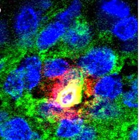
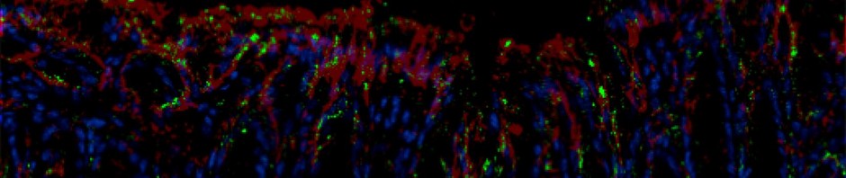
Photo : ©Serimedis/Inserm

Despite important progress in the treatment including new drugs, new medical devices and innovative disease management programmes, the diagnosis of heart failure is often difficult in older adults with co-morbidities. Screening tests are usually based on blood pressure, glycaemic and cholesterol control. Although they are useful to detect high risk patients, they are limited regarding their sensitivity and specificity. During the past decade, promising biomarkers such as natriuretic peptides have been identified to diagnose heart failure, but their predictive value remains relatively poor. The HOMAGE consortium is willing to validate more specific and more sensitive biomarkers which should facilitate an early detection of the disease in patients at risk.

To achieve this goal, the consortium agreed on the use of an ‘omic-based’ approach. This approach aims to validate promising biomarker candidates by crossing a large volume of data (genomics, proteomics, miRNomics, transcriptomics and metabolomics). This would permit scientists to understand new pathophysiological mechanisms, signaling pathways and identify new therapeutic targets to prevent heart failure.

The HOMAGE consortium will manage cohorts for a total of 30 000 patients. The European researchers will firstly identify biomarker candidates in blood and study their predictive value for heart failure and common co-morbidities associated with ageing (renal impairment, cognitive disorders…). Subsequently, HOMAGE will lead a clinical trial to look for novel treatments of heart failure that can be targeted specifically to those patients at risk.

This trial will allow identifying patients’ omics based biomarker profiles most likely to predict response to treatment with the better benefit/risk ratio, an attempt into personalized medicine.


[1] Fédération Française de Cardiologie

The 17 partners of HOMAGE project : www.homage-hf.eu (en ligne le 21 février)

Inserm, France : https://www.inserm.fr/

Inserm U942/ Biomarqueurs et maladies cardiaques, France

Inserm UMR 1048/ Equipe 7, France

Inserm U744/ Recherche des Déterminants Moléculaires des Maladies Cardiovasculaires, France

Inserm Transfert, France : https://www.inserm-transfert.fr/

European Drug Development Hub, France : https://www.fondationtransplantation.org

ACS Biomarker, Pays-Bas : https://acsbiomarker.com/

Randox Testing Service, Royaume-Uni : https://www.randoxtestingservices.com/

Medical University of Graz, Autriche : https://www.meduni-graz.at/en/

University of Manchester, Royaume-Uni : www.manchester.ac.uk

Fundación para la Investigación Médica Aplicada, Espagne : www.cima.es

University College Dublin, Irlande : www.ucd.ie

University of Hull, Royaume-Uni : www.hull.ac.uk

Maastricht University, Pays-Bas : https://www.maastrichtuniversity.nl/

Istituto di Ricerche Farmacologiche ‘Mario Negri’, Italie : https://www.marionegri.it/mn/en/

Hannover Medical School, Allemagne : https://www.mh-hannover.de

University of Leuven, Belgique : www.kuleuven.be/english/

London School of Hygiene, Royaume-Uni : https://www.lshtm.ac.uk/

Emory University, Etats Unis : https://www.emory.edu

University of Glasgow, Royaume-Uni : https://www.gla.ac.uk/

Collaborative institutions:

The Trustees of Boston University, National Heart, Lung, and Blood Institute’s Framingham Heart Study, Etats Unis

Imperial College, Royaume-Uni

Steno Diabetes Center (Novo Nordisk), Danemark

History of stroke and coronary heart disease – a fatal combination

The cardiology service team at the Hôpital Bichat and the Mixed INSERM Unit 698 (AP-HP, Université Paris Diderot), in collaboration with international teams of researchers, studied a cohort of patients suffering from coronary disease. The study showed that those patients with a history of stroke or transient ischæmic attack (TIA) are not only at higher risk of cardio-vascular episodes but also of haemorrhagic events, stressing the therapeutic challenge involved in treating such patients. The research is published online in Circulation*.


© Serimedis/Inserm

Heart and cerebro-vascular disorders represent the two leading causes of death throughout the world. They are sometimes combined in a single patient and their combination represents both a considerable risk to the patient and a therapeutic challenge

Today’s anti-thromobotic, blood-thinning medication provides effective treatment for coronary heart disease. Several randomised tests to assess new anti-thrombotic treatments in coronary patients have identified the fact that a history of stroke or TIA constitutes a marker for increased risk of intracranial bleeding that could prove fatal for the patient. The study was conducted by the Cardiology Service Team of the Bichat Hospital and INSERM Unit 698, working with several international teams, and consisted in monitoring and analysing 26,389 coronary patients from REACH, the international register of athero-thrombic patients, over a four-year period, in order to very accurately assess the ischaemic and haemorrhagic risk associated with a history of stroke or TIA in coronary patients.

The results of this study highlight the frequency of the phenomenon. It showed that 4,460 patients, 17% of the cohort of coronary patients, had a history of stroke or TIA. This stroke or TIA antecedent was associated, moreover, with an approximate 50% increase (in relation to coronary patients without a history of stroke) of dying from a heart attack or stroke, with increased risk of ischæmic and hæmorrhagic stroke.

This study also aims at investigating the therapeutic challenge of treating such patients. In fact, higher doses of anti-platelet or anti-coagulant are associated with a particularly high increased risk of hæmorrhage.

The conclusions of the study are important since they emphasise the difficulty in treating this type of patient, a type that is frequent but whose prognosis is particularly serious. It also shows how important it is to provide the right anti-thrombotic treatment. Additional clinical trials are currently being conducted to test new treatment strategies for these patients”, explained Professor Steg of the Cardiology Department at the Hôpital Bichat.

Stroke is where there is a blockage or rupture of a blood vessel that transports blood inside the brain. The result is a lack of oxygen to the brain, which put one or more areas of the brain in danger.

A stroke occurs when the blood circulation to or within the brain is interrupted by a blocked blood vessel (an ischæmic stroke), the commonest form, or a blood vessel ruptures (a hæmorrhagic stroke), the latter occurring in less than 20% of cases.

Ischæmic stroke. Where the artery becomes blocked by a high-cholesterol platelet (atherosclerosis), the patient suffers an ischæmic stroke (40 to 50% of strokes), or where a blood clot blocks the artery, in which case it is a cerebral embolism (30% of AVCs).

Hæmorrhagic stroke (20% of cases). This is most frequently due to a ruptured aneurism. Tumours, hypertensive crisis and miscellaneous blood-clotting disorders can also cause hæmorrhagic strokes.

Transient Ischæmic Attack (TIA) When the obstruction in the cerebral artery reabsorbs itself and produces no sequel, the event is described as a transient ischæmic attack. The symptoms are the same as those of a stroke, but they only last a few seconds or minutes before a return to normal. An AIT may even pass unnoticed and be mistaken for mere discomfort. Yet it indicates an important risk of a more serious AVC.

The role of the innate immune cells in the development of Type 1 diabetes

Julien Diana and Yannick Simoni of the “Immune Mechanisms in Type 1 Diabetes (Inserm/Université Paris Descartes), directed by Agnès Lehuen, have just published the results of their work on type 1 diabetes in the Nature Medicine journal. This is a disease characterised by the self-destruction of the p pancreatic cells that produce insulin. The researchers reveal the role of the innate immune cells, especially the dendritic cells, that cause the activation of the killer T-lymphocytes whose action is directed against the p pancreatic cells. The results obtained in mice make it possible to consider new ways of regulating the auto-immune reaction generated by the innate immune cells.

Type 1 diabetes, or insulin-dependent diabetes, is an auto-immune disease characterised by the destruction of insulin-producing pancreatic β cells that are present in the Islets of Langerhans which are themselves in the pancreas. The peculiarity of this type of diabetes lies in the fact that the cells are destroyed by T lymphocytes that kill the patient’s immune system. This is an auto-immune reaction. Much of the research has highlighted the role of auto-reactive T lymphocytes in the pancreatic β cells. Yet the mechanisms involved in the initial activation of the immune system that triggers the sequence of events leading to the death of the cells are still ill-defined.

The work of the team working on “Immune mechanisms of Type 1 diabetes” (Inserm/Université de Paris Descartes) in NOD (Non-obese diabetic) mice, the model used for studying Type 1 diabetes, reveals the essential role played by the cells of the innate immune system[1] that were not hitherto considered to be involved in diabetes. These cells were known to play a role in other auto-immune diseases such as lupus and psoriasis. In this study, researchers managed to describe the mechanisms initiating the activation of T lymphocytes attaching themselves to the pancreatic β cells.

The innate immune system is normally activated when an infection occurs. This is when the mobilisation and activation of neutrophils and dendritic cells is observed, constituting the first stages of the immune response. Abnormally in NOD diabetic mice, the natural physiological death of pancreatic β cells, occurring as the process progresses, involves an innate auto-immune response in the pancreas. Researchers have provided details of the introduction of a sequence of activation events in these innate immune cells in the Islets of Langerhans.

Activation of the immune system leading to death of the p cells

 ©Inserm / Agnès Lehuen – Juliette Hardy

 The natural deterioration of the β cells (1) leaves cell debris in the tissues that abnormally activate the neutrophils (2). These alarm cells in the immune system warn the dendritic cells (pDC) (3) which in turn cause the production of IFN α interferon, an alarm molecule (4). The interferon α then stimulates the T lymphocytes which, by recognising functional pancreatic β cells, cause these cells to die (5).

Innate immune cells and killer T-cells in the Islets of Langerhans

 ©Inserm / Yannick Simoni

A-B) Fluorescent photographs of a pancreatic islet (green) and close-up (B) of a neutrophil  (yellow) producing an activating molecule (red)
C) Photograph of the Islets in untreated mice in which the killer T-cells (white arrows) can be observed.

“We have observed in mice that treatment prevents activation of the innate immune cells, neutrophils and dendritic cells, warning of the onset of diabetes by inhibiting the appearance of auto-immune T responses aimed at the pancreas” explains Agnès Lehuen, head of the Inserm team.

These results show, for the first time, the important role played by innate immune cells in the sequence of events leading to the onset of Type 1 diabetes. Researchers continue to strive to understand how to regulate the auto-immune reaction produced by dendritic cells without compromising the innate immune system, an essential one in cases of infection. Several routes are being taken to attempt to regulate the production of the INF α alarm molecule that precedes activation of the killer T-cells, for example, by specifically targeting certain activation routes for the pDC dendritic cells.

These therapeutic approaches are currently being tested in other auto-immune diseases such as lupus and psoriasis. Such innovative treatments could be useful in the prevention of Type 1 diabetes. It will first be necessary, however, to perform studies in diabetic and pre-diabetic patients to be able to better understand how the innate immune cells function, something that has not been studied until recently in auto-immune diabetes,” concludes Agnès Lehuen.

The research benefited from a grant from LabEx INFLAMEX as part of investment for the future and for the Ile-de-France region.


[1] System present from birth that makes it possible to initiate an immune response to infection, regardless of the infectious agent involved. It is distinguished from the “acquired” or so-called “adaptive” immune system that is a specific response involving recognition of the infectious agent and the memorising of the infectious event.


Adipocyte inflammatoire de l’obèse : une cellule en perte de contrôle

Intestinal bacteria an aggravating factor in obesity-related illnesses

Researchers from INRA and Inserm have discovered that intestinal bacteria (“microbiota”) cause metabolic complications in the liver related to obesity. This is the first time such a link has been established using intestinal bacteria transplanted in mice. The results, published online in the journal Gut on 29 November 2012, pave the way towards preventive and therapeutic treatments based on controlling intestinal microbiota to cure type-2 diabetes and obesity-related liver conditions.

© Fotolia

Certain types of liver disease are linked to a condition called metabolic syndrome and develop in obese individuals in particular. The severity of these illnesses varies, from steatosis, in which lipids accumulate abnormally in the liver (a condition present in 80% of obese patients), to cirrhosis (present in 20 to 30% of all cases).

No two metabolisms react in exactly the same way to the same high-fat diet.  Based on that premise, the INRA team, working with a team from Inserm, decided to study the role played by the billions of bacteria present in the digestive tract – known as intestinal microbiota – in the appearance or worsening of obesity-related liver diseases. To establish the precise effects of the bacteria, researchers transplanted microbiota into mice initially devoid of them. One group received intestinal microbiota from a mouse suffering from insulin resistance and steatosis, while another group received bacteria from a healthy mouse. Fed an excessively fat-rich diet for 16 weeks, all the mice became obese. Those which had been transplanted with microbiota from the insulin resistant and steatosis-afflicted mice, however, developed hyperglycaemia and hyperinsulinaemia, while the second group did not. Analysis of the liver tissue also revealed a more severe degree of steatosis in the first group, as confirmed by a higher concentration of lipids in the livers of these mice (see photos, below). Sequencing of the composition of the microbiota revealed the presence of different bacteria species in the two groups.

These results show that the composition of intestinal microbiota (determined by a wide variety of factors, including diet, genetic predisposition, and environment) plays a role in the development of metabolic syndrome-related illnesses. While the exact mechanisms of these bacteria remain unknown, the study suggests that controlling or transplanting intestinal microbiota could be a new way to prevent and/or treat type-2 diabetes and obesity-related liver disease.

Sections of liver in mice transplanted with microbiota from other mice presenting metabolic disorders (left) and with microbiota from healthy mice (right).

Only the mice transplanted with “bad” microbiota (left) developed severe steatosis when exposed to an excessively fat-rich diet (as evidenced by the clearly visible white fat droplets).

© Stephan Bouet, Histiology Workshop, GABI, INRA

(1) Metabolic syndrome is a medical condition known to precede the development of several serious illnesses, including type-2 diabetes, heart disease and stroke.

(2) Insulin resistance is typically present in cases of obesity. During periods of insulin resistance, the pancreas of an obese person secretes insulin which no longer has an effect on its receptors, a phenomenon known as desensitisation. Despite the presence of insulin, glucose enters cells less effectively and accumulates in the bloodstream, increasing blood sugar levels (hyperglycaemia). This in turn provokes a hypersecretion of insulin (hyperinsulinaemia).

METACARDIS: a European project that deciphers the genes of the gut microbiota responsible for cardiometabolic diseases

METACARDIS (Metagenomics in Cardiometabolic Diseases) is a European project coordinated by Inserm that aims to study the role of the gut microbiota in the development of cardiometabolic diseases (CMDs). Thanks to the support of the European Commission, fourteen partners in six European countries will work together over a five-year period to translate the results obtained with novel CMD diagnosis and treatment methods and pave the way for personalised medicine in CMDs. Researchers and clinicians from the Institute of Cardiometabolism and Nutrition (ICAN) will also be involved in the METACARDIS project.

The aim of the METACARDIS project is to develop and implement personalised treatments for patients with CMDs in order to meet an urgent need for improved patient care and relieve the socioeconomic burden on the health care system. CMDs – which comprise cardiovascular diseases, metabolic diseases and obesity – are a major cause of mortality and morbidity around the world. Their socioeconomic cost is substantial; it is estimated that they alone cost the EU economy more than €192 billion a year (57% due to direct treatments, 21% due to production losses and 22% due to indirect treatments[1]).

The aim of METACARDIS is to study the impact of changes in the gut microbiota on the onset and progression of CMDs and their associated conditions. These conditions manifest in highly different ways and in all likelihood are interlinked by initial common channels and dysfunctions. As a result, it is essential to thoroughly understand the physiopathological mechanisms involved, make detailed early diagnoses and deliver treatments that are tailored to each disease. In other words:

  • Find novel shared biological targets and channels that play in role in the progression of CMDs.
  • Validate the gut microbiota targets and biomarkers.
  • Refine the analysis of patients’ clinical profiles through molecular phenotyping.
  • Develop new systems (software) for the integration of environmental data and of patients’ clinical and biological information.

METACARDIS is the first systemic study designed to correlate the gut microbiota with CMD in people.

It brings together groups of European researchers from various disciplines with biotech and industry experts. The teams will have access to state-of-the-art technologies to accelerate the development of novel preventive and therapeutic strategies for CMDs. Amongst these teams are those directed by Karine Clément and Dominique Gauguier of the Cordeliers Research Centre (Inserm Unit 872) and of the university hospital and research complex ICAN, by Serge Hercberg of the Nutritional Epidemiology unit (Inserm Unit 557) and by Hervé Blottière, Joel Dore and Dusko Ehrlich at INRA (France’s National Institute of Agricultural Research).

The preliminary studies forming the basis of the METACARDIS project

Work conducted in recent years to characterise the genome of the gut microbiota, i.e. the entire genome of the bacteria in the intestine, has opened up new opportunities in in-depth understanding of the possible channels shared by the various conditions that comprise CMDs.

In oncology, biological signatures that make it possible to predict the characteristics of tumours have been identified. Likewise, research has shown that gene expression models of the gut microbiota vary from one cardiometabolic condition to another. Obese patients presenting insulin resistance, inflammatory syndrome, dyslipidaemia and/or glucose dysregulation are characterised by an alteration in the diversity and composition of their gut microbiota. Some species of gut bacteria have even been found to be associated with coronary vascular diseases. Imbalances in gut intestinal flora therefore may play a role in the development of cardiometabolic conditions.

Patients included in the project in late 2012

Once the METACARDIS project begins in late 2012, its teams will conduct a first series of studies on an already established patient cohort (MetaHIT and Micro-Obes projects; > 200 subjects). This cohort will provide the teams with immediate access to a wide range of clinical, medical and environmental data as well as biological specimens. The teams will thus be able to conduct initial research on CMD biomarkers by means of a metabolomic approach and metagenomic profiling. A second patient cohort with CMDs at various stages will be set up during the project in three countries (France, Denmark, Germany). Enrolment is scheduled to end in mid-2015. More than 2000 patients, nearly half of the cohort, will come from the Heart and Metabolism Centre of Pitié-Salpêtrière Hospital.

Information on the patients’ lifestyle (nutrition, physical activity and psychosocial factors) will be integrated with their clinical and biological data in order to evaluate how they influence the gut microbiota and the progression of associated diseases. This data will be combined with interventional studies that are recognised to reduce the risks of CMDs.

To find out more


[1] Leal J. et al, 2012, Economic Costs In: European Cardiovascular Disease Statistics

New bacteria to fight against intestinal inflammation

Nathalie Vergnolle, director of research at Inserm, and her team at the Centre for Physiopathology at Toulouse Purpan (CPTP Inserm / Université Toulouse III – Paul Sabatier /CNRS), with Philippe Langella director of research at INRA and his team at the Institut Micalis[1], in collaboration with the Institut Pasteur, have recently succeeded in producing “beneficial bacteria” capable of protecting the body against intestinal inflammation. This protection is provided by a human protein, Elafin, which is artificially introduced into dairy produce bacteria (Lactococcus lactis and Lactobacillus casei). In time, this discovery could be useful for individuals suffering from chronic inflammatory diseases such as Crohn’s disease or ulcerative colitis.

The results of this research were published in the Science Translational Medicine review on 31 October 2012.

InFrance, nearly 200,000 individuals suffer from chronic inflammatory bowel disease, known as IBD, (specifically Crohn’s disease and ulcerative colitis).  The occurrence rate of this type of disease continues to rise (8,000 new cases diagnosed per year). During inflammatory outbreaks, IBDs are chiefly characterised by abdominal pain, frequent diarrhoea (sometimes with bleeding) or even disorders in the anal area (fissure, abscesses). These symptoms mean that taboos are associated with these diseases.

Different avenues are being explored to explain the origin of IBDs, including the role of genetic or environmental factors. The intestinal flora seems to play an important role in the outbreak of inflammation, although little is known about it. Identifying an effective treatment is also at the heart of the investigations.

Researchers are focussing on a human protein, known for its anti-inflammatory proprieties: Elafin. Although this protein is found naturally in the intestine to protect it against attacks, it disappears in patients suffering from IBDs.

Their hypothesis? Administering Elafin directly into the intestine could protect against inflammatory attacks and restore intestinal equilibrium and its functions.

Using non-pathogenic bacteria found naturally in the intestine and food, scientists from Inserm and Inra have designed modified bacteria to produce Elafin. To this end, the human Elafin gene, isolated in collaboration with a team from the Institut Pasteau, was introduced in Lactococcus lactis and Lactobacillus casei, two food-grade bacteria found in dairy products.

Results in mice…

When administered orally to mice, the human Elafin-producing bacteria are found a few hours later on the surface of the intestine where they deliver the anti-inflammatory protein. In different mice models of chronic or acute intestinal inflammation, oral treatment using these Elafin-producing bacteria provided significant protection of the intestine and decreased inflammatory symptoms.

… and in humans

Elafin expressed by these bacteria also protects cultured human intestinal cell lines from inflammatory outbreaks similar to those observed in chronic inflammatory bowel diseases. Elafin produced in this way restores the equilibrium of intestinal mucus by reducing inflammation and accelerating cell healing processes.

Potential clinical applications

These results may result in a clinical application where Elafin would be administered to patients suffering from IBDs using beneficial bacteria (probiotic), which are already commonly found in food (yoghurt, cheese), thus protecting the patients from inflammatory symptoms. According to the researchers “This kind of secure treatment could even be used over the long-term, to treat inflammatory diseases”.

This research is protected by a patent and an exclusive licence assigned to an industrial partner, managed by Inserm Transfert.   


[1]  Institut MICrobiologie de l’ALImentation au Service dela Santé Humaine (INRA/AgroParisTech) in Jouy-en-Josas

Infertility: how can the ovulation function be restored?

It is well known that breast feeding increases the secretion of the prolactin hormone and inhibits ovulation in women. This prevents the onset of a new pregnancy too soon, and so breast feeding was used in the past as a method of contraception. In addition to this physiological condition, there are many other pathological conditions in which the production of prolactin is increased. One of the most frequent is the existence of tumours that induce an over-secretion of this hormone. These women present with chronic infertility due to anovulation. Thanks to the work of the Inserm researchers from unit 693 “Steroid receptors: endocrinian and metabolic physiopathology”, the intimate mechanism of the hyperprolactinaemia alterations affecting reproduction in mice has been discovered.

This work has been published in the journal JCI.

Hyperprolactinaemia is a major cause of anovulation and is responsible for menstruation disorders and infertility.  However, not much was know in detail of the mechanisms that cause this pathology.

All that was known was that an increase in prolactin in women disturbed one of the most important hormones affecting reproduction and fertility: GnRH[1].

Up until now, we had been unable to understand this inhibition of prolactin in the GnRH neurons, because most of these neurons did not express the prolactin receptor.

So the researchers put forward another hypothesis: what if it was due to the indirect action of other molecules?

The team led by Jacques Young and Nadine Binart from Inserm unit 693 “Steroid receptors: endocrinian and metabolic physiopathology” at the Bicêtre hospital, discovered that prolactin had an indirect effect on GnRH. Using mice as models, they demonstrated that prolactin effectively inhibits the secretion of neurons situated upstream the GnRH neurons and that are essential to their functioning. They secrete a neurohormone known as kisspeptin.

Kisspeptin: the key to infertility?

In mice, hyperprolactinaemia directly inhibits the secretion of kisspeptin and by preventing the secretion of GnRH, effectively blocks ovarian cyclicity. By administering kisspeptin, we can restore the release of GnRH and restart ovarian cyclic functioning and ovulation despite hyperprolactinaemia.

Retentissement de l’hyperprolactinémie sur le cycle ovulatoire

The effect of hyperprolactinaemia on the ovulation cycle (credit: J Young/Inserm)

This is both a physiopathological discovery that for the first time explains the link between infertility and hyperprolactinaemia, and a new approach opening the way to an original therapy. On-going studies are aiming to validate the concept in women, so that we can provide a therapeutic alternative when the subject is resistant to the available medication.


[1] GnRH  is a hypothalamic hormone released in a pulsatile manner. It regulates what is known as the pituitary gonadotropins LH and FSH that control ovarian cyclicity

Un traitement novateur pour régénérer les vaisseaux sanguins

(French) Une mutation d’un gène en cause dans des formes familiales d’anévrysme de l’aorte

Sorry, this press release is only available in French.

The ‘appetite-suppressing’ effect of proteins explained

Frequently recommended in weight-loss diets, dietary proteins have proven effectiveness thanks to their appetite-suppressing effects. A team led by Gilles Mithieux, Director of Inserm’s Unit 855 “Nutrition and the Brain” in Lyon, has managed to explain the biological mechanisms behind these properties. The researchers describe in detail the chain reactions triggered by digesting proteins, sending a ‘satiety’ message to the brain long after a meal. Their results, published on 5 July in the Cell review, will make it possible envisage improved care for obese or overweight patients.

The team of researchers from Inserm, CNRS(2) and the Université Clause Bernard Lyon 1 has managed to shed light on the sensation of fullness experienced several hours after a protein-rich meal. This sensation is explained by messages exchanged between the digestive system and the brain, initiated by the dietary proteins that are mainly found in meat, fish, eggs or even some cereal-based products.

In previous studies, researchers proved that consuming dietary proteins triggers glucose synthesis in the intestine, after periods of food assimilation (a function known as gluconeogenesis). The glucose that is released in the blood circulation (portal vein) is detected by the nervous system, which sends an “appetite-suppressing” signal to the brain. Best-known in the liver and kidneys from which it supplies other organs with sugar, gluconeogenesis in the intestine sends an “appetite-suppressing” message after meals, characteristic of the sensation of “fullness”.

In this new study, the researchers managed to accurately describe how digesting proteins triggers a double-loop of chain reactions involving the ventral (via the vagus nerve) and dorsal (via the spinal cord) peripheral nervous systems.

The in-depth study of biological mechanisms identified the specific receptors (μ-opioid receptors [1]) found in the portal vein nervous system, at the outlet of the intestine. These receptors are inhibited by oligo-peptides, produced during protein digestion.

In an initial phase, the oglio-peptides act upon the μ-opioid receptors, which send a message through the vagus nerve and the spinal chord to areas of the brain specially-designed to receive these messages.

During a second phase, the brain sends a return-message that triggers gluconeogenesis via the intestine. The intestine then sends the “appetite-suppressing” message to areas of the brain that control food intake, such as the hypothalamus

1 Consumption of dietary proteins

2 Protein residues (oligo-peptides) travel to the intestine in the portal vein

3 Recognition of oligo-peptides by μ-opioid receptors

4 Receipt of peripheral signals

5 Gluconeogenesis induction

6 “Appetite-suppressing” message sent to brain

Identifying these receptors and their role in intestinal gluconeogenesis paves the way to explore new avenues for the treatment of obesity. The challenge is now to determine how to act on the μ-opioid receptors to control the fullness sensation over long periods. According to Gilles Mithieux, the leading author in the study: “If used too intensely, these receptors may become insensitive. A means of inhibiting them ‘moderately’ must be found, thus retaining their long-term beneficial effects on controlling food intake”.


[1]Receptors recognized in the brain for their role in the nervous system in terms of feelings of pleasure, particularly the pain relief effects of morphine.

[2]annotation 2

fermer