Menu

Researchers Warn About Mixtures of Endocrine Disruptors During Pregnancy

© Fotolia

A new study led by Inserm researchers from Irset, the Research Institute for Environmental and Occupational Health,[1] shows for the first time in humans that simultaneous exposure to endocrine disruptors exacerbates the effects observed from exposure to each chemical independently. This study focused principally on the human fetal testes and the potential consequences of these mixtures on development of the reproductive system, as the selected chemicals inhibited testosterone production. These results were published in Environmental Health Perspectives.


The increased use of new materials, products, and industrial/agricultural processes characteristic of a “modern” way of life has led to environmental contamination (including domestic, professional, and food-related environments) with multiple chemicals. Many of them have been identified as having endocrine-disrupting effects, and in particular as antiandrogens (=antitestosterone). It now appears clear that continuing to focus research on these “individual” chemical products would underestimate the risk linked to their simultaneous exposure, particularly in pregnant women. 


Experimental data, notably across a range of different animal species and on cell lines in culture, supports the idea of a “mixture” or “cocktail” effect. Paradoxically, however, in view of the stakes for human health, it has not yet been proven that these “cocktail effects” are present in humans. The authors of this new article have developed mathematical models to predict these combined effects based on the individual toxicology profiles of the chemicals. These mathematical models are the first stage in evaluating the risk of exposure to mixtures of endocrine disruptors in humans, and in particular in pregnant women. The study had two objectives:

  • to expand the known list of endocrine-disrupting chemicals in humans; and
  • to verify that experimental data are in line with mathematical predictions for the mixtures.


The Irset researchers – with the support of colleagues from the Rennes University Hospital, and Professor Andréas Kortenkamp and Dr. Martin Scholze from Brunel University in London – undertook an entirely new experimental approach, screening 27 chemicals, including 7 drugs, 14 industrial chemicals (pesticides) and 6 so-called sociocultural chemicals (alcohol, caffeine, etc.). Eleven endocrine-disrupting chemicals were identified in this way, some for the very first time in humans. 


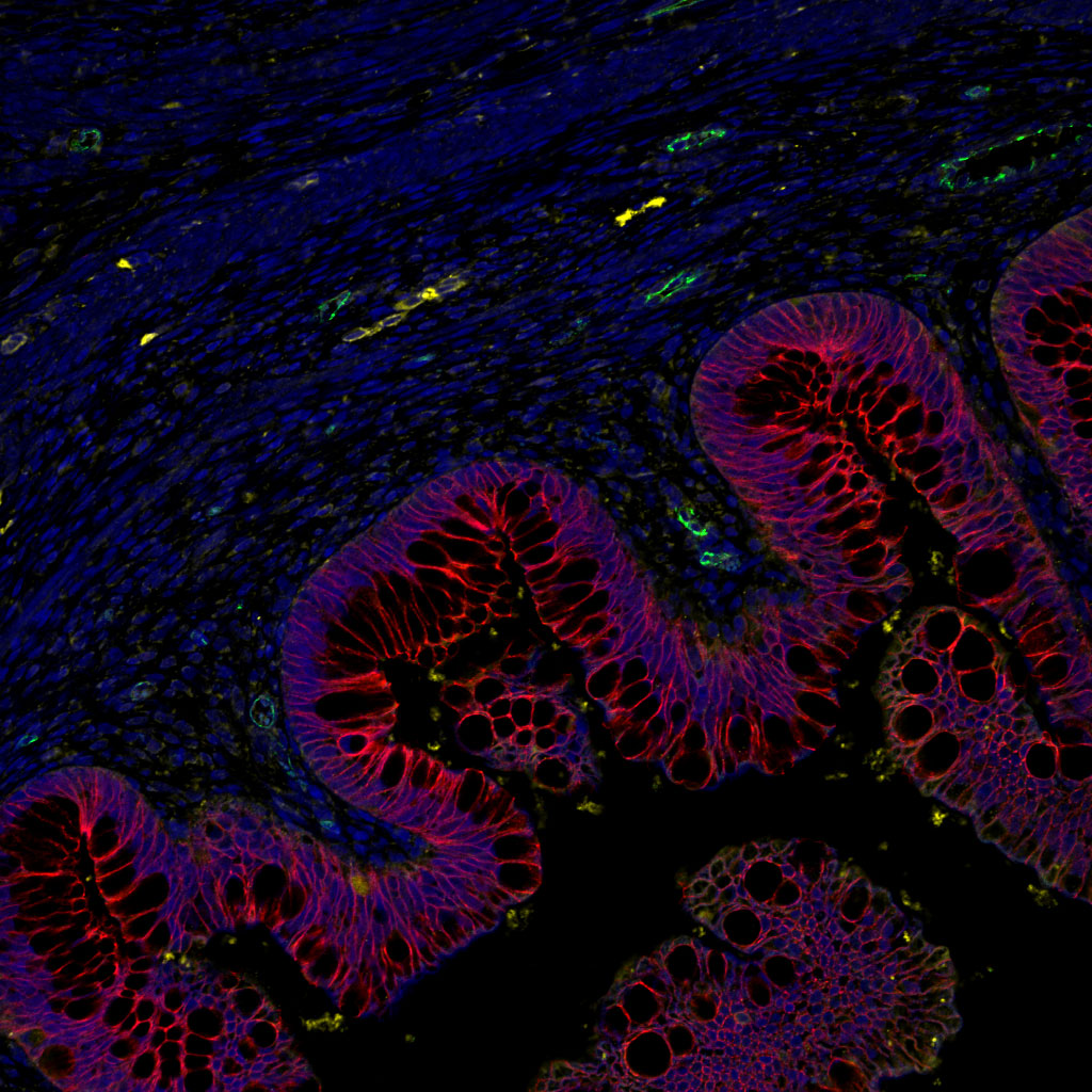
From these 11 chemicals, four mixtures were developed and tested on human fetal testes. The experimental results for these mixtures corroborated the mathematical predictions, for a number of components greater than 3. This demonstrates that the model developed by the authors of the article is capable of illustrating cocktail effects, for the first time in a human organ, and that the combined effects observed can be predicted mathematically.


Finally, the authors of this article were able to measure the exacerbation of the individual effects of each of the chemicals in the mixture. In other words, they were able to provide a response to the question “how much more powerful is the chemical in a mixture than alone?” by showing that this exacerbated potency varies from a factor of 10 to 1,000, depending on the chemical in question. 


According to Bernard Jégou, Irset director, Inserm researcher, research director of the EHESP-School of Public Health and the lead on this study, Pierre Gaudriault, pharmacist and doctor at the Université de Rennes 1, and Inserm researcher Séverine Mazaud-Guittot, the conclusions of this work, which was supported by the French Agency for Food, Environmental and Occupational Health & Safety (ANSES), must be taken seriously: “there is a very precise critical window during the 1st trimester of fetal development during which simultaneous exposure to weak doses of multiple endocrine disruptors may represent a risk to the development of the child’s genitals and reproductive system. It is particularly concerning as the individual potency of these chemicals can be exacerbated by up to a factor of 1,000. All the experimental data from different models lead us toward the same conclusions. This experimental proof of concept study shows that intensifying research into identifying the real mixtures to which individuals are exposed, and testing the effects on appropriate models, is a necessity.”

[1] Research Institute for Environmental and Occupational Health; Inserm; EHESP-School of Public Health, Université de Rennes 1.

A New Avenue to Explore in the Fight Against Sepsis

©Fotolia

Inserm researchers have succeeded in producing a human protein in laboratory conditions and to use it against bacterial infections and for the treatment of sepsis. Sepsis is a systemic inflammatory response by the body to a serious infection. Should the inflammation reach a critical stage (septic shock), the condition becomes life-threatening. Sepsis kills one person every 3 to 4 seconds worldwide. This research, published in Scientific Reports, therefore represents a serious avenue to explore in the fight against a condition that currently remains a medical emergency.


In developed countries, there are 377 cases of sepsis per 100,000 inhabitants. Each year, it is responsible for the deaths of 6 million infants. In France, the mortality rate for sepsis is 27%, which rises to 50% for the most severe form of the infection. The number of cases is expected to double in 50 years’ time, mainly as a result of population aging (source:  Institut Pasteur). On the global scale, the situation is so serious that WHO delegates decided to recognize sepsis as a major public health concern at the World Health Assembly held in Geneva in May 2017. The next World Sepsis Day, an initiative of the Global Sepsis Alliance, will take place on September 13, 2017.

In the majority of cases, sepsis involves an infection caused by Gram-negative bacteria naturally present in the body (generally in the intestine) that become toxic in susceptible individuals. The toxic part of the bacterium is found on its outer membrane in the form of a lipopolysaccharide complex, known as an endotoxin.

The specific chemical aspect of these endotoxins formed the starting point of the study conducted by the Inserm researchers.  Indeed, previous studies have shown that plasma phospholipid transfer protein (PLTP) is able to bind to endotoxins located on the outer membranes of bacteria and even transport them to the liver. When infection is present, this protein therefore appears to play a role in eliminating the endotoxins.

The research team, with the help of an American team, studied a genetically engineered mouse model which had the particularity of no longer expressing the PLTP gene to verify this hypothesis. When these mice were injected with bacterial endotoxins, the researchers observed that they died without being able to fight the infection generated. Hence their hypothesis that PLTP presents previously unknown and potentially major importance in the domain of innate immunity.

The challenge was then to obtain sufficient quantities of human PLTP in order to conduct therapeutic tests to demonstrate its ability to counteract the effects of these endotoxins. For this, the researchers approached the “Development and Reproduction Biology” joint research unit at Inra, which is able to produce this protein in the milk of transgenic female rabbits.

Once obtained, the researchers tested the ability of PLTP to fight the inflammatory response in mice with sepsis. Quite small quantities of PLTP were all it took to obtain considerable improvements in the health of these mice. “However, our ultimate objective was to understand how it all works,” summarizes Laurent Lagrost.

Continuing their work, the researchers demonstrated that PLTP can block the proliferation of bacteria by weakening their outer membranes. They also observed that PLTP, in addition to being able to neutralize the activity of these endotoxins, can also disintegrate them before transferring them to lipoproteins. These lipoproteins, usually simple cholesterol transporters, have the ability to transform into emergency vehicles to escort the endotoxins to the liver for elimination via the biliary route.

“Unless we endogenously neutralize, within the body of the individual patient, the bacterial endotoxins that will be responsible for the inflammatory response and the entire resultant cascade of harmful effects, we cannot definitively resolve the problem. However, it appears that PLTP is able to neutralize these endotoxins and detoxify the blood, at least in mice,” conclude the researchers.

This approach is part of an original concept of biomimetics in which “the more we copy nature, the closer we get to the truth,” they add.

An antidiabetic drug moves a step forward

In humans, apelin is able to regulate blood sugar levels and increase the sensitivity of cells to insulin. These two observations have paved the way for a clinical trial led by Inserm researchers from Toulouse, and represent a promising step forward for the development of a new treatment for diabetes, in particular type 2 diabetes.

This work has been published in the journal Diabetes, Obesity and Metabolism


 It is a journey that began over 10 years ago. A classic research story that demonstrates the long road between discovering a therapeutic molecule and its possible use in humans. The potential use of apelin was demonstrated in 2008 by university professor Philippe Valet and his Inserm team. This ubiquitous molecule (it is found throughout the body) can, if necessary, regulate the body’s blood sugar level instead and in place of insulin. However, this rescue pathway is only activated if the main pathway does not function properly.

Normally, sugar from food is stored in the liver, muscle, and adipose tissue, and is released as and when the body requires. This process is however dependent on the action of insulin, which “captures” sugar for storage. If insulin does not function properly, it leads to diabetes (increased blood sugar levels). Either it is not produced by the body at all: this is type 1 diabetes. Or the insulin receptors located on the surface of the liver, muscle, and adipose tissue cells become desensitized: this is type 2 diabetes. This results in two problems: the levels of circulating glucose are too high, and in time this becomes harmful to the body.

After discovering this alternative pathway, which enables another way of absorbing sugar, the researchers soon had the idea of stimulating this natural pathway and producing synthetic apelin.

Today, the researchers report the positive results of a clinical trial in 16 patients that was carried out within the Diabetology Department headed by Professor Pierre Gourdy. Healthy but overweight men were recruited to take part in a study that aimed to prove the efficacy and tolerability of two different doses of intravenously-administered apelin. The first group received a dose equivalent to 9 nmol/kg, and the second group received 30 nmol/kg. The patients’ glycemia was measured before and after the injection.

The results show that the injection of the smallest dose led to better absorption of circulating blood glucose, while administration of the highest dose also led to a demonstrable increase in cell insulin sensitivity. No side effects were observed.

“This is what we call a ‘proof of concept’ study”, explains Philippe Valet. “Although the sample is a small one, the results that we have just obtained encourage us to move on to larger studies in order to confirm them on a larger scale and be able to consider proper marketing authorization.”

This work could notably contribute to research into the treatment of diabetes, which affects over 400 million people around the world.

FGF 19: a New Factor to Fight Skeletal Muscle Wasting

Skeletal muscle wasting, a process commonly associated with aging but also seen with various chronic diseases (obesity, cancer, kidney failure), situations of immobilization (accidents, post-operative periods) or prolonged weightlessness (astronauts), strongly impacts quality of life. Researchers from the Cardiovascular, Metabolism, Diabetology and Nutrition (CarMeN) laboratory (Inserm/Inra/Université Claude Bernard Lyon 1/Insa Lyon) in Lyon (France), led by Hubert Vidal, Inserm Research Director, in collaboration with the team of Dr. Jérome Ruzzin from the Department of Biology of the University of Bergen (Norway), have discovered that a hormone produced by the intestine called fibroblast growth factor 19 (FGF19), is able to increase skeletal muscle mass in mice and increase the size of human muscle cells in culture. The researchers have also shown that FGF19 protects from muscle wasting in various experimental mouse models, highlighting its potential therapeutic value. These results were published in Nature Medicine on June 26, 2017.


Fibroblast growth factor 19 (FGF19), an enterokine (hormone secreted by the intestine) well known for its effects on biliary acid metabolism in the liver, is also able to target other tissues and play a glucose-regulation and lipid-homeostasis role. When investigating the therapeutic potential of FGF19 in metabolic diseases such as type 2 diabetes and obesity, the researchers showed that mice treated with FGF19 for 7 days gained less weight and adipose tissue despite eating more than the untreated mice. Under these conditions, the researchers have shown that skeletal muscle mass and muscle strength increases in the treated animals, thereby identifying for the first time a new role of FGF19.

At the molecular level, the researchers have identified a signaling pathway that mediates the hypertrophic effects of FGF19 in muscle and show that this effect is the consequence of enlarged muscle fiber size, irrespective of the type of fiber.

The researchers then demonstrated the therapeutic potential of FGF19 using various mouse models of muscle wasting, including glucocorticoid-treated mice, genetically-obese mice and aged mice. In each model, they demonstrated the capacity for treatment with FGF19 to maintain or increase muscle mass and strength. “For the first time, this demonstrates a potential use of FGF19 to fight skeletal muscle wasting and possibly also in agronomics to increase livestock muscle mass,” concludes Hubert Vidal and his collaborators. They envisage setting up clinical trials to validate these observations in humans.

Healing a mother’s skin lesions using her fetal cells?

Schwangere Frau liegend betrachtet Ultraschallbilder

©Fotolia

Since the end of the 20th century, it has been proven that women who have been pregnant (including those who have miscarried or aborted) store the fetal cells of their children in their bone marrow for at least 50 years. The team of Professor Selim Aractingi – Paris Descartes Faculty of Medicine, Cochin Hospital (Paris hospital group) Dermatology Department, “Saint Antoine Research Center” Inserm joint research unit, and Université Pierre et Marie Curie – have recently proven that it is possible, in mice, to mobilize these fetal cells to accelerate the healing of chronic skin wounds. These results were published in Nature Communications on May 18, 2017.

Fetal cells transferred to mothers have the particularity of being potentially beneficial in the event of a health problem. For example, research teams have observed that thyroid or liver problems in pregnant women caused this cell type to participate in the regeneration of these organs. This is known as microchimerism because the mother mobilizes cells which, although not her own, are contained in her body in very small quantities (non-self cells) to accelerate the repair process of a damaged organ.

However, the signals sent by the mother to the fetal cells nested in the bone marrow, enabling them to mobilize and aid maternal healing, had remained unelucidated until now. Professor Selim Aractingi and his team recently made a discovery in this respect: “we have identified a molecular signaling pathway called Ccl2/Ccr2”, he explains. The next objective was then to try to potentiate this effect by artificially activating this signaling pathway.

This research has shown that injecting small quantities of this molecular pathway into the chronic wounds of a mouse that had long since given birth, results in healing at the same rate as that of a normal wound. This is made possible by mobilizing a specific population of fetal progenitor cells by Ccl2/Ccr2.

“Our concept involves natural fetal stem cell therapy”, continues Professor Aractingi. In that respect, it differs from more traditional cell therapy techniques in which cultured cells are injected into diseased tissue. The one restriction of this technique is that it can only benefit mice that have given birth. The same methods have been tested in mice that had never gestated and they were not shown to be effective.

The prospects offered by this study are very interesting because we can hope to ultimately reproduce this type of therapy in women. “Some testing is still required, but we are hopeful that this will lead to beneficial treatments for women who have been pregnant relatively soon” concludes Professor Aractingi.

Microbiota and food contaminants : a mycotoxin amplifies the genotoxic action of a gut bacterium

Darmflora

©Fotolia

Researchers at the French National Institute for Agricultural Research (Inra) and their partners[1] have done animal studies on the consequences of having a certain group of microbiota bacteria and a common food contaminant, deoxynivalenol (DON), present in the gut simultaneously. They show that the presence of this mycotoxin enhances the genotoxicity of the bacteria, i.e. it increases the number of DNA strand breaks in intestinal cells, a phenomenon that can lead to the emergence of malignant cells. This work raises the question of synergy between food contaminants and the intestinal microbiota with respect to the process of colorectal carcinogenesis.

The gut microbiota in humans contains some 100,000 billion highly diverse bacteria. One species, Escherichia coli, is very common, and contains different groups. E. coli group B2 bacteria produce a genotoxic substance, i.e. a product that damages the DNA of intestinal cells, known as colibactin. An increase has been noted in the number of group B2 bacteria in the gut microbiota of populations from industrialised countries.

Mycotoxins are the most common natural contaminants present in human and animal food. One of these, deoxynivalenol (DON), is produced by moulds from the Fusarium family, which mainly develop in cereals. The human populations in Europe and North America are widely exposed to it in their food. In France and Europe, exposure of some fractions of the population, especially children, exceeds the toxicity reference values for this toxin.

The Inra researchers and their partners conducted in vitro and in vivo animal studies to see what happened when colibactin-producing Escherichia coli and DON were simultaneously present in the gut.

In animals colonised with colibactin-producing bacteria and exposed to DON in their food, the DNA damage to intestinal cells was significantly greater, compared with animals not producing colibactin. They thus show that the presence of the mycotoxin enhances the genotoxicity of group B2 E. coli.

These first results provide new data regarding possible synergy between food contaminants and the gut microbiota. The researchers will continue to work to elucidate the mechanism involved in this enhanced genotoxicity in the presence of DON, and studies are planned to extend the observations up to an advanced stage of colorectal carcinogenesis.

[1] Inra’s partners in this work: (Inserm / Toulouse III – Paul-Sabatier University, National Veterinary School of Toulouse [ENVT])

Effects of the gut microbiota revisited

cp-serino-synopsis-1

© Matteo Serino

Matteo Serino, Inserm Research Fellow at the Digestive Health Research Institute (IRSD, an Inserm/University of Toulouse III Paul Sabatier/National Veterinary College of Toulouse/Inra joint research unit), and his collaborators show that alteration of the intestinal microbiota, whether nutritional or genetic in origin, may have beneficial effects on liver metabolism. These results contradict results previously obtained in the area, which showed that transferring an altered microbiota inevitably had negative consequences for health. They have just been published in Molecular Systems Biology.

The gut microbiota is involved in the occurrence of metabolic diseases such as obesity and liver diseases. However, the molecular mechanisms are still incompletely known, and its role as a cause or consequence of these diseases remains a matter of debate.

To understand this, Matteo Serino and his collaborators transferred the gut microbiota from obese mice to wildtype mice fed a normal diet and unexposed to any antibiotic treatment. Indeed, it has been shown that antibiotic treatment prior to transplantation of the gut microbiota could limit the occurrence of metabolic disease, and affect the efficacy of gut microbe transfer.

The researchers observed that on a normal diet, mice that received a microbiota from obese mice underwent a sharp reduction in liver glucose production and fasting blood glucose. Transfer of the microbiota from obese mice not only modified the gut microbiota, but also the function of the bacteria (or microbiome) of the recipient mice.

Unexpectedly, when mice were transplanted with a microbiota from obese mice, and then fed a high-fat diet, their liver metabolism and overall carbohydrate metabolism seemed to be protected. Moreover, their fat mass was less developed and their fat cells were smaller in size.

Conversely, transferring a microbiota from lean mice did not affect liver glucose production.

These research results show that alteration of the microbiota, subsequent to a high-fat diet, is not always harmful. Insofar as the intestinal barrier is intact and the immune system functional, an altered gut microbiota may even protect against the harmful effects of a high-fat diet. “Our observations have opened a new debate on the role of alteration of the gut microbiota in the occurrence of metabolic diseases,says Matteo Serino.

A molecule to regenerate insulin-producing cells in type 1 diabetic patients

illustration-en

Inserm researchers led by Patrick Collombat at Unit 1091,“Institute of Biology Valrose” (Inserm/CNRS/Nice Sophia Antipolis University), show that GABA, a neurotransmitter that is sometimes used as a dietary supplement, can induce the regeneration of insulin-producing cells. This discovery, confirmed in mice and partially validated in humans, gives new hope to patients with type 1 diabetes.

This research is published in the journal Cell.

Type 1 diabetes is a disease characterised by the selective loss of cells that produce insulin, a hormone that lowers blood sugar levels upon sugar intake. These cells are called pancreatic beta cells. Discovering how to regenerate these cells is a major research challenge, as current treatments are not always sufficient in preventing (serious) complications.

Scientists have shown in previous studies that it is possible to regenerate pancreatic beta cells by genetically transforming cells that resemble them: the glucagon-secreting alpha cells. This approach involved the forced activation of the Pax4 gene in all alpha cells. The results also proved that these alpha cells were continuously regenerated and again converted into beta cells, leading to a massive increase in the number of beta cells. “This advance is significant, but it is not possible to carry out this approach in humans,” explains Patrick Collombat, Inserm Research Director. To eventually translate their findings to human, the scientists therefore initiated a search for compounds mimicking the effects of the Pax4 gene.

In this new study, the research team demonstrates that this effect can be induced with no genetic modification using GABA, a neurotransmitter that is naturally present in the body and also available as a dietary supplement.

In mice, GABA induces the continuous, yet controlled, regeneration of pancreatic alpha cells and their transformation into insulin-producing beta cells. The regenerated cells are functional and can cure chemically-induced diabetes multiple times.

With regards to humans, researchers observe that in pancreatic islets (which contain both alpha- and beta-cells) treated with GABA, the number of glucagon-producing alpha-cells is decreased by 37% while a 24% increase in insulin-producing cell count is noted, suggestive of a conversion of alpha-cells into beta-cells.

Finally, by transplanting the equivalent of 500 human islets to mice, the same results are obtained after supplementing the animals for 1 month with GABA (daily). These results are truly encouraging for a putative application in humans. Accordingly, a pilot clinical trial will soon be initiated to determine whether GABA may effectively help patients with type 1 diabetes.

These studies received financial support from ERC and the Juvenile Diabetes Research Foundation.

The cause of uncombable hair syndrome identified

Surprised disheveled preschooler girl with long hair

In 1973, the rare syndrome of uncombable hair or ‘pili trianguli et canaliculi‘ was described by a Toulouse dermatologist. More than 40 years later, Michel Simon, Inserm research director his colleagues at the ‘Epidermal Differentiation and Rheumatoid Autoimmunity’ Unit [UDEAR] (Inserm/CNRS/Toulouse III – Paul Sabatier University) have identified its genetic cause. These results are published in The American Journal of Human Genetics.

Uncombable hair syndrome is a rare disease of the hair, the prevalence of which is unknown. It generally begins during childhood between 3 months and 12 years. Dry and unruly, the hair of affected children becomes gradually silver-blond or straw coloured. Hairs stand up on the scalp and grow in all directions. It is impossible to comb it or to flatten it with a comb. In detail, scanning electron microscopy reveals a longitudinal groove running their entire length, with a triangular or kidney-shaped cross-section. However, this syndrome is not disabling and undergoes spontaneous at the end of childhood.


uncombable-hair

he researchers, working with a team from the Human Genetics Institute at Bonn University and dermatologists or geneticists from 7 different countries, have discovered that the disease is due to recessive mutations of a trio of genes that contribute to forming the hair: the gene coding for one of its structural components, trichohyalin (TCHH); or two genes coding for enzymes that take it in turns as target: peptidyl-arginine deiminase 3 (Pad3) and transglutaminase 3 (TGase3).

Furthermore, the researchers have also shown, in mice, that inactivating the Pad3 gene alters the shape of the fur and whiskers of animals, as had already been reported in TGase3-deficient mice.

In conclusion, the absence of TCHH or failure of the biochemical cascade that results in stiffening the hair stem are responsible for the hair formation abnormalities characteristic of uncombable hair syndrome or ‘pili trianguli et canaliculi‘.

These results, as well as describing the molecular cause of the disease and enabling better diagnosis, provide knew knowledge about the hair and the mechanisms of its formation£ concludes Michel Simon, Inserm research director.

For further information

Although extremely rare, the syndrome has long been known. It was brought to public awareness by the famous literary figure ‘Struwwelpeter’ created by children’s author Heinrich Hoffmann in 1845. The book was subsequently translated into English by Mark Twain as ‘Slovenly Peter’. Although he never said so, one might even think that it inspired director Tim Burton to make his film Edward Scissorhands.

Functional human intestine grown from stem cells

photo-mahe

Cross-section of human intestinal organoid with enteric nervous system after transplantation in mice.*

(Copyright CCHMC – Wells/Helmrath labs)

American researchers at the Cincinnati Children’s Hospital Medical Center and French researchers from Inserm (joint research Unit 913 “Neuropathies of Enteric Nervous System and Digestive Diseases”, Nantes) have succeeded in generating a functional human intestine using pluripotent human stem cells. This significant breakthrough was achieved by cultivating human intestinal tissue using nerve cells. Details of their work are published online on 21 November 2016 in Nature Medicine.


The gut is an essential organ of the human body. It is the main interface for exchanges with the external environment and has a surface area equivalent to two tennis courts. Given its importance, the gut has its own nervous system and has been commonly known as “the second brain”. The enteric nervous system controls many functions, which include mixing and propelling food along the digestive tract, hormone secretion and epithelial permeability. Disruptions of this system are the cause of many diseases. Poor gut function can actually affect intestinal muscle contraction. This plays a role in triggering abdominal pain, diarrhoea, constipation and, in serious cases, leads to functional obstruction (intestinal occlusion) that requires surgery.

Researchers have developed an innovative approach to engineering tissue involving the use of stem cells to create a functional human intestine that can be used to study intestinal diseases.

First in vitro studies — finding the right balance

To do this, the scientists successively added a cocktail of molecules designed to promote the differentiation of human pluripotent stem cells into intestinal tissue. The process was virtually the same used in 2010 and 2014 by the same laboratory that succeeded in developing the first generation of human intestinal tissue. However, the intestinal tissue did not possess an enteric nervous system through this approach alone, which is essential for absorbing nutrients and eliminating waste through the digestive tract.

At the same time and in order to develop a functional nervous system, researchers created embryonic nerve cells known as neural crest cells. These cells were manipulated to form cells that are enteric neural cell precursors. “The difficulty with this step was to identify how and when to incorporate the neural crest cells into the developing gut that was previously created in vitro”, explains Maxime Mahé, first co-author of this work and recently recruited as a junior research associate by Inserm.

The co-culture of intestinal tissue and enteric neural cell precursors resulted in human tissue that resembled a developing fetal intestine. This led to the first generation of complex and functional “mini-intestines” (known as intestinal organoids) created entirely from human pluripotent stem cells.


Conclusive in vivo trials

The next challenge was to transplant these functional organoids into a living organism, in this instance, lab mice that lacked an immune system. This step allowed scientists to observe tissue development and function in vivo. Data from this study show that the tissues function and are structured in a way that is remarkably similar to the human intestine. They develop and ensure intestinal functions, such as the treatment of nutrients. Finally, these tissues exhibit motor functions similar to peristalsis, i.e. the series of muscle contractions that move food through the digestive tract.

Researchers then used this technology to study a rare bowel disease — Hirschsprung Disease — a disorder where the rectum and colon do not develop a nervous system, which leads to constipation and intestinal occlusion. A lethal form of Hirschsprung disease is caused by a mutation of the PHOX2B gene. In vitro tests in mice have made it possible for researchers to show that the mutated PHOX2B gene causes significant deleterious changes in the innervated intestinal tissues.

“Our work marks an essential step in understanding human digestive diseases where few models exist. This new technology offers a screening platform for new intestinal treatments. This technology is still in its infancy. However, an approach towards regenerative and personalised medicine is possible, particularly with regards to patient-specific intestinal transplantation”, explains Maxime Mahé.

This discovery yields two significant research possibilities. Firstly, intestinal disorders can be modelled and studied using three-dimensional and functional human tissue involving patient-specific cells. Secondly, new treatments can be tested using this functional human intestine before proposing clinical trials in humans.


In this image we can see the intestinal epithelium in red (E-cadherin) with lumen opening. The tissue underlying the epithelium is rich in blood vessels (green, CD31) and has neurons (yellow, TUBB3) that are also derived from human pluripotent stem cells. The observed tissue is incredibly similar to normal human tissue.

Rituximab effective in the treatment of membranous glomerulonephritis

A national trial on membranous glomerulonephritis in 80 patients was coordinated by Prof. Pierre Ronco, of the Department of Nephrology and Dialysis at Tenon Hospital AP-HP, of the Inserm Unit “Rare and common kidney diseases, matrix remodelling and tissue repair” [1] and Pierre and Marie Curie University, and by Dr Karine Dahan, of the Nephrology Day Hospital at Tenon Hospital AP-HP. This serious autoimmune disease is the most common cause of nephrotic syndrome in adults. In 30% of cases, it progresses to very severe renal failure. Conducted in collaboration with Prof. Tabassome Simon, from the Department of Clinical Pharmacology and East Paris Clinical Research Centre at Saint Antoine Hospital, AP-HP, this trial shows, for the first time, the efficacy and safety of rituximab in treating the disease.

This work was published on 27 June 2016 in the Journal of the American Society of Nephrology.


Membranous glomerulonephritis is a rare autoimmune disease (one new case recorded per year per 100,000 inhabitants), in most cases caused by antibodies directed against a protein (PLA2R) located in the renal filter (the glomerulus). The immunosuppressive treatments – aimed at attenuating this immune reaction of the body – that have been used until now have shown some efficacy, associated, however, with considerable toxicity: risks of infection, fertility problems, subsequent development of cancer or impaired renal function.

Rituximab is a monoclonal antibody specifically directed against the B lymphocytes that produce the toxic antibodies. Until now, its safety and efficacy had not been demonstrated.

In this context, Prof. Pierre Ronco and Dr Karine Dahan conducted a study in 80 patients with a severe form of membranous glomerulonephritis at Tenon Hospital AP-HP. The patients were enrolled from January 2012 to July 2014 in 31 nephrology departments throughout France, including 9 departments of Paris public hospitals (AP-HP) [2], with annual follow-up for two years.

This randomised study made it possible to compare the efficacy of the standard treatment, known as “antiproteinuric,” with the same treatment combined with 2 intravenous infusions of rituximab (375 mg/m2) given at a one-week interval. Patients were observed for the occurrence of immunological remission (disappearance of antibodies), clinical remission (reduction or disappearance of proteinuria) and adverse effects of the treatment.

Results showed that rituximab had a positive effect on immunological remission (50% from 3 months) and clinical remission (with 64% of patients entering remission before the end of the study), with corresponding values of only 12% and 34% respectively in patients given the antiproteinuric treatment alone.

The percentage of remission was similar to that obtained with other immunosuppressive treatments, but with a much lower therapeutic risk, since the number of adverse events was the same in both treatment groups (with or without rituximab).


“This study contributes a very important element to the debate surrounding immunosuppressive treatments in membranous glomerulonephritis,” explains Prof. Pierre Ronco.

“In clinical terms, it favours the use of rituximab as a first-line treatment in severe forms, with very regular monitoring of the level of anti-PLA2R antibodies in these patients.”

This study will provide a basis for other protocols aimed at increasing the percentage of clinical and immunological remission without increasing the rate of adverse effects. It is likely that some patients did not respond to the treatment because the rituximab leaked into the urine. These protocols will therefore include the use of higher or more frequent doses, and further IV infusions in patients who maintain high antibody levels.

fermer