Menu

(French) Légionellose : reprogrammation inédite des cellules hôtes à l’avantage de la bactérie Legionella pneumophila

The surprising ability of blood stem cells to respond to emergencies

A research team of Inserm, CNRS and MDC lead by Michael Sieweke of the Centre d’Immunologie de Marseille Luminy (CNRS, INSERM, Aix Marseille Université) and Max Delbrück Centre for Molecular Medicine, Berlin-Buch, today revealed an unexpected role for hematopoietic stem cells: they do not merely ensure the continuous renewal of our blood cells; in emergencies they are capable of producing white blood cells “on demand” that help the body deal with inflammation or infection. This property could be used to protect against infections in patients undergoing bone marrow transplants, while their immune system reconstitutes itself. The details of the research is published in Nature on april 10th 2013.

Macrophages

Cells in our blood feed, clean and defend our tissues, but their lifespan is limited. The life expectancy of a red blood cell rarely exceeds three months, our platelets die after ten days and the vast majority of our white blood cells survive only a few days.

The body must produce replacement cells in a timely manner. This is the role of hematopoietic stem cells, more commonly called blood stem cells. Nestled in the core of the bone marrow (the soft tissue in the center of long bones such as the chest, spine, pelvis and shoulder), they dump billions of new cells into the bloodstream every day. To accomplish this strategic mission, they must not only multiply but also differentiate, i.e. to produce specialized white blood cells, red blood cells or platelets.

For many years, researchers have been interested in how this process of specialization is triggered in stem cells. Michael Sieweke and his team previously discovered that the latter do not engage randomly in a particular differentiation pathway but “decide” their fate under the influence of internal factors and signals from the environment.

An important issue remains: how do stem cells manage to respond appropriately to emergencies? For example, are they able to meet the demand by producing white blood cells like macrophages to eat microbes during infection?

Until now, the answer was clear: the stem cells could not decode such messages and were content to differentiate randomly. Michael Sieweke’s team has demonstrated that, far from being insensitive to these signals, stem cell perceive them and in return manufacture the cells that are most appropriate for the danger that is faced.

“We have discovered that a biological molecule produced in large quantities by the body during infection or inflammation directly shows stem cells the path to take,” said Dr. Sandrine Sarrazin, Inserm researcher, co-author of the publication. “As a result of this molecule, called M-CSF (Macrophage Colony-Stimulating Factor), the switch of the myeloid lineage (the PU.1 gene) is activated and the stem cells quickly produce the cells that are best suited to the situation such as macrophages.”

Now that we have identified this signal, it may be possible in the future to accelerate the production of these cells in patients facing the risk of acute infection,” said Dr. Michael Sieweke, CNRS Research Director. “This is the case for 50,000 patients worldwide each year* who are totally defenseless against infections just after bone marrow transplantation. Thanks to M-CSF, it may be possible to stimulate the production of useful cells while avoiding to produce those that can inadvertently attack the body of these patients. They could therefore protect against infections while their immune system is being reconstituted”.

About the discovery

This seemingly simple discovery is quite original, both in its approach and by the technology it required. To reach their conclusions the team had to measure the change of state in each cell. This was a double challenge: the stem cells are not only very rare (there is only one stem cell per 10,000 cells in the bone marrow of a mouse), but they are also completely indistinguishable from their descendants.

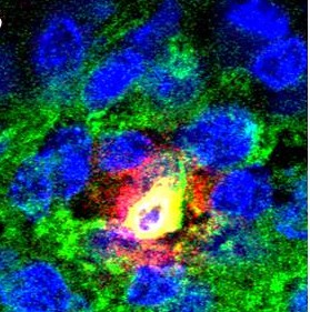
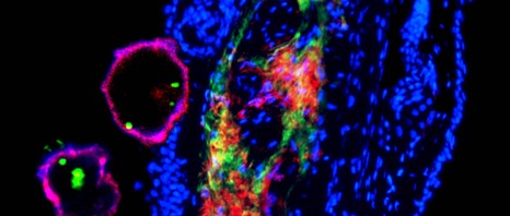
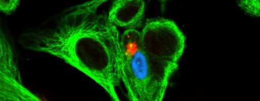
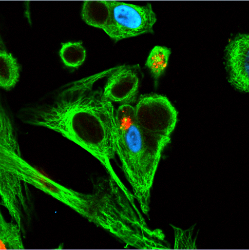
“To differentiate the protagonists we used a fluorescent marker to indicate the status (on or off) of the myeloid cell switch: the protein PU.1. First in the animal, then by filming the accelerated cell differenciation under a microscope, we showed that stem cells “light-up” almost immediately in response to M-CSF,” said Noushine Mossadegh-Keller, CNRS assistant engineer, co-author of this publication. “To be absolutely sure, we recovered the cells one by one and confirmed that the myeloid genes were activated in all the cells that had turned green: once they perceived the warning message, they changed identity.”

Is the mother’s immune system capable of protecting the foetus from cytomegalovirus infection?

An infection from the cytomegalovirus (CMV), a virus belong to the herpesviridae family, usually goes unnoticed in a healthy subject, but in pregnant women, it can be harmful to the foetus. Nabila Jabrane-Ferrat, research officer of the CNRS Physiopathology Centre at Toulouse Purpan (Inserm unit / université Toulouse III – Paul Sabatier / CNRS), working with The “Immunity, Gestation, Therapy” research team led by Philippe Le Bouteiller, an Inserm research director, has demonstrated that certain immune cells from the mother located at the foeto-maternal interface are capable of fighting the infection in order to protect the foetus. This work was published in the review Plos Pathogens on April 4, 2013.

Pregnancy induces major modifications in the uterus that are necessary to provide the needs of the foetus. Following the implantation of a fertilized egg, the wall of the uterus (the endometrium) is massively infiltrated with a specific population of immune cells known as decidual Natural Killer cells (dNK), that come from the mother.

The functions of these immune cells are finely regulated during pregnancy. In the endometrium, their function is not to kill, but to protect to allow successful implantation of the embryo. dNK cells release soluble factors that help the embryo to implant into the maternal tissue. They contribute to exchanges between the mother and the fœtus and are in direct contact with the placenta. By providing an enriched microenvironment, dNK cells play a major part in pregnancy.

The authors of the study published this month wondered whether these cells that are “armed to kill” could have their gregarious instinct aroused in the event of an attack by a pathogenic agent. To this end, they studied the action of the dNK cells when the mother was infected by CMV during pregnancy.

Congenital infection by the CMV is a major public health problem that affects between 0.2 and 0.5 percent of newly-borns in France and is a known cause of foetal fatality. CMV contamination occurs by close contact with affected persons or healthy carriers. Therefore, pregnant women must respect the classic rules of hygiene to avoid infection during their pregnancy, since the risk of foetal contamination is between 30 to 50%.

There are no clinical signs of CMV in a healthy adult; but it is dangerous for the foetus if the mother becomes infected. The virus passes into the mother’s bloodstream, crosses the placental barrier and infects the cells in the foetus, causing serious damage, even death to the foetus. Congenital CMV infection induces placental development problems and remodelling of the uterine arteries that can be detected by ultrasound scanning.

Researchers have observed phenotype and functional changes in maternal dNK cells. In addition, analysis of tissues from terminations of pregnancy due to CMV has shown that dNK cells are capable of migrating to the actual site of the infection in the placenta.

Quite unexpectedly, the researchers found that dNK cells become cytotoxic in order to kill off infected cells and fight infection.

For Cover

© N. Jabrane-Ferrat / Inserm. In red and greed above: the NK cells attempting to make their way to the infected cell (in glue and green).

These results suggest that dNK cells could protect the foetus from maternal CMV infection. They open the way for the development of new treatments. The next step is to figure out how to trigger a massive cytotoxic reaction by the dNK cells when faced with the virus.

Characterisation of the immune cells capable of controlling HIV infection

How is it that certain patients infected by HIV have immune cells that are capable of controlling the infection? Victor Appay, INSERM Research Director (Mixed INSERM  Research Unit 945 “ Immunity and Infection” / Université Pierre and Marie Curie / Hôpital Pitié-Salpêtrière (AP-HP)) and his colleagues have succeeded in identifying the molecular characteristics of certain T lymphocytes that have the specific property of being able to detect and control HIV and its mutant versions. Their work was published in 22 March 2013 in the Immunity journal and has received support from the ANRS. 

Today, according to the OMS, 34 million people are infected with HIV, yet only 1.3 million sufferers in the poorest countries are being treated and 6,800 people a day are infected by HIV.

The Human Immunodificiency Virus (HIV) attacks the cells of the immune system and destroys them or renders them ineffectual. Acquired Immunodeficiency Syndrome (AIDS) is the final stage in HIV infection. It may appear after 10 to 15 years.

The inherent danger in a virus lies very much in its ability to develop multiple strategies to escape detection by the immune system. HIV is no exception to the rule. HIV is known to mutate and adapt in each individual and his/her major histocompatibility complex (MHC).

HIV is a good example of how a pathogenic agent can evolve and mutate by creating what is known as a new variant, in order to circumvent its host’s immune system.

In certain cases, however, the immune system is able to react to control these mutant pathogens.

The molecular basis of the process has not been clearly established hitherto, but this is what researchers have now been able to do. Their work makes it easier to under the mutual adaptation mechanism between the immune system and HIV.

In order to better understand how HIV works, Victor Appay, INSERM Research Director, in collaboration with his colleagues at Cardiff University (United Kingdom), Kumamoto University (Japan) and Monash University (Australia), and with the support of the ANRS, studied the cells of patients whose immune response proved effective against the virus and its variants. These patients, all of them from the ANRS Primary Infection cohort, were monitored from the time primary infection with HIV was diagnosed. The detailed study focused on the response caused by the T CD8+ lymphocytes of patients infected by HIV and in whom these cells were particularly well preserved and functional.

In other words, these cells not only managed to control and reduce the replication of HIV but even of its variants, something that is not the case in patients who develop the full-blown disease.

This protective immune response is possible due to certain T CD8+ lymphocytes, that are known as “cross reactive”. They have a receptor on the surface whose structure gives then the special ability to recognise not only the non-mutant virus but even its mutant variants. This discovery is the first to really pinpoint how HIV can be controlled by certain T CD8+ lymphocytes.

The study highlights the complexity of the forces and mechanisms that lead to the development of the virus and the way it alters the immune system during the course of HIV infection. A better understanding of immunological determinants as the basis for control of replication of HIV is essential for the development of effective vaccines. In fact, the choice between immunogens and adjuvants in the development of vaccines against HIV needs to be rationalised in order to encourage the production of T CD8+ lymphocytes that are particularly skilled at recognising the wilder and mutant forms of the virus.

COMBACTE: A new step in the fight against resistance to antibiotics

Antimicrobial resistance represents is a growing problem in public health due the increasing rarity of antibiotics capable of combating resistant bacteria. The COMBACTE project, that has just obtained 195 million euros worth of finance from the Innovative Medicines Initiative (IMI), aims to work to develop new antibiotics and introduce a successful clinical trials platform combining private and public research. 

Developing innovative clinical trials for antibiotics

The COMBACTE (Combatting Bacterial Resistance in Europe) project, resulting from the sixth Call for proposals issued by the IMI, is one of the projects that is part of the “New Drugs For Bad Bugs” (ND4BB) programme. It is the result of the initial association between industry and two academic consortia, the Eu-ACT and INCRAID, run respectively by Marc Bonten of the University of Utrecht and Bruno François of Limoges University Hospital, both being responsible for overall project coordination along with representatives of the EFPIA, Scott White (GlaxoSmithKline) and Seamus O’Brien (Astra Zeneca).

The project will last for seven years and will bring together about 20 partners from all overEurope. It is designed to generate innovative trials to facilitate the registration of new anti-bacterial agents, mainly through the creation of a network of experienced investigators. It will also design and validate tests to support the diagnosis of patients, identify the most suitable treatments and monitor the treatment response.

Much of the project will be devoted to performing clinical trials of anti-infectious medication currently being developed by the pharmaceutical companies involved in the programme. The first antibiotic to be subjected to clinical trials under the COMBACTE programme has been developed by GlaxoSmithKline’s laboratories.

For these purposes, the total budget for the COMBACTE project amounts to nearly 195 million euros, a scale of finance hitherto unequalled in private-public clinical research.

French partners in the European COMBACTE Project

Of the various partners, several French entities are involved in the COMBACTE project.

Dr Bruno François, under the aegis of Limoges University Hospital, will be responsible for coordinating the clinical trials in collaboration with all the European investigator centres and the GSK Group and GSK France (GSK Medical Management France) Research Groups. Dr François will also participate in the overall project management.

INSERM and its Midi-Pyrénées/Limousin directorate headed by Armelle Barelli will be responsible for budget management for all the project’s clinical trials.

ECRIN (European Clinical Research Infrastructures Network) coordinated by INSERM and headed by Professor Jacques Demotes, is an infrastructure whose purpose is to facilitate the setting up of international trials in Europe. ECRIN will be responsible for the management of the project’s clinical trials through its European partners, ensuring coordination between the various national networks.

Dr Laurent Abel (INSERM U980 “Human genetics of infectious diseases”), another French member of the consortium, will participate in the identification in humans of genetic markers affecting the  susceptibility/resistance to bacterial infection and the response to their treatment, along with the other  partners.

Two French networks will also participate in clinical trials  for the COMBACTE project, the Réseau National de Recherche Clinique en Infectiologie (RENARCI) coordinated by Professor Bruno Hoen (Besançon University Hospital), with the support of the Institut Thématique Multi-Organismes “Microbiologie et Maladies Infectieuses” (IMMI) directed by Professor Jean-François Delfraissy, and the CRICS (Clinical Research in Intensive Care and Sepsis) network, headed by Dr Bruno François and Professor Pierre-François Dequin at the Tours University Hospital.  The Groupe pour la Recherche et l’Enseignement en Pneumo-Infectiologie (a Working Party emanating from the Société de Pneumologie de Langue Française) coordinated by Professor Anne Bergeron at the AP-HP Saint-Louis with the collaboration of Dr Muriel Fartoukh at the APHP Tenon will be associated with the CRICS network.

COMBACTE, a unique excellence project with international visibility

COMBACTE is the European public/private partnership set up for the development of drugs.

The development of new antibiotics represents a challenge that justifies the association of several of the major players. By bringing together experts in the various fields (research bodies, universities, hospitals and the pharmaceutical industries) specialising in microbiology, epidemiology, the development of drugs and clinical trials, the aim of COMBACTE is to improve and accelerate the development of antibiotics.

Unique in its scale, ambitious, with benefits that can be expected for patients, public health and research in Europe, COMBACTE has the potential to become the leading light in Europe in the antimicrobial drug development.

The fight against anti-microbial resistance –  the “New Drugs For Bad Bugs” programme

Antimicrobial resistance represents a serious and growing world threat to human and animal health. According to the World Health Organization, “resistance to antibiotics is about to become a public health emergency of yet unknown proportions”. In Europe, resistance to antibiotics is responsible for more than 25,000 deaths annually and the cost of treatment is estimated at 1.5 billion euros annually. New forms of resistance emerge every day, leaving clinicians increasingly devoid of solutions to fight infections. Despite the recognised need to develop new antibiotic weapons, only two new classes of medication have entered the market in the last thirty years.

In November 2011, the European Commission, as part of its Action Plan to combat the increasing threat of antimicrobial resistance, called for “unprecedented collaborate research and development effort to bring new antibiotics to patients” including the launch of the sixth IMI Call for proposals in May 2012 as part of the programme entitled “ New Drugs For Bad Bugs”.

IMI : a unique public-private partnership programme

IMI (Innovative Medicines Initiative) is a unique European public-private partnership between the European Commission and the EFPIA (European Federation of Pharmaceutical Industries and Associations), each contributing one billion euros to finance various projects through issuing tenders.

The aim of the IMI is to propose a coordinated approach to promote the development of safer and more effective treatments for patients by encouraging collaboration between various academic and industrial partners, the public authorities and patient associations and by increasing European competitiveness.

The research leading to these results has received support from the Innovative Medicines Initiative Joint Undertaking (www.imi.europa.eu) under Grant Agreement n°115523, resources of which are composed of financial contribution from the European Union’s Seventh Framework Programme (FP7/2007-2013) and EFPIA companies’ in kind contribution.

(French) Infection VIH par voie sexuelle : élucidation de mécanismes-clé chez l’homme

The HOMAGE (Heart OMics in AGEing) project, coordinated by the French National Institute of Health and Medical Research (INSERM), has been awarded a grant by the European Commission for a 6 year period. The project aims to identify and validate specific biomarkers of heart failure in order to prevent the development of the disease affecting elderly population. 17 research groups from 10 countries will collaborate to investigate new ways of preventing heart failure. The project will use an innovative ‘omic-based’ approach which investigating simultaneously a huge amount of genes, proteins and metabolites.

The 17 partners will meet at Nancy on February 22nd for the kick off meeting of HOMAGE.

Professor Faiez Zannad, Head of the Centre d’Investigation Clinique Pierre Drouin Inserm U9501 and Inserm Unit 1116 based in Nancy, is the project coordinator.  The 12 million euros grant of the European Commission will be dedicated to the HOMAGE consortium for research on heart failure, a serious illness altering myocardial activity which affects more than 6.5 million persons in Europe. Indeed, the prevalence of heart failure is increasing worldwide due to an ageing population as well as a rising trend of risk factors for heart disease such as diabetes, obesity and hypertension.

Heart failure is a major cause of mortality and morbidity in the world and remains the most frequent cause of hospitalization for patients over 65 years old.

The costs related to heart failure have been estimated around 1.5 billion euros per year in France[1].

Photo : ©Serimedis/Inserm

Despite important progress in the treatment including new drugs, new medical devices and innovative disease management programmes, the diagnosis of heart failure is often difficult in older adults with co-morbidities. Screening tests are usually based on blood pressure, glycaemic and cholesterol control. Although they are useful to detect high risk patients, they are limited regarding their sensitivity and specificity. During the past decade, promising biomarkers such as natriuretic peptides have been identified to diagnose heart failure, but their predictive value remains relatively poor. The HOMAGE consortium is willing to validate more specific and more sensitive biomarkers which should facilitate an early detection of the disease in patients at risk.

To achieve this goal, the consortium agreed on the use of an ‘omic-based’ approach. This approach aims to validate promising biomarker candidates by crossing a large volume of data (genomics, proteomics, miRNomics, transcriptomics and metabolomics). This would permit scientists to understand new pathophysiological mechanisms, signaling pathways and identify new therapeutic targets to prevent heart failure.

The HOMAGE consortium will manage cohorts for a total of 30 000 patients. The European researchers will firstly identify biomarker candidates in blood and study their predictive value for heart failure and common co-morbidities associated with ageing (renal impairment, cognitive disorders…). Subsequently, HOMAGE will lead a clinical trial to look for novel treatments of heart failure that can be targeted specifically to those patients at risk.

This trial will allow identifying patients’ omics based biomarker profiles most likely to predict response to treatment with the better benefit/risk ratio, an attempt into personalized medicine.


[1] Fédération Française de Cardiologie

The 17 partners of HOMAGE project : www.homage-hf.eu (en ligne le 21 février)

Inserm, France : http://www.inserm.fr/

Inserm U942/ Biomarqueurs et maladies cardiaques, France

Inserm UMR 1048/ Equipe 7, France

Inserm U744/ Recherche des Déterminants Moléculaires des Maladies Cardiovasculaires, France

Inserm Transfert, France : http://www.inserm-transfert.fr/

European Drug Development Hub, France : http://www.fondationtransplantation.org

ACS Biomarker, Pays-Bas : http://acsbiomarker.com/

Randox Testing Service, Royaume-Uni : http://www.randoxtestingservices.com/

Medical University of Graz, Autriche : http://www.meduni-graz.at/en/

University of Manchester, Royaume-Uni : www.manchester.ac.uk

Fundación para la Investigación Médica Aplicada, Espagne : www.cima.es

University College Dublin, Irlande : www.ucd.ie

University of Hull, Royaume-Uni : www.hull.ac.uk

Maastricht University, Pays-Bas : http://www.maastrichtuniversity.nl/

Istituto di Ricerche Farmacologiche ‘Mario Negri’, Italie : http://www.marionegri.it/mn/en/

Hannover Medical School, Allemagne : http://www.mh-hannover.de

University of Leuven, Belgique : www.kuleuven.be/english/

London School of Hygiene, Royaume-Uni : http://www.lshtm.ac.uk/

Emory University, Etats Unis : http://www.emory.edu

University of Glasgow, Royaume-Uni : http://www.gla.ac.uk/

Collaborative institutions:

The Trustees of Boston University, National Heart, Lung, and Blood Institute’s Framingham Heart Study, Etats Unis

Imperial College, Royaume-Uni

Steno Diabetes Center (Novo Nordisk), Danemark

Harmful effects of bisphenol A proved experimentally

Weak concentrations of bisphenol A are sufficient to produce a negative reaction on the human testicle. This has just been shown experimentally for the first time by René Habert and his colleagues (UMR Cellules souches et Radiations [UMR Stem Cells and Radiation], Inserm U 967 – CEA – Paris Diderot University) in an article that appeared in the journal entitled  Plos One.

Bisphenol A (BPA) is a chemical compound that is included in the composition of plastics and resins. It is used, for example, in the manufacture of food containers such as bottles and  babies’ feeding bottles. It is also found in the protective films used inside food and drink cans and on till receipts where it used as a discloser. Significant levels of BPA have also been found in human blood, urine, amniotic fluid and placentas. Recent studies have shown that this industrial component has harmful effects on reproductive ability, development and the metabolism of laboratory animals. BPA is strongly suspected of having the same effects on humans.

As a precautionary measure, the manufacture and sale of babies’ feeding bottles containing bisphenol A have been banned in Europe since January 2011. This ban will be extended in France to all food containers from July 2015. It will also be important to ensure that in the future, bisphenol A is not replaced by substitutes that have the same action.

Crédit photo : R habert/Inserm

System for culturing fœtal testicles developed by René Habert’s team. The testicles are sliced into tiny fragments then deposited on a filter in the centre of a petri dish. In an appropriate atmosphere, they are able to survive thus for several days, while secreting testosterone.

In an article published in Plos One, René Habert and his colleagues provide the first experimental proof that weak concentrations of bisphenol A are sufficient to produce a negative reaction on the human testicle.

No experimental study has shown hitherto that bisphenol A has a deleterious effect on male human reproduction and the few epidemiological studies that exist remain contradictory.

In collaboration with the Antoine-Béclère Hospital, Clamart1, researchers kept petri dishes of human fœtal testicles alive in the presence of bisphenol A or in the absence thereof, using an original procedure developed by this team. In 2009, this procedure made it possible to show for the first time, that phtalates (a different category of endocrine disruptors2  that are found in PVC, plastics, synthetic materials, sprays, etc.) inhibit the development of future spermatozoa in the human fœtus.

In this new study, researchers observed that exposure of human fœtal testicles to bisphenol A reduces the production of testosterone, and of another testicular hormone that is necessary for the testicles to descend into the sacs in the course of fœtal development. A concentration equal to 2 micrograms per litre of bisphenol A in the culture medium was sufficient to produce these effects. This concentration is equal to the average concentration generally found in the blood, urine and amniotic fluid of the population.

Testosterone is known to be produced by the testicle during the life of the fœtus, imposing masculinisation of the internal and external genitals which, in the absence of testosterone, develops spontaneously in the female direction. Furthermore, it is likely that testosterone also plays a role in the development of the testicle itself. Consequently, the current exposure of pregnant women to bisphenol A may be one of the causes of congenital masculinisation defects (of the hypospadia and cryptorchidism types) the frequency of which has doubled overall in the past 40 years. According to René Habert, “it is also possible that bisphenol A contributes to a reduction in the production of sperm and  the increase in the incidence of testicular cancer in adults that have been observed in recent decades”.

Furthermore, researchers have compared the response to bisphenol A in human fœtal testicles to those in the fœtal testicles of rats and  mice. “We have observed that the human species is far more sensitive to bisphenol A than the rat and the mouse. These results should encourage greater caution in regulatory toxicology in the extrapolation of data obtained on animals to define tolerable exposure thresholds in human health”, explains René Habert.

Finally, the researchers show in this article that Bisphenol A acts through a mechanism that is non-standard and that remains unknown but that it is important to identify in order to better understand the action of endocrine disruptors.

©Fotolia

The role of the innate immune cells in the development of Type 1 diabetes

Julien Diana and Yannick Simoni of the “Immune Mechanisms in Type 1 Diabetes (Inserm/Université Paris Descartes), directed by Agnès Lehuen, have just published the results of their work on type 1 diabetes in the Nature Medicine journal. This is a disease characterised by the self-destruction of the p pancreatic cells that produce insulin. The researchers reveal the role of the innate immune cells, especially the dendritic cells, that cause the activation of the killer T-lymphocytes whose action is directed against the p pancreatic cells. The results obtained in mice make it possible to consider new ways of regulating the auto-immune reaction generated by the innate immune cells.

Type 1 diabetes, or insulin-dependent diabetes, is an auto-immune disease characterised by the destruction of insulin-producing pancreatic β cells that are present in the Islets of Langerhans which are themselves in the pancreas. The peculiarity of this type of diabetes lies in the fact that the cells are destroyed by T lymphocytes that kill the patient’s immune system. This is an auto-immune reaction. Much of the research has highlighted the role of auto-reactive T lymphocytes in the pancreatic β cells. Yet the mechanisms involved in the initial activation of the immune system that triggers the sequence of events leading to the death of the cells are still ill-defined.

The work of the team working on “Immune mechanisms of Type 1 diabetes” (Inserm/Université de Paris Descartes) in NOD (Non-obese diabetic) mice, the model used for studying Type 1 diabetes, reveals the essential role played by the cells of the innate immune system[1] that were not hitherto considered to be involved in diabetes. These cells were known to play a role in other auto-immune diseases such as lupus and psoriasis. In this study, researchers managed to describe the mechanisms initiating the activation of T lymphocytes attaching themselves to the pancreatic β cells.

The innate immune system is normally activated when an infection occurs. This is when the mobilisation and activation of neutrophils and dendritic cells is observed, constituting the first stages of the immune response. Abnormally in NOD diabetic mice, the natural physiological death of pancreatic β cells, occurring as the process progresses, involves an innate auto-immune response in the pancreas. Researchers have provided details of the introduction of a sequence of activation events in these innate immune cells in the Islets of Langerhans.

Activation of the immune system leading to death of the p cells

 ©Inserm / Agnès Lehuen – Juliette Hardy

 The natural deterioration of the β cells (1) leaves cell debris in the tissues that abnormally activate the neutrophils (2). These alarm cells in the immune system warn the dendritic cells (pDC) (3) which in turn cause the production of IFN α interferon, an alarm molecule (4). The interferon α then stimulates the T lymphocytes which, by recognising functional pancreatic β cells, cause these cells to die (5).

Innate immune cells and killer T-cells in the Islets of Langerhans

 ©Inserm / Yannick Simoni

A-B) Fluorescent photographs of a pancreatic islet (green) and close-up (B) of a neutrophil  (yellow) producing an activating molecule (red)
C) Photograph of the Islets in untreated mice in which the killer T-cells (white arrows) can be observed.

“We have observed in mice that treatment prevents activation of the innate immune cells, neutrophils and dendritic cells, warning of the onset of diabetes by inhibiting the appearance of auto-immune T responses aimed at the pancreas” explains Agnès Lehuen, head of the Inserm team.

These results show, for the first time, the important role played by innate immune cells in the sequence of events leading to the onset of Type 1 diabetes. Researchers continue to strive to understand how to regulate the auto-immune reaction produced by dendritic cells without compromising the innate immune system, an essential one in cases of infection. Several routes are being taken to attempt to regulate the production of the INF α alarm molecule that precedes activation of the killer T-cells, for example, by specifically targeting certain activation routes for the pDC dendritic cells.

These therapeutic approaches are currently being tested in other auto-immune diseases such as lupus and psoriasis. Such innovative treatments could be useful in the prevention of Type 1 diabetes. It will first be necessary, however, to perform studies in diabetic and pre-diabetic patients to be able to better understand how the innate immune cells function, something that has not been studied until recently in auto-immune diabetes,” concludes Agnès Lehuen.

The research benefited from a grant from LabEx INFLAMEX as part of investment for the future and for the Ile-de-France region.


[1] System present from birth that makes it possible to initiate an immune response to infection, regardless of the infectious agent involved. It is distinguished from the “acquired” or so-called “adaptive” immune system that is a specific response involving recognition of the infectious agent and the memorising of the infectious event.


(French) L’action du vaccin, dépendante de son mode d’administration : nouvelles pistes

The HOIL1 gene – the cause of a new rare disease

The researcher Capucine Picard, working with the team from Inserm unit 980 “Human genetics and infections diseases”/Université Paris Descartes under the leadership of Jean-Laurent Casanova, along  with researchers from a CNRS/Institut Pasteur laboratory headed by Alain Israël have succeeded in identifying the part played by the HOIL1 gene in cases of paradoxal association of an immune deficiency with a chronic autoinflammatory deficiency and a muscular deficiency in 3 children from 2 different families. This study once more highlights the importance of genetics in the body’s response to infectious agents. These works were published on line in the review Nature Immunology, of 28.10.12.

The science of genetics of infectious diseases arose from the observation that there is a wide variability of resistance to diseases from one person to another; that the same pathology could be fatal to one person, while benign or asymptomatic in others. The study also demonstrated that the predisposition to an infection is due to genetic particularities that result in variations in the molecular mechanisms of the immune response.

The 3 children, 2 of whom were monitored at the Hôpital Necker sick children’s hospital, suffered simultaneously from 3 pathologies: invasive bacterial infections (pneumococcus or other), an autoinflammatory disease (inherited recurring fevers) and amylopectinosis (a muscular deficiency that can affect the cardiac muscles in particular). The fact that 2 siblings suffered from the same symptoms drew the researchers’ attention to the hereditary genetic cause of this disease.

The team then carried out in-depth genetic studies in an attempt to identify the genetic defect responsible for these 3 observed pathologies: mutations of the HOIL1 gene.

Incomplete expression of this gene causes a dysfunction of the immune system. However, what makes this pathology unique is the fact that the genetic defect does not express in the same way, depending on the type of cells involved in the immune response. On the one had, this mutant gene is responsible for an over-reaction of leucocytes, which explains the autoinflammatory disease. On the other hand and quite to the contrary, this same genetic defect inhibits a response from other cells, which explains the susceptibility of these children to bacterial infections.

The HOIL1 molecule, derived from the gene of the same name, is responsible for an instability of the LUBAC complex that plays an important part in transmitting the signal received by the immune system cells in case of infection. This suggests that the genetic defect on HOIL1 in humans is responsible for a knock-on defect in the LUBAC complex, and that the LUBAC complex controls the immune response differently depending on the cell types involved.

Previously, the LUBAC complex had only been studied in mice. This is the first time that this deficiency has been detected in a human. For the moment, only 3 patients inFranceand inItalyhave been identified with this HOIL-1 deficiency. The discovery of this new genetic defect may allow us to identify new patients in other regions of the world.

fermer