Menu

What if the severity of our seasonal influenza were related to our genetic background?

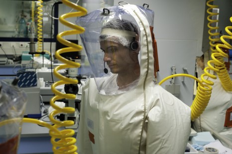
While most of us recover from influenza after a week, it can be a very severe disease, and even fatal in rare cases, with no reason for physicians to have expected such an outcome. By analysing the genome of a little girl who contracted a severe form of influenza at the age of two and a half years, researchers at the Laboratory of Human Genetics of Infectious Diseases (a joint French-American international laboratory), which brings together researchers from Inserm, Paris Descartes University, and physicians from the Paris public hospitals (AP-HP; Necker Hospital for Sick Children), working at the Imagine Institute, and from The Rockefeller University in New York, have discovered that she has a genetic mutation, unknown until now, that causes a subtle dysfunction in her immune system. More generally, these results show that genetic mutations could be the root cause of some severe forms of influenza in children, and indicate in any event that immune mechanisms missing in this little girl are needed for protection against this virus in humans. These results are published in the journal Science Express.
Woman coughing and blowing her nose in autumn

©fotolia

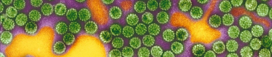
Seasonal influenza is an acute viral infection caused by the influenza virus. It is characterised by high fever, headaches, sore muscles, etc. Apart from vaccination, there is no treatment for it other than symptomatic (pain) treatment. In most cases, patients recover after a week, but in more vulnerable people influenza can cause acute respiratory distress, which is potentially fatal.

The main known risk factors for severe forms of influenza are some acquired comorbidities, such as chronic lung disease. However, the cause of most fatal cases remains unexplained, especially in children.

The absence of cases of severe influenza in patients with known acquired immunodeficiencies, which usually increase susceptibility to infections, is also surprising.

Given these different observations, the researchers at Jean-Laurent Cassanova’s and Laurent Abel’s laboratory, in Paris and New York, therefore formulated a hypothesis whereby severe influenza in healthy children might be the result of genetic errors.

To test this hypothesis, they sequenced the entire genome of a 7-year-old child who had contracted a severe form of influenza (influenza A virus strain H1N1), requiring her admission to a paediatric intensive care unit in January 2011, at the age of two and a half years. At the time, she showed no other known pathology that might have suggested greater vulnerability to the virus than that of other children.

This analysis, combined with analysis of her parents’ genomes, made it possible to show that the little girl had inherited a mutated allele of the gene encoding interferon regulatory factor (IRF7) from both of her parents. The latter is a transcription factor known to amplify the production of interferons in response to viral infection in mice and humans.

In contrast to her parents, in whom the mutation of a single allele of the gene is of no consequence, in the little girl, mutation of both alleles of the gene encoding IRF7 has led to its inactivation. The result: failure to produce interferons, disrupting her system of defence against influenza virus infection in a cascading manner.

By carrying out a comprehensive series of experiments on blood cells, particularly dendritic cells, and by generating lung cells from stem cells taken from the young girl, the researchers provided proof that the mutations observed in this little girl explain the development of severe influenza. Furthermore, this discovery demonstrates that interferon amplification dependent on IRF7 expression is needed for protection against influenza virus in humans. They now need to search for mutations in this or other genes in other children recruited following an episode of unexplained severe influenza.

Based on these initial observations, the researchers at Inserm believe that therapeutic strategies based on recombinant interferons, available in the pharmacopoeia, could help to combat severe forms of influenza in children.

This multidisciplinary study required multiple collaborations in Europe and the United States.

Targeting a host receptor instead of the virus: a new experimental approach against hepatitis C virus.

An international collaboration led by Professor Thomas Baumert (Inserm/University of Strasbourg Joint Research Unit 1110, “Institute for Viral and Liver Disease”) has shown that a monoclonal antibody directed specifically against claudin-1, a liver protein essential for infection by the hepatitis C virus (HCV), enables the prevention and treatment of chronic infection by this virus in an animal model. It turns out that this antibody, which was known to inhibit HCV entry and thereby prevent the initiation of infection, can also eliminate infected cells. This discovery, published in a letter in the Nature Biotechnology issue of 23 March 2015, opens the way to developing an approach to hepatitis C that is not only preventive, but therapeutic as well.

Infection with hepatitis C virus (HCV) leads to cirrhosis of the liver and liver cancer, the second leading cause of cancer death in the world. These complications are major indications for liver transplantation, but HCV reinfection of the transplant is a challenge. To date there is no vaccine, and the new treatments developed recently can be accessed by only a minority of patients worldwide because of their high cost. The development of new preventive and therapeutic strategies therefore continues.

The team directed by Prof. Thomas Baumert (Inserm/University of Strasbourg Joint Research Unit 1110, “Institute for Viral and Liver Disease”), in collaboration with international teams, decided to target a liver protein essential for viral infection instead of targeting the virus. They chose claudin-1, a molecule that is important in the initial steps of HCV infection, and involved in cell-cell contacts.

Using mouse models with humanised liver, the researchers show that a monoclonal antibody directed against claudin-1 can prevent HCV infection by blocking the entry of the virus into liver cells. Surprisingly, the researchers also observed that this antibody enables the treatment of chronic HCV infection by inhibiting the activation of intracellular signalling pathways needed by the virus for survival. As a result, the infected cells disappear and are gradually replaced by uninfected cells.

The advantage of this strategy is that it does not need to be combined with an antiviral agent.

 Moreover, by using different viral strains, the researchers show that it is difficult for the virus to escape from this antibody and develop resistance.

“Claudin-1” is a protein that is usually localised in the tight junctions that are the points of contact between adjacent cells. It is interesting to note that tight junction proteins constitute receptors for other pathogens, such as dengue virus and Shigella species. This innovative approach, employing injection of a monoclonal antibody directed against a protein on the host cell, makes it possible to foresee the development of a vaccine strategy and new therapeutic approaches against HCV, and also against other pathogens that use similar infection mechanisms.

This study received support from the European Union (ERC, INTERREG-IV-Upper Rhine [ERDF], FP7), ANRS (French National Agency for Research on AIDS and Viral Hepatitis), the HepSYS and netRNA Laboratories of Excellence of the French National Research Agency (ANR), ARC Foundation for Cancer Research, IHU Strasbourg MIX-Surg, Wilhelm Sander Foundation, Alsace Region, French National Cancer Institute (INCa), French National Institute of Health and Medical Research (Inserm), French National Scientific Research Centre (CNRS), University of Strasbourg, Ghent University (GOA 01G01712), Flanders Research Foundation (FWO) and Cardiex (Nantes).

An antihypertensive drug improves corticosteroid-based skin treatments

Basic research on blood pressure has led researchers from Inserm (Inserm Unit 1138, “Cordeliers Research Centre”) to obtain unexpected results: drugs used to treat hypertension (high blood pressure) reduce side effects from corticosteroid-based creams used to treat certain skin diseases.

This work is published in the Journal of Investigative Dermatology.

To switch the subtitles of the movie on or off during playback check/uncheck the Subtitles enabled option.

Corticosteroid-based dermatological creams are indicated for the symptomatic treatment of inflammatory skin conditions, such as atopic dermatitis and psoriasis, for example. However, they have frequent side effects, such as a slight burning sensation, and very often end by inducing skin atrophy (thinning of the skin, which becomes fragile), which is inconvenient for the patient, and for which there is presently no treatment.

The researchers from Inserm formulated a hypothesis whereby this harmful effect might be related to the inappropriate activation by these creams of mineralocorticoid receptors located in the epidermis. These receptors, which are present in the kidney, heart, eye, and certain neurons in particular, reacted with aldosterone, a hormone that regulates the blood pressure. Moreover, previous studies also showed them to be highly sensitive to corticosteroids.

Application of corticosteroids to cultured skin causes it to become thinner: in 6 days, the thickness of the epidermis was reduced by one-third. The researchers then induced a pharmacological blockade of the receptors by adding specific antagonists to the corticosteroid treatment. The inability of the corticosteroid to bind to the mineralocorticoid receptors restores proliferation of the epidermal cells, and partially corrects epidermal atrophy.

From the clinical point of view, it turns out that spironolactone, a drug used for a very long time as an antihypertensive drug (and which has marketing authorisation), is an antagonist of the mineralocorticoid receptor. The researchers therefore tested a treatment based on spironolactone for 28 days in 23 healthy volunteers. Creams of different composition were applied to 4 areas of their arms:

– a cream containing a corticosteroid used in dermatology

– a cream containing spironolactone

– a combination of both drugs

– a placebo

The results obtained show that adding spironolactone to the corticosteroid significantly limits skin atrophy.

For Nicolette Farman, “This is a highly original piece of work, at the crossroads between endocrinology and dermatology, and brings together researchers in basic science and clinicians. Now it remains to reformulate this old drug for a new application, and test this product in patients with various skin diseases in order to confirm the reduction in side effects from corticosteroids without loss of efficacy.”

Female hands using skin cream

© fotolia

Preliminary results of the JIKI clinical trial to test the efficacy of favipiravir in reducing mortality in individuals infected by Ebola virus in Guinea.

Preliminary data from the JIKI clinical trial, which is testing the efficacy of favipiravir[1] in reducing mortality associated with Ebola, provide two important pieces of information:

• absence of efficacy in individuals who arrive at treatment centres with a very high level of viral replication and who already have serious visceral involvement,

• and encouraging signs of efficacy in individuals arriving at treatment centres with a high or moderate level of viral replication, who have not yet developed overly severe visceral lesions.

With this classification into two groups, we have a much better understanding of Ebola virus disease, and can redefine the role of antiviral monotherapies in the therapeutic arsenal used against the disease.

The trial, sponsored by Inserm and funded by the European Commission from the Horizon 2020 Initiative under the project title REACTION, is supported by two NGOs, Médecins Sans Frontières/Doctors Without Borders (MSF) and Alliance for International Medical Action (ALIMA); two laboratory networks, Belgian First Aid and Support Team (B-FAST) and European Mobile Laboratory (EMLab); the French Red Cross, and the French Military Health Service.

These preliminary data are being presented on Wednesday 25 February as a late-breaking abstract at the CROI international conference (Conference on Retroviruses and Opportunistic Infections) in Seattle.

Essai clinique JIKI Inserm – Ebola treatment centre in Guékédou Guinea © X Anglaret & D Sissoko /Inserm

Given the high mortality associated with Ebola virus despite high-quality symptomatic treatment, study of specific innovative therapeutic agents is essential. Potentially useful drugs against the virus include favipiravir (T-705), an antiviral drug already tested against influenza virus in adult humans (and well tolerated). The latter (no more than other potential treatments) has never been tested in humans for treating Ebola, but its efficacy has been demonstrated in vitro and in mice.

As part of the mission given to Aviesan to organise the research as a matter of urgency, the JIKI clinical trial, a phase II multicentre noncomparative trial, began in Guinea on 17 December 2014, to test the ability of favipiravir to reduce mortality in individuals infected by Ebola virus.

Sponsored by Inserm, and jointly funded by the European Commission, the JIKI trial is being conducted in partnership with MSF, ALIMA, the French Red Cross, EMLab, B-Fast and the French Military Health Service, and is taking place in four Ebola treatment centres in Guékédou (MSF), Nzérékoré (ALIMA), Macenta (French Red Cross) and Conakry (carers’ treatment centre).

In these centres, adults and children over one year of age with a positive Ebola PCR test who agree to take part (parental consent in the case of minors) receive treatment with favipiravir for 10 days along with basic care. Favipiravir is provided by FUJIFILM Corporation/Toyama Chemical Co., Ltd. Favipiravir comes in the form of 200 mg tablets (the tablets can be dissolved in a drink) and is administered according to the following dose regimen :

– Adults: Day 0: 2,400 mg at H0, 2,400 mg at H8 and 1,200 mg at H16, then 1,200 mg twice a day for 9 days;

– Children: doses adjusted to body weight.

The JIKI trial is being followed by an independent monitoring committee, which met on 11 December 2014, and on 5 January, 14 January and 26 January 2015. At this last meeting, the committee authorised the investigators to publish the interim data, which they judged to contain messages that should be quickly shared with the international community. These messages, obtained from the first 80 participants (69 adolescents or adults, and 11 children) are as follows:

– 42% of participants arrived at the treatment centres with a strongly positive PCR test (cycle threshold value, CT, < 20), reflecting a very high viral load . Of these patients, 81% had refractory renal failure and 93% died. In the three months preceding the trial, mortality among individuals presenting with the same features was 85%. Comparison of the trial and pretrial data shows that it is highly unlikely that favipiravir monotherapy will ultimately be proven to reduce mortality in this population with advanced disease.

– 58% of participants arrived in the treatment centres with a cycle threshold (CT) ≥ 20, reflecting a high or moderate viral load. Of these patients, 42% had renal failure, but only 15% died. In the three months preceding the trial, mortality among individuals presenting with a CT ≥ 20 was 30%. Comparison of the trial and pretrial data therefore leads us to hope that favipiravir monotherapy may reduce mortality in this population with less advanced disease.

For the researchers, these preliminary data encourage us:

– to continue the trial while trying to provide favipiravir treatment as soon as possible after the symptoms appear, so as to treat patients in whom viral multiplication can be controlled, and who have not yet developed visceral lesions (especially renal lesions);

– to explore other therapeutic options for patients who come to the treatment centres when their disease is too far advanced.

Yves Levy, the chairman and CEO of Inserm said: “The results of this non-comparative trial have to be confirmed using a larger number of patients. However, they open up other therapeutic opportunities in drug combinations, in particular for the treatment of patients suffering from more advanced stages of this disease. They also clearly show that research plays an essential role in tackling such epidemics. I would also like to stress that without the excellent Guinean-French cooperation, the pioneering role of the Médecins Sans Frontières (MSF) in this research, the fruitful partnerships with all NGOs involved, and the European Commission’s responsiveness, this progress could not have been accomplished.”

European Commissioner for Research, Science and Innovation Carlos Moedas said: “I am excited about the encouraging results of one of our EU-funded projects to tackle Ebola. We have preliminary evidence that the antiviral drug ‘favipiravir’ may be effective against early Ebola disease. If these results are confirmed by the ongoing clinical trial, it will be the first-ever treatment to be deployed against this deadly disease during the current outbreak. These results show the success of the European Commission’s quick reaction to the Ebola outbreak to support urgent research on several potential treatments and vaccines against Ebola with funding from our Horizon 2020 research programme. This is an astounding example of what the best brains can achieve with EU support when there is so much at stake. It shows how EU funding can lead to discoveries that save people’s lives and which are the result of rapid EU, international and industry cooperation.”

According to Agustin Augier, Secretary-General of Alima, “Those positive results will reinforce the confidence between affected populations and the treatment center. This therapeutic solution, even if partial, will significatively attract ebola patients to the treatment center. It is a significative step towards tackling the outbreak in the villages where it still goes on.”

“MSF is pleased to see that favipiravir seems to have a positive effect for certain patients suffering from EVD. But it also seems that the most vulnerable patients, the people that are most likely to die from the disease, don’t benefit at all from favipiravir. That fact, and the fact that these are only preliminary results, show that it is really too soon to start using favipiravir outside a trial environment. Research into favipiravir, and into other potential treatments for EVD, must be continued, and MSF is willing to play a role in these clinical trials,” says Dr. Bertrand Draguez, medical director of MSF.

[1] “Favipiravir is provided by FUJIFILM Corporation/Toyama Chemical Co., Ltd.”

This project has received funding from the European Union’s Horizon 2020 research and innovation programme


Immune cells commit suicide to prevent allergy

Scientists from the CNRS, INSERM and Université de Limoges, working in the Laboratoire Contrôle de la Réponse Immune B et Lymphoproliférations (CNRS/Université de Limoges)[1] have demonstrated that the production of type E immunoglobulins (IgE)[2]by B lymphocytes induces a loss in their mobility and the initiation of cell death mechanisms. These antibodies, present in small quantities, are the most powerful “weapons” in the immune system and can trigger extremely violent immune reactions or immediate allergies (asthma, urticaria, allergic shock) as soon as their levels rise, even slightly. These findings, published online in Cell reports on 12 February 2015, thus elucidate how our bodies restrict the production of IgE in order to prevent an allergic reaction.

image1

B lymphocytes under a confocal microscope (x1000). The cytoskeleton (actin molecules is labeled with a green fluorescent probe (phalloidin FITC). B lymphocytes with an IgM on their surface display protuberances (pseudopods) which testify to their mobility, while IgE+ B lymphocytes lose theses structures and become immobile.
© CNRS Laboratoire Contrôle de la réponse immune B et lymphoproliférations 

Immunity is based on cells, B lymphocytes, which carry or secrete antibacterial or antiviral “weapons”, the immunoglobulins (IgG, IgM, IgA, IgE) or antibodies. Although these weapons of immunity offer protection, they can also sometimes turn on us. This is the case for the most effective of antibodies, IgE, where even infinitesimal traces (these IgE are 100,000 times less abundant than other antibodies) can trigger extremely violent allergic reactions.

The lymphocytes that produce IgM, IgG or IgA are numerous, easily identifiable and persistent (as “memory cells”). For hitherto unexplained reasons, the cells that produce IgE are rare and have thus been the subject of very little study. In order to understand the mechanisms controlling IgE, the scientists first of all used genetic engineering to force cells to produce these antibodies in large numbers. They then succeeded in demonstrating two principal control mechanisms. They showed that as soon as a B lymphocyte carries an IgE on its membrane, it “freezes”, swells, loses its pseudopods[3] and becomes incapable of moving, although lymphocytes are generally highly mobile. The scientists also revealed that the lymphocyte activates several mechanisms leading to apoptosis, or programed cell death. This self-destruction causes the rapid elimination of lymphocytes carrying IgE, while other cells in the immune system are able to survive for up to several years.

During evolution, our bodies have thus developed several self-restriction mechanisms around one of their most powerful immune “weapons”, IgE. Because a cell carrying IgE can no longer move, it can only survive for a brief period — just long enough to play a short-lived protective role against parasites, toxins and poisons. It then self-destructs by committing a sort of “hara-kiri” which strongly reduces IgE production and hence the triggering of allergies. The scientists now wish to explore in more detail the different molecular pathways governing this self-restriction. Indeed, these may constitute numerous new therapeutic targets whose pharmacological activation could block allergies, or even permit the reduction of other pathological B lymphocytes, such as those involved in lymphomas.

image2

Internalization of membrane IgE (spontaneous endocytosis) contributing to weak IgE expression and the death of these cells. Visualization under a confocal microscope (x1000) of IgE+ B lymphocytes at 37 °C with anti-IgE antibodies. This labeling is able to demonstrate the internalization of membrane IgE indicated in blue (right). 
© CNRS Contrôle de la réponse immune B et lymphoproliférations

[1] In collaboration with an immunologist from the Laboratoire Microenvironnement et Cancer (INSERM/Université de Rennes 1).
[2] Immunoglobulins, or antibodies, are proteins secreted by type B lymphocytes in reaction to introduction into an organism of a foreign substance (antigen).
[3]  Deformations of the membrane that allow a cell to eat and “crawl”.

Elucidating the origin of MDR tuberculosis strains

A study has focused on the evolutionary history of the mycobacterium that causes tuberculosis, and more specifically on the Beijing lineage associated with the spread of multidrug resistant forms of the disease in Eurasia. While confirming the East-Asian origin of this lineage, the results also indicate that this bacterial population has experienced notable variations coinciding with key events in human  history. They also demonstrate that two multidrug resistant (MDR) clones of this lineage started to spread concomitantly with the collapse of the public health system in the former Soviet Union, thus highlighting the need to sustain efforts to control tuberculosis. Finally, this work has made it possible to identify new potential targets for the treatment and diagnosis of this disease. This study was carried out by scientists at the Centre d’Infection et d’Immunité de Lille (CNRS/Institut Pasteur de Lille/Inserm/Université de Lille) and the Institut de Systématique, Evolution, Biodiversité (CNRS/Muséum national d’Histoire naturelle/UPMC/EPHE), working in collaboration with a large international consortium[1]. Its findings were published on 19 January in Nature Genetics.
PhotoCP web

Tuberculosis bacilli visualized through scanning electron microscopy © Jean-Pierre Tissier (INRA, Villeneuve d’Ascq) et Franco Menozzi (Institut Pasteur de Lille)


Tuberculosis remains a major public health problem. The disease is  responsible for nearly one million-anda-half deaths each year, and strains of the infective agent that are increasingly resistant to antibiotics continue to emerge. The lineage of so-called Beijing strains is closely associated in particular with the spread of multi- and ultra-resistant tuberculosis in Eurasia. By studying the genetic fingerprints[2] of almost 5,000 isolates from this lineage, originating from 99 countries (i.e. the largest collection analyzed to date[3]), and then analyzing around a hundred bacterial genomes in more detail, the authors of this study were able to identify its original source and track the main stages in its spread.

The results of these genetic analyses indicate that the Beijing lineage emerged nearly 7,000 years ago in a region lying between northeastern China, Korea and Japan, whence it radiated throughout the world in successive waves associated with historical movements of human populations from east to west. In the modern era, the bacterial population first saw its size increase during the industrial revolution and the First World War. These phases of growth were probably linked to increases in human population density and deprivation respectively associated with these events. The only phase of  decline observed thereafter was concomitant with the widespread use of antibiotics during the 1960s. This decline ended in the late 1980s, and was  related to the HIV/AIDS epidemic and the onset of multidrug resistance.

This study has also shown that the more recent epidemic spread of the two  strains most closely associated with MDR in Central Asia and Eastern Europe coincided with the collapse of the public health system in the former Soviet Union. These findings underline the importance of maintaining highly  efficient disease control systems and developing new and more effective methods for  diagnosis and treatment.

In this context, the scientists identified a series of mutations and genes that might be connected with epidemic episodes and multidrug resistance. These genes constitute potential targets for treatment and for the development of new and more rapid diagnostic methods for multidrug resistance, based on  genomic sequencing.

[1] Consortium also led by the Borstel Research Center (Germany).
[2] Based on an internationally standardized molecular method, these analyses were performed by the team at the Centre d’Infection et d’Immunité de Lille, working in collaboration with the company Genoscreen.
[3] This collection was built up thanks to an international consortium, which includes the US Center for Disease Control.

Inserm is launching a development programme for a new generation vaccine strategy against the Ebola virus

A clinical trial project, coordinated by Inserm, involving the testing of a preventive vaccine against Ebola has been selected by the European Commission. The protocol plans to include participants throughout Europe and Africa to evaluate immune response and tolerance to a vaccine strategy named “prime boost”, based on the use of two candidate vaccines developed by Janssen, a pharmaceutical company of Johnson & Johnson.

Microscopic view of the ebola virus

© Fotolia


The development and rapid access to treatment and candidate vaccines are among the WHO’s recommendations to stop the transmission of the Ebola virus and prevent the international spread of infection. Since the beginning of the epidemic, the French and international scientific community has been actively working towards these objectives.

Inserm has established an academic partnership with the London School of Hygiene and Tropical Medicine (LSHTM), as well as an industrial partnership (Crucell Holland BV; one of Janssen’s companies), The University of Oxford and the Centre Muraz to develop a vaccination from Phase 1 to Phase 3 that combines two vaccines derived from viral vectors (Ad26.ZEBOV et MVA-BN-Filo[1]). This project has been selected for funding by the European Commission under the second call for IMI2 (Innovative Medicines Initiative) projects. As part of this programme, Inserm will conduct Phase 2 trials in Europe and Africa, coordinated by Prof. Rodolphe Thiébaut.

These Phase 2 trials will evaluate the tolerance and quality of the immune response to the vaccine strategy. These tests will supplement data from Phase 1 trials that are currently in progress in the United States, England and are soon to be conducted in Africa. The data will be available in March 2015 and will be critical to evaluating effectiveness in areas at risk.

“This strategy, unlike conventional immunization protocols that involve one or more administrations of the same vaccine, is based on the concept of a vaccination involving several steps with two different vectors that will expose the organism to the same antigens in several ways. This is a new approach in the development of a vaccine against Ebola” stated Prof. Yves Levy, Inserm CEO.

The Grant Agreement is currently being finalised. Definitive information on the project, including the budget, will be published once the agreement has been signed.

[1] Both viral vectors used are conventional vectors that are already widely used in humans in several vaccines for other infectious diseases.

Action on malaria: 16 new mosquito genomes sequenced

The complete genomes of 16 anopheline mosquito species from the five continents have just been sequenced. Ten years of research have enabled an international consortium, coordinated by the University of Michigan and University of Notre Dame (United States) and including researchers from the French Institute for Development Research (IRD) and Inserm, to publish in the 27 November 2014 issue of the journal Science the DNA sequence for these mosquitoes, which are vectors of malaria. These results lead the way to the development of comparative genomic studies, and will make it possible to improve strategies for vector control, which are essential to controlling the disease in the absence of a vaccine.Moustique

© IRD/ M. Dukhan : female mosquito : Anopheles sundaicus

Malaria causes over 600,000 deaths a year, mainly in Sub-Saharan Africa. The parasites, from the genus Plasmodium, are transmitted by mosquitoes of the genus Anopheles. Of the 450 Anopheles species on the planet, only a dozen are responsible for most of the transmission to humans.

In order to improve vector control strategies, researchers have devoted many years to decoding the genome of Anopheles species. That of the African mosquito, Anopheles gambiae, a major vector of malaria, has been available since 2002. Those of its South American (Anopheles darlingi) and Indian (Anopheles stephensi) homologues have been the subject of recent publications, in 2013 and 2014 respectively. Until now, the lack of knowledge on the genetic resources of other Anopheles species has restricted comparisons that would enable the identification the key features determining the ability of some mosquitoes to transmit the parasites.

10 years of North/South partnership

Led by the University of Michigan’s Broad Institute and the University of Notre Dame’s Eck Institute, over a period of 10 years the international consortium enlisted over a hundred researchers from 50 research institutes from the northern (United States, Europe) and southern (Africa, Asia, South America, Oceania, Australia) hemispheres.

The researchers studied Anopheles specimens from many regions in the world: Africa, Asia, Asia Minor, Central America and Oceania. They combined the most recent techniques for sequencing, assembly of genomes and of expressed genes, and chromosome mapping, using innovative methodologies for sequence analysis and genome comparison. Using this multidisciplinary approach, the researchers succeeded in sequencing and annotating the complete genomes of 16 new Anopheles species.

Strong genetic evolution in Anopheles

The sequenced genomes turned out to be very different, both in their composition and their general organisation. Thus their size varies from 135 to 275 million base pairs. Between 10,000 and 16,000 genes were identified per species. The first comparative analyses showed strong genetic peculiarities, including a high rate of molecular evolution compared with other insects (particularly Drosophila), and substantial genomic plasticity, with much gain and loss of genes (or entire groups of genes) in the course of evolution.

Analyses carried out on certain groups of genes involved in key elements of anopheline biology—such as reproduction, immune system, insecticide resistance, composition of the cuticle or saliva, odour perception or hormonal communication—enabled the researchers to identify a certain number of specific genes and traits acquired during evolution, which underpin the emergence of anthropophily[1] and parasite transmission in anophelines.

The knowledge of this genetic material improves the understanding of the mechanisms by which the vectors adapt to humans and their environment. The researchers now have new avenues of research for making their vector control strategies more effective, and controlling the transmission of malaria.

[1] Describes organisms (plant or animal) that live in contact with humans or in places they frequent.

Ebola virus: update on research in France

The Ebola epidemic is continuing to spread, particularly in West Africa. According to the latest report by the World Health Organisation (WHO) dated 17 October, 9,216 cases of Ebola have been recorded and 4,555 people have died of the virus.

Laboratoire P4 Jean Mérieux/Inserm.

F.Guenet/Inserm

With the current situation of the Ebola epidemic, it quickly became necessary for French research to be mobilised rapidly.

In August 2014, the French Minister of Health and Secretary of State for Higher Education and Research made Aviesan responsible for preparing and organising the response of French research to infectious emergencies.

With extensive past experience and recognised for coordinating research during the H1N1 epidemic in 2009 and more recently in response to the emergence of the Chikungunya virus in the Caribbean in 2013, the Aviesan Institute of Microbiology and Infectious Disease benefitted from the expertise of the REACTing[1] network to launch the Ebola research programme.

This network relies on existing teams, research centres and platforms in France and southern countries.

The implementation of these projects is coordinated mainly by teams from Inserm, CEA, the Pasteur Institute, IRD, Universities, not forgetting a close partnership with teams in Guinea, Ivory Coast and Senegal. The network of ANRS sites and researchers in West Africa have also been mobilised.

Human papillomavirus linked to auto-immune disease

Erosive oral lichen planus (OLP) is an auto-immune disease affecting skin and mucous membranes which results in an abnormal immune response against mucocutaneous cells. Today, scientists at the Institut Pasteur, Inserm, Paul Sabatier University (Toulouse) and the CNRS have proven that the immune cells involved in OLP are the same as those activated during an immune response to human papillomavirus (type HPV-16). This suggests a link between OLP and HPV. The results of this study were published in the Journal of Investigative Dermatology.

Erosive oral lichen planus (OLP) is an inflammatory disease which is considered auto-immune because of the abnormal immune response it triggers against other cells in the body. The condition affects mucous membranes around the mouth and genitals causing lesions and the destruction of skin cells called keratinocytes. Because current treatments are only partially effective, the condition is chronic and, although rare (between 0.1 and 4% of the overall population is affected), OLP can have serious side effects such as pain, difficulty eating, and cancer.

Papillomavirus

Papillomavirus© Institut Pasteur

Little was known about the underlying biological mechanisms of OLP until teams led by Marie-Lise Gougeon (Institut Pasteur), Nicolas Fazilleau (Inserm, Paul Sabatier University, CNRS) and Hervé Bachelez (Sorbonne Paris Cité, Paris Diderot University) demonstrated that the immune response which leads to the destruction of mucosal cells involves the same lymphocytes responsible for the immune response to human papillomavirus (HPV). This would suggest a link between OLP and infection by the HPV-16 strain, a virus known to be responsible for genital warts and cervical cancer.

Initial analyses of lesional tissue and blood samples from OLP patients revealed the presence of cytotoxic lymphocytes around the destroyed cells. This prompted scientists to try to characterize the specific role and origin of these lymphocytes.

For the ten patients enrolled in a study by Manuelle Viguier (Institut Pasteur, Sorbonne Paris Cité, Paris Diderot University), analyses showed an abnormally high population of a particular type of lymphocyte specific for HPV-16: T CD8 (type Vβ3).

The proportion of this type of lymphocyte was then measured during the different phases of OLP (outbreaks or remission stages). This allowed the scientists to show that the number of these cells decreased during periods of clinical remission and multiplied during outbreaks.

One of the theories put forward by the scientists was that keratinocytes in OLP patients might express an autoantigen (an endogenous normal tissue constituent) very similar to the HPV-16 antigen. This could be a source of confusion for T lymphocytes which, having already been exposed to HPV, might mistake the surface antigen on keratinocytes of OLP patients for the HPV antigen and trigger a cytotoxic immune response against keratinocytes.

This research indicates that the auto-immune disease OLP could involve T CD8 lymphocytes specific to HPV-16. This is the first time a link has been established between infection by HPV-16 and an auto-immune disease.

These results open up new therapeutic possibilities for treating severe forms of OLP. The Institut Pasteur has filed to patent this research in hopes that use of the HPV vaccine will be expanded to include OLP.

This study was financed by the French Dermatological Society, the Fondation ARC (Cancer Research Foundation), the French Cancer League, the French National Cancer Institute, the Midi-Pyrénées Regional Council and the Marie Curie International Re-integration Grant.


Key step in allergic reactions revealed

By studying the mode of action of the interleukin-33 protein, an alarmin for white blood cells, a team at the Institut de Pharmacologie et de Biologie Structurale (IPBS – CNRS/Université Toulouse III – Paul Sabatier) has been able to evidence truncated forms of the protein that act as potent activators of the cells responsible for triggering allergic reactions. This breakthrough in the understanding of the mechanisms underlying allergy could have important applications in the treatment of asthma and other allergic diseases such as eczema and allergic rhinitis. Co-directed by CNRS researcher Corinne Cayrol and INSERM senior researcher Jean-Philippe Girard, this work is published in PNAS on 13 October 2014.


Interleukin-33 (IL-33), discovered in 2003 by Jean-Philippe Girard’s team, is a protein in the family of interleukins, soluble messengers that enable communication between cells in the immune system and play a crucial role in tissue inflammation. This protein, which is stored in the blood vessels, lungs, skin or intestine, is only released in the event of cell damage. It acts as an alarmin that warns the body of trauma or infection by stimulating numerous cells in the immune system.

For several years, researchers have been trying to understand how the activity of interleukin-33 is regulated. They have now discovered that the protein is released by damaged cells and is then truncated by enzymes secreted by mastocytes, white blood cells that are key factors in allergy. By amplifying the danger signal to the immune system, these truncated forms have been shown to be 30 times more potent than the original form of interleukin-33.

The scientists have demonstrated that this highly potent signal is able to alert cells related to lymphocytes, group 2 innate lymphoid cells (ILC2). By triggering the chain reactions responsible for the allergic symptoms of asthma, eczema or allergic rhinitis, these cells have an essential role in allergy.

For the research team, preventing production of the truncated forms of interleukin-33 in order to reduce the allergic reactions triggered by ILC2 represents a promising strategy for the treatment of asthma and other allergic diseases.

This work notably received funding from the French National Research Agency (ANR), the Fondation ARC for cancer research and the French Medical Research Foundation (Fondation pour la Recherche Médicale – FRM)

fermer