Menu

Syndecan-1: A Candidate to Activate Neural Stem Cells and Regenerate the Brain

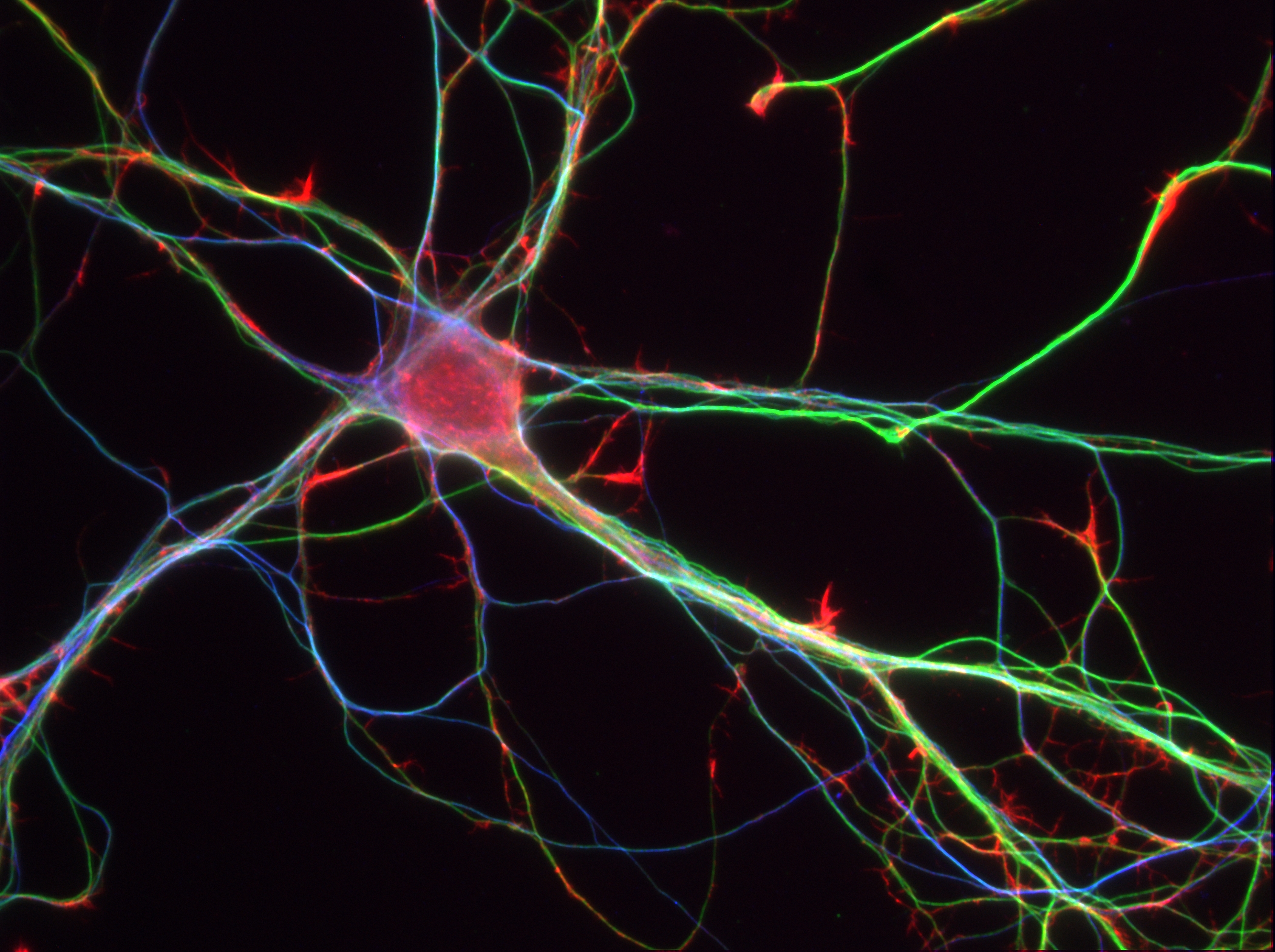
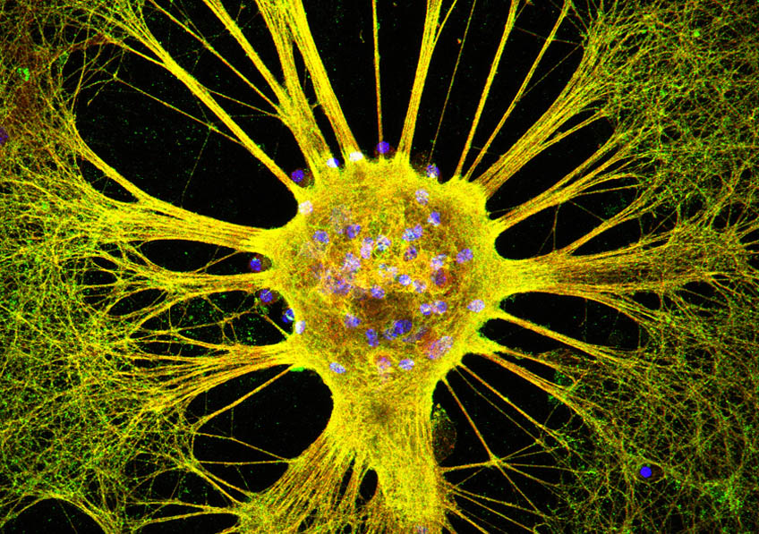
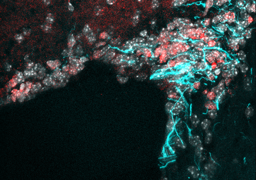
Crédits: M-A.Mouthon et al.

The adult brain has a very limited capacity for regeneration. However, researchers have updated the role of a key membrane molecule in the proliferation of stem cells in the adult mouse brain: Syndecan-1. CEA, Inserm and the Paris-Sud and Paris Diderot universities have joined forces to demonstrate that Syndecan-1, present in the membrane of the neural stem cells, permits their proliferation. Syndecan-1 therefore becomes a possible tracer of brain regeneration, opening up new perspectives in the field of regenerative medicine. This research, for which a patent is pending, was published on August 14 in Stem Cell Reports.

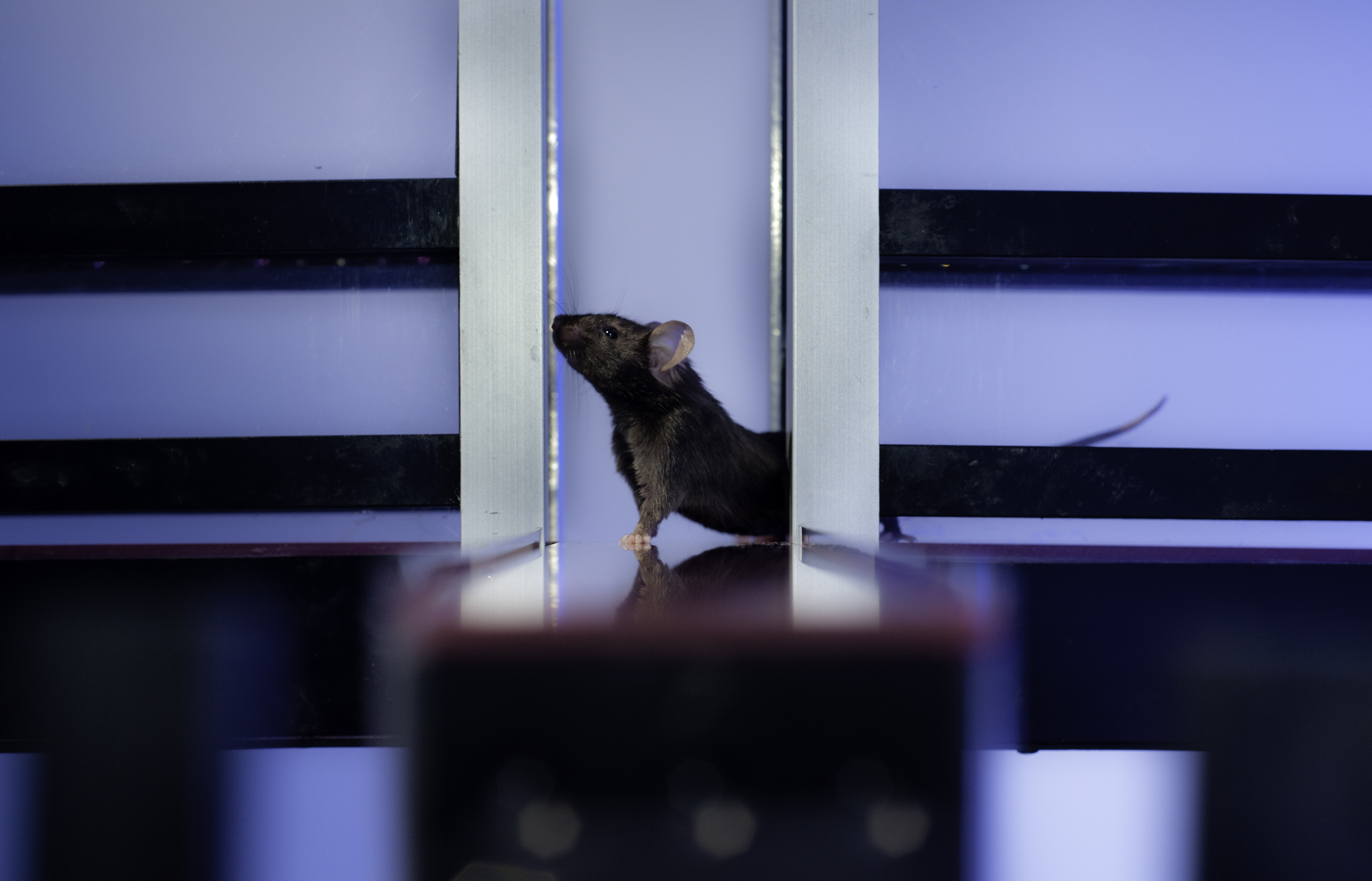
The adult brain has a very limited capacity for regeneration. It contains a small number of neural stem cells in specialized niches. These cells, which have the capacity to produce new functioning neurons, lie dormant for the most part but can be activated in response to stress.

Thanks to an innovative cell sorting technique and comparative transcriptome analysis, researchers at the Institute of Cellular and Molecular Radiobiology (IRCM)[1] have revealed some of the molecular modifications that accompany the activation of dormant neural stem cells, in response to the brain’s exposure to ionizing radiation. Through comparative mapping of the genes expressed, they identified the membrane molecule Syndecan-1 as being specifically expressed in the neural stem cells activated. By means of functional studies, the team also demonstrated that this molecule played a direct role in the proliferation of these cells.

Knowledge of the molecular signals that regulate the activity of the neural stem cells present in the adult brain could make it possible to stimulate the regenerative capacities of the brain and have many therapeutic applications.  Syndecan-1, as well as the various molecular pathways identified in this research, open up new perspectives in this field of medical research.

[1] Joint Research Unit 967, François Jacob Institute, CEA Paris Saclay; Inserm; Université Paris-Sud / Paris Saclay; Université Paris Diderot.

Adolescent depression: early signs and changes in brain development underscore the need for new preventive approaches

Photo by Abbie Bernet on Unsplash

Teams of AP-HP, Inserm and Université Paris Sud, studied as part of an international research group, depressions called “subsyndromal” among young teenagers as they have a high risk to progress to depression in adolescence and later in adulthood.

This research demonstrates the existence of deviations of the microstructure of white matter in the prefrontal beams that provide connections between brain regions. The study in nearly 100 adolescents from 14 years of school, with only some depressive symptoms without apparent seriousness, and compared to a control population of more than 300 adolescents without symptoms recruited at the same time is published in the journal The American Journal of Psychiatry.

This variation of the normal maturation individual predictive value of a diagnosis of depression two years later. The identification of these adolescents at risk could improve the prevention of depression.

Adolescence is a particularly vulnerable period for the onset of depressive disorders. Authentic depressive episodes can occur, affecting about 12% of teenagers, but about 20% of teens will present depressions called subclinical or subsyndromal, that is to say that do not have visible symptoms.

Brain areas rearrangements such as cerebral cortex and white matter occur at this age, but the predictive factors of cerebral transition to depression in adolescents are not known.

Recently changes in prefrontal gray matter associated with the risk of depression in adolescence have been reported. The Child Psychiatry Service of the research teams and Adolescent to Pitie-Salpetriere Hospital, AP-HP, INSERM, Université Paris Descartes and Paris-Sud University (unit Neuroimaging and psychiatry 1000) have investigated the changes in the white matter underlying the subsyndromal emotional states, commonly observed in young characterized without psychiatric disorders.

Comparing a group of teenagers from 14 years of school, with only some depressive symptoms without apparent gravity (96 individuals) to a control group (336 individuals), the researchers demonstrate the existence of deviations of the microstructure of white matter bundles prefrontal among teenagers in the first group.

These deviations relate to areas usually involved in major depressive episodes involved in the regulation of emotions and motivation.

In detail, the results suggest a delayed development of the myelin and a different maturation in these adolescents compared to control teenagers.

In addition, the research team found that these deviations have individual predictive value of a diagnosis of depression two years later.

These deviations from normal adolescent development constitute a vulnerability factor. Through these results, the authors encourage the development of preventive strategies for adolescents at risk.

A brain mechanism involved in mediated learning is identified

Neurone d’hippocampe Crédits: Inserm/Peris, Leticia

Imagine that you are eating a Granny apple under a red parasol on the terrace of a public garden. The next day you eat another Granny apple at home in your kitchen, but soon afterwards find yourself ill. The next time you go back to the public garden, you avoid sitting under that red parasol. While there may not seem to be a link between the parasol and the fact that you were ill, there actually is! This is an example of the mediated learning process, and researchers from INRA and INSERM have just identified the brain mechanism involved in it. Their results, published online on 30 August 2018 in Neuron, show that cannabinoid receptors in the hippocampus play a key role in establishing these types of associations.

Direct learning, which implies a specific link between information and positive or negative consequences, influences our future choices. However, our behaviour is most often guided by mediated learning, which is based on associations between seemingly insignificant information. This explains why we are often put off or attracted by people, places and things we have never directly associated with aversive (or positive) situations, but which were previously encountered along with other information that acquired an aversive or positive meaning. The apple and red parasol case is an example of this.

Receptors, neurons and the brain structure involved in mediated learning have been identified

While the neurobiological bases of direct learning have been thoroughly studied, understanding of mediated learning mechanisms remains rather limited. Researchers from INRA and INSERM began by establishing mediated learning behaviour models in mice in laboratory experiments. They repeatedly and simultaneously exposed mice to an odour (banana or almond) and a taste (sweet or salty) without a particular effect on the animal. They then associated the taste with a gastric malaise (similar to food poisoning). When the researchers later exposed the mice to the odour initially associated with this taste, they observed that the animals specifically avoided this odour, which showed a transfer of the aversive value between the taste and the odour. The scientists were able to demonstrate similar results with light and sound and the transfer between these sensory inputs of an attractive value (by giving the animals a reward), and thereby generalising the phenomenon.

The scientists were able to pinpoint the mechanism in question: the mediated learning process (between an odour and a taste or a light and a sound) involves the hippocampus and a major neuromodulatory system within this brain structure, called the endocannabinoid system. More specifically, this particular form of learning requires the involvement of type 1 cannabinoid receptors (CB1R) in hippocampal GABAergic neurons.

These unprecedented results will help researchers evaluate whether CB1R are also involved in other brain structures during mediated learning. This could lead to new research to understand certain pathologies (schizophrenia or psychosis) in which mediated learning is altered.

Novel brain network linked to chronic pain in Parkinson’s disease

©Inserm/U746, 2011

Scientists have revealed a novel brain network that links pain in Parkinson’s disease (PD) to a specific region of the brain, according to a report in the journal eLife.


The research reveals why a subset of neurons in part of the brain called the subthalamic nucleus is a potential target for pain relief in PD, as well as other diseases such as dementia, motor neuron disease and Huntington’s, and certain forms of migraine.


People with PD often report unexplained pain such as burning, stabbing, aching, itching or tingling sensations that are not directly related to their other PD symptoms. Treatment with deep brain stimulation in the subthalamic nucleus can help with the movement-related symptoms of PD, but recent studies have shown it also reduces pain. The way it does this, however, is currently unclear.


“In this study, we set out to determine whether the subthalamic nucleus is involved in translating a harmful stimulus such as injury into pain, and whether this information transmission is altered in PD,” explains lead author Arnaud Pautrat, PhD Student at University Grenoble Alpes, France.


The team started by using electrophysiology to measure the firing of electrical signals in nerve cells in the subthalamic nucleus of rats given a shock to their back paw. Nerve cells were indeed temporally activated by this stimulation. They also found that the neurons fell into three response groups, showing an increase, decrease or no change in their baseline firing rate.


They next looked at whether these responses caused a change in brain function. Rats with a damaged subthalamic nucleus took much longer to show signs of discomfort than healthy rats. When they expanded their study to rat models of PD, the team found that nerve cells in the subthalamic nucleus had higher firing rates and the responses to pain were bigger and longer than the healthy animals. Taken together, this suggests that dysfunctional pain-processing pathways in the subthalamic nucleus are the cause of PD-related pain.


To understand where the pain signals to the subthalamic nucleus were coming from, the team looked at two brain structures known to be important in relaying damage signals from the spinal cord: the superior colliculus and the parabrachial nucleus. Blocking their activity revealed that both structures play a crucial role in transmitting pain information to the subthalamic nucleus, and that a direct communication pathway exists between the parabrachial nucleus and the subthalamic nucleus. As a result, the team believes this pathway is likely to be involved in the beneficial effects of deep brain stimulation on pain in PD and that these novel insights could help to target stimulation to specific parts of the brain to make it more effective as a pain reliever.


“We have found evidence that the subthalamic nucleus is functionally linked to a pain-processing network and that these responses are affected in Parkinsonism,” concludes senior author and INSERM researcher Veronique Coizet, PhD. “Further experiments are now needed to fully characterise the effects deep brain stimulation on this brain region in our experimental models, with a view to finding ways to optimise it as a treatment for pain caused by Parkinson’s and other neurological diseases.”

Metformin Improves Motor Skills in Patients with Steinert Myotonic Dystrophy, the Most Common Adult Neuromuscular Disease

Cellules musculaire de patient atteint de dystrophie myotonique. Crédits: Inserm/IGBMC

Inserm researchers at I-Stem – the Institute for Stem Cell Therapy and Exploration of Monogenic Diseases – report encouraging results with metformin, a known diabetes drug, for the symptomatic treatment of Steinert myotonic dystrophy. A phase II trial conducted in 40 patients at Henri-Mondor Hospital AP-HP has shown that, after 48 weeks of treatment at the highest dose, patients treated with metformin (versus placebo) gain in motor skills and recover a more stable gait. The results of this trial, which received 1.5 million euros in funding from AFM-Téléthon, are published today in Brain.

Steinert myotonic dystrophy (DM1) is the most common form of muscular dystrophy in adults. Of genetic origin, its prevalence is estimated at 1/8,000, or about 7 to 8,000 patients in France. It affects the muscles, which weaken (dystrophy) and have difficulty to relax after contraction (myotonia) which disrupts the movements (unstable walking for example). It also affects other organs (heart and respiratory system, digestive system, endocrine system and nervous system). To date, this muscular dystrophy does not benefit from any curative treatment.

The findings published in Brain are the result of research conducted for several years at I-Stem. Indeed, thanks to the development of cellular models from pluripotent stem cells, in 2011 Cécile Martinat’s team identified new mechanisms at the origin of Steinert myotonic dystrophy (Cell Stem Cell – March 31, 2011). In 2015, Sandrine Baghdoyan, a research engineer at I-Stem, succeeded in correcting certain splice defects in embryonic stem cells and myoblasts from people with DM1 using metformin, a well-known diabetes drug identified as effective in this new indication thanks to high-throughput screening (Mol.Therapy – Nov 3, 2015).

Encouraged by these results, I-Stem launched a phase II, single-center, double-blind, randomized controlled clinical trial in 40 patients, in collaboration with teams from Henri Mondor Hospital AP-HP, that of Dr. Guillaume Bassez of the Ile-de-France Reference Center for Neuromuscular Diseases, and that of the Clinical Investigation Center coordinated by Prof. Philippe Le Corvoisier. In this placebo-controlled study, metformin was administered three times daily, orally, with a 4-week increase in dose (up to 3 g/day), followed by 48 weeks at the highest dose. The evaluation of treatment efficacy was based on the “6-minute walk” test. At the end of the study, after one year of treatment, patients who received metformin gained an average walking distance of about 33 meters on their initial performance whereas the group receiving placebo remained stable (average gain of 3 meters). This motricity, analyzed in depth using the Locometryx tool developed by Jean-Yves Hogrel’s Hogrel’s Laboratory of Physiology and Neuromuscular Evaluation at the Institute of Myology at the Pitié-Salpêtrière Hospital AP-HP, is closely linked to the fact that metformin improves the overall posture of patients who, in fact, go from walking in an unstable “broad-based” manner before treatment, to a straight, faster and therefore more efficient gait”.

These results demonstrate, for the first time, the efficacy of a pharmacological treatment on motor function in Steinert myotonic dystrophy and, to our knowledge, the therapeutic efficacy of a molecule identified on the basis of the modeling of a pathology with pluripotent stem cells.

Mechanism of Repression: Can a Person’s Memories be Altered Without Their Knowledge?

Photo by Christophe Hautier on Unsplash

In recent years, the cognitive neurosciences have shown that it is possible to use conscious effort to alter memories. Researchers from the Inserm Center for Psychiatry and Neuroscience, Sainte Anne Hospital and Université Paris Descartes now show that it is possible to unconsciously alter memories. This experimental demonstration of the unconscious manipulation of memories, which is similar to the psychoanalytical concept of repression, has been published in the journal Cognition.

Researchers have known for several years that by making a conscious effort to suppress a specific memory, it is possible to alter it, i.e. reduce our ability to recall it. This effect is expressed by the deactivation of the cerebral hippocampi, brain structures involved in encoding memory. Can the same be done unconsciously?

Led by Professor Raphaël Gaillard, researchers from the Inserm Center for Psychiatry and Neuroscience, Sainte Anne Hospital and Université Paris Descartes put their expertise in consciousness and unconscious processes to good use to test this hypothesis.

In order to recreate in a laboratory setting the conditions of an unconscious recall mechanism, volunteers learned related word pairs (e.g. candle-champagne, walk-hill), and then were trained, when the first word was presented to them, to either think of the second word in the pair or to avoid thinking about it, according to a visual cue. A similar experimental system had in the past made it possible to demonstrate the alteration of a memory by conscious effort.

The originality of this study lies in the fact that these visual cues giving th e instruction to think of or try to avoid thinking of the second word in the pair were sometimes presented subliminally, i.e. too briefly to access the consciousness. When this occurred, the volunteers had to determine as quickly as possible whether the first word was masculine or feminine. While these cues were not perceived at the conscious level, the researchers revealed an alteration in the capacity to recall the second word.

Consequently, the cue associated with the instruction not to think of the second word decreased the ability to recall that word, and the cue associated automatically with the instruction to think of it increased it. This study therefore demonstrates that it is possible to unconsciously manipulate the memory of a word.

This research opens up new perspectives in the understanding of unconscious psychological phenomena. If, within the environment of a laboratory and a two-hour experiment time, it is possible to demonstrate such a phenomenon of repression, in daily life the repetition of elements altering a memory could have major effects.

More generally, this research once again shows that memory is vulnerable to distortion –and even manipulation– thereby representing a challenge for witness accounts and memories of one’s own life.

Mice’s individuality is influenced by their relations

Inserm/Latron Patrice, 2009, Inserm images

Individuality exists in all animals, and a number of factors shape it over time. For mice, one of those factors is the social environment, as researchers at CNRS, INSERM and Sorbonne Université have just shown. In this species, some stable character traits may even be inscribed in an individual’s neuron activity and change when the group’s composition changes. These results are published on August 6, 2018 in Nature Communication.

Individuality is not exclusive to humans. Though this idea was previously rebutted by biologists, today it is accepted that individuality is found in all animal species. It is defined as all the behavior differences between individuals of a single species that are relatively stable over time. Though the process called individuation is supported by genetic and development components, researchers have just demonstrated in mice that the social environment and activity of some neurons also participate in determining the emergence of distinct individuals.

To reach this conclusion, teams at Laboratoire Neuroscience Paris-Seine (CNRS/INSERM/Sorbonne Université), Laboratoire Adaptation Biologique et Vieillissement (CNRS/Sorbonne Université)1 and at Sorbonne Université’s Institut de la Longévité at Hôpital Charles Foix (AP-HP) studied the life of mice living in “Souris City” (Souris is the French word for mouse), an innovative experimental device having two common living spaces for the animals, and an option to make them take a test one by one, without human intervention. It is by means of this test that the researchers have identified different “personalities” among the mice. This device includes a T-shaped maze where the mice had to choose one of the two arms, where one led to normal water and the other led to sweetened water. These two positions were alternated regularly. When faced with this problem, two radically different strategies emerged: some mice varied their choice very often, others hardly ever.

The first thing that the authors observed was that the type of behavior adopted by each individual was correlated to the activity of dopamine-producing neurons, which are especially involved in decision making. For example, the mice that alternated the most had lower dopaminergic activity. Therefore, one can say that mice have biologically inscribed individuality.

To understand the role of mice’s social environment on how these different individualities develop, the researchers continued their experiments by changing the composition of the groups in Souris City. They grouped the individuals who adopted the same test strategy, those who rarely alternated on one side, and those who alternated often on the other. Surprise! After a few weeks, roles had redistributed in both groups! Some mice who had rarely varied their choice had become the more adventurous ones in their new group, and vice versa.

What was even more surprising, this behavior change was correlated with a change in dopamine pathway activity in the mice.

These results suggest that decision-making mechanisms, behavioral repertoires and activity levels in the nervous systems of each individual are far from being set and adapt according to the social structure in which the animals evolve.

The fact that the social environment contributes to differences between the individuals has implications for sociology, psychology, biology and medicine. Social factors also participate in the development of psychiatric pathologies such as addiction. This is a field that the researchers are going to investigate, studying the influence of social environment on vulnerability to drugs.

1 These laboratories are members of the Institut de Biologie Paris-Seine

Resistance to Antidepressants: the Ability of Neurons to Self-regulate

Why are some depressed patients more or less totally resistant to the most commonly-prescribed antidepressants? This question was addressed by researchers from Inserm and Sorbonne Université at the Fer à Moulin Institute who were able to reveal the major role of neurons that secrete serotonin – the preferred target for antidepressants – in regulating their own activity. Implicated is a serotonin receptor carried by these neurons whose deficiency could be decisive in the absence of response to the most commonly prescribed antidepressants. This research, published in Neurospychopharmacology, will help elucidate the role of serotonin in psychiatric disorders.

Serotonin is a neurotransmitter – a chemical substance produced by some neurons in order to activate others – which is implicated in a number of psychiatric disorders, such as depression, addiction, impulsiveness and psychosis. It is secreted by specific neurons known as serotonergic neurons.

Releasing serotonin outside the neuronal cell activates neurons which possess receptors specific to this neurotransmitter. When these receptors detect sufficient serotonin in the extracellular environment, they send a message to activate or inhibit the neuron that expresses them. The serotonergic neurons also possess several types of serotonin receptors – called autoreceptors – making it possible to self-regulate their activity.

Researchers from Inserm and Sorbonne Universités/UPMC at the Fer à Moulin Institute (Inserm, UPMC) studied the role of one of the serotonergic neuron autoreceptors – known as 5-HT2B – in the regulation of their activity, in order to elucidate the lack of efficacy of some antidepressant treatments.

Usually, when a serotonergic neuron secretes serotonin in the extracellular environment, it is capable of recapturing some of that serotonin which it will release again later on.  This mechanism, ensured by a specific transporter, enables it to regulate the level of serotonin present in the extracellular environment. This transporter is the preferred target of the antidepressant drugs used to treat psychiatric disorders involving serotonin.  These drugs are called selective serotonin reuptake inhibitors (SSRIs) because they prevent this recapture by the transporter. In the context of depression in which serotonin secretion is too reduced, SSRIs make it possible to maintain normal levels of serotonin in the extracellular environment.

The research team took as their starting point the observation that, in the mouse, when the serotonergic neuron does not carry any 5-HT2B autoreceptors, there is lower than usual serotonergic neuron activity and that the molecules blocking the activity of the transporter, such as SSRIs, have no effect on extracellular serotonin levels. The researchers therefore showed that in order to have an effect, these molecules needed the presence and normal expression of the 5-HT2B serotonin receptor.

They also discovered that when a neuron secretes serotonin, its 5-HT2B autoreceptor detects the quantity present in the extracellular environment and sends a signal to the neuron for it to secrete more serotonin.  To avoid the excessive secretion of serotonin, the serotonergic neuron possesses a negative regulator: the 5-HT1A autoreceptor which also detects the level of extracellular serotonin and sends a signal to the serotonergic neuron to inhibit the secretion. In order to maintain normal neuronal activity, 5-HT2B makes it possible to maintain a certain level of activity by acting as a positive self-regulator.

These findings, which remain to be confirmed in human subjects, reveal a fine serotonergic neuron self-regulation mechanism balanced between the activator autoreceptors and the inhibitor autoreceptors.  They constitute a step forward in identifying new drug targets, in elucidating the role of serotonin in certain psychiatric disorders and in understanding the inefficacy of certain antidepressants.

Hypertension at Age 50 is Said to Increase the Risk of Developing Dementia

©JORGE LOPEZ on Unsplash 

And what if, depending on the age at which it develops, hypertension had more or less significant consequences on maintaining our cognitive function? This was suggested by a study conducted by an Inserm team in partnership with the Department of Epidemiology and Public Health at University College London which has been monitoring changes in blood pressure and the onset of dementia in more than 10,000 volunteers since 1985. This research, published in the European Heart Journal, suggests that, at the age of 50, high blood pressure, although still below the diagnostic threshold for hypertension, could be linked to a higher risk of developing dementia later in life, even for individuals with no other cardiovascular disorders.

Although studies linking blood pressure to an increased risk of dementia at an advanced age already exist, they focus on blood pressure values in a large population segment ranging from age 35 to 68, and have never been conducted in specific age groups.

With the Whitehall II study, Inserm researchers, in partnership with the Department of Epidemiology and Public Health at University College London (UCL), embarked upon the long-term follow-up of a population of 10,000 volunteers aged 35 to 55 when the study began in 1985, in order to study the link between age, hypertension, and dementia. The researchers measured the participants’ blood pressure in 1985, 1991, 1997, and 2003. Participants were monitored until 2017 in order to detect the possible onset of dementia.

Less than 5% of participants developed dementia as they aged, and the mean age at diagnosis was approximately 75 years.

The research team studied two different types of blood pressure values: systolic pressure – measured as the heart contracts to eject blood into the arteries (systole) – and diastolic blood pressure – measured as the heart relaxes and fills with blood (diastole).

While diastolic pressure does not appear to have an impact on the risk of developing dementia, the researchers nonetheless observed that fifty-year-olds with a systolic pressure of 130 mmHg or over (according to the European Society of Cardiology, the limit value for diagnosing hypertension is 140 mmHg) had a 45% higher risk of developing dementia compared to individuals with a lower systolic pressure at the same age. No increase in this risk was observed among individuals with hypertension at age 60 or 70.

Furthermore, the higher risk associated with blood pressure above 130 mmHg is also observed among individuals not developing cardiovascular disorders during the follow-up period: their risk was 47% higher compared to individuals without cardiovascular disorders, with a systolic pressure below 130 mmHg.

According to Archana Singh-Manoux, Inserm research director in charge of the research project and professor emeritus at UCL, these analyses “suggest that the impact of blood pressure on brain health is dependent on the duration of exposure; hence, individuals with high blood pressure at the age of 50 would be more likely to develop dementia than those who develop hypertension at 60 or 70.”

This could be explained by the fact that high blood pressure causes ministrokes which, although often undetected, are harmful to the brain and may ultimately lead to a decline in function.

“In this study, we were able to evidence different patterns of association according to the age groups studied,” clarified Jessica Abell, the lead author of the article, postdoctoral researcher at Inserm and associate researcher at UCL, who adds that “these results could thus help redefine the age groups to be studied in order to assess the impact of hypertension on health.” She concludes: “it is important to emphasize that these results originated from an observational study on a population sample, and cannot be directly used as predictive instruments for each individual. Defining the optimum limit value for diagnosing hypertension is currently the focus of the debate.”

A Computer Program Able to Automatically Detect and Identify Brain Lesions

 ©Emmanuel Barbier – Inserm/Inria/Univ. Grenobles Alpes  – Figure d’IRM chez l’homme obtenue ici en présence d’une tumeur cérébrale. En gris, des IRM classiques, en couleur, des IRM quantitatives.

Will the radiology of the future come from machine learning? That is the view of Inserm and Inria researchers working in collaboration at the Université Grenoble Alpes who have developed a program able to localize and diagnose various types of brain tumors via MRI image analysis. These analyses have produced highly reliable results, with tumor localizations and tumor-type diagnoses accurate in 100% and over 90% of cases, respectively. This innovative method and its results are the subject of a study published in IEEE-TMI.

MRI – or magnetic resonance imaging – with its ability to reveal various brain tissue characteristics is the medical imaging technique of reference when it comes to obtaining highly-detailed images of the brain. It can produce what is known as “quantitative” images, which each map a measurable brain parameter (such as blood flow or blood vessel diameter). Although the quality of these quantitative images is less dependent on the calibration of the measuring apparatus than that of the standard images obtained with MRI – and so is more reliable – this type of technique is still infrequently used in the clinical MRI setting.

Inserm researchers have been working in conjunction with a research team from Inria on the analysis protocols of these quantitative images at the Université Grenoble Alpes.  The researchers combined various innovative mathematical tools in order to teach a computer program how to analyze quantitative brain MRI images and diagnose any tumors present.

First of all, the program learned how to recognize the characteristics of a healthy brain. Then, when it was shown images of brains with cancer, it became able to automatically localize the regions whose characteristics diverge from those of healthy tissues and to extract the distinguishing characteristics.

Finally, in order to teach the artificial intelligence how to discriminate between the different types of tumor, the researchers then gave it the diagnosis associated with each of the pathological brain images which had been presented to it.

In order to test the ability of the program to differentiate healthy from diseased tissue, the research team provided it with images that it had not seen before – sometimes of healthy brains, sometimes of pathological brains. The program had to indicate whether a tumor was present in these images and, if so, be able to characterize it. And, by succeeding in localizing the lesions perfectly (100%) and diagnosing them very reliably (over 90%), the artificial intelligence turned out to be a very quick study.

“At present, the acquisition of quantitative images does not correspond to what is happening in routine clinical practice in the MRI departments”, specifies Emmanuel Barbier, Inserm researcher leading the study. “But this research shows the value of acquiring these types of images and informs radiologists of the analytical tools that could be available to them in the near future to aid their interpretations. “

In the meantime, the research team will focus on the most relevant images to acquire in order to diagnose brain tumors as precisely as possible and with the greatest possible reliability. It will therefore continue to develop mathematical tools with the aim of improving the program’s self-learning abilities, with the ultimate objective being to extend the diagnostic potential of this artificial intelligence to other brain diseases, such as Parkinson’s.

These quantitative MRI machine learning tools applied to brain tumors are currently being evaluated as part of the Cancer Plan driven by Inserm, within the Tumor Heterogeneity and Ecosystem program.

Their development in the context of Parkinson’s disease diagnosis is also underway via the NeuroCoG multidisciplinary project funded by the Université Grenoble Alpes IDEX.

Our Food Choices Predicted by our Brain Anatomy

©Katherine Chase – Unsplash 

Do you tend to go for cake or vegetables? If you sometimes find it difficult to eat healthily, a study conducted by a team of researchers from Inserm, CNRS and Sorbonne Université, including Liane Schmidt and Hilke Plassmann, at the Brain & Spine Institute (ICM) has established a link between the anatomy of certain regions of our brain and our ability to control our food choices. These results were published in The Journal of Neuroscience on June 4, 2018.

For many people, eating healthily is no easy feat. Individuals differ greatly in their ability to maintain a balanced diet and make healthy food choices.

From the cognitive perspective, making a choice involves two principal mechanisms – the first of which consists of attributing a value to each option. In the case of food, its taste and nutritional quality can, for example, determine its value. The second mechanism for our brain consists of analyzing the value given to each option in order to choose the most suitable – i.e. the food item to which we attribute the highest value.

How are these decisions expressed in the brain? To answer these questions, Liane Schmidt, Inserm researcher, Hilke Plassmann, and their colleagues Anita Tusche from the California Institute of Technology (USA), Cendri Hutcherson from the University of Toronto (Canada) and Todd Hare from the University of Zurich (Switzerland), pooled brain imaging data taken from four studies on food choices.

In three of these studies, the participants performed the same task which involved evaluating their appetite for a specific food according to three criteria: its taste, health benefits and their own natural preference. As such, the participants could base their choice either on taste or on nutritional value.

In the fourth study, the participants were asked to use a method of their choice (save money, eat organic, or lose weight) to reduce their desire for tasty products devoid of nutritional value. This last study “involved a more flexible control strategy not specifically focused on attributes of taste or health but on the various means of distancing oneself from a food or resisting desire” specifies Liane Schmidt, lead author of the study and Inserm researcher.

The researchers studied the variations in the quantity of gray matter in the brains of the participants using imaging data from the first three studies.

As such, they revealed a correlation between food choices and the quantity of gray matter in two regions of the frontal lobe: the dorsolateral prefrontal cortex (dlPFC), which governs the regulation of decisions, and the ventromedial prefrontal cortex (vmPFC), which is in charge of value attribution. They observed that people with more gray matter in these regions had a greater appetite for the foods they considered healthy.

The research team then sought to predict the food choices of the participants in the fourth study based on the quantity of gray matter detectable in the two regions identified previously. “The idea here was to see whether the correlations established in a context in which the control strategies are very clear – focus on taste or health – can be generalized to a situation in which the control strategies are more vague. ” continues Hilke Plassmann.

The researchers confirm these results, thereby establishing for the first time that neuroanatomical differences in the dlPFC and vmPFC regions play a role in individual food choices. These results open up avenues which should ultimately lead to the treatment of eating disorders associated with disrupted food control, such as bulimia nervosa or anorexia nervosa.

fermer