Menu

Towards Understanding the Origin of the Most Common Form of Female Infertility

Researchers from Inserm and Université de Lille have discovered that the most common female infertility disorder – polycystic ovary syndrome (PCOS) – may be caused by overexcitation of brain neurons. The culprit? A hormone produced by the ovaries, called “anti-Müllerian hormone” (AMH), which is produced in excess in women with PCOS. Research conducted by the team in mice show the importance of in utero exposure to abnormally high levels of AMH in the occurrence of the disease. These findings, published in Nature Medicine, pave the way for new concepts linked to the embryonic origin of the disease as well as new avenues for the development of a treatment.

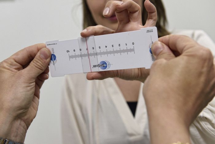
One in ten women of childbearing age suffers from polycystic ovary syndrome (PCOS), in which the ovaries produce androgens (male hormones) in excess, disrupting the growth mechanisms of the ovarian follicles. A greater number of these follicles will stagnate – hence the (erroneous) term “polycystic ovaries” – leading to ovulation dysfunction responsible for infertility.

While we currently know how to diagnose the disease, its cause remains unknown. Current therapeutic options aim to reduce symptoms and prevent complications but no preventive or curative treatment exists.

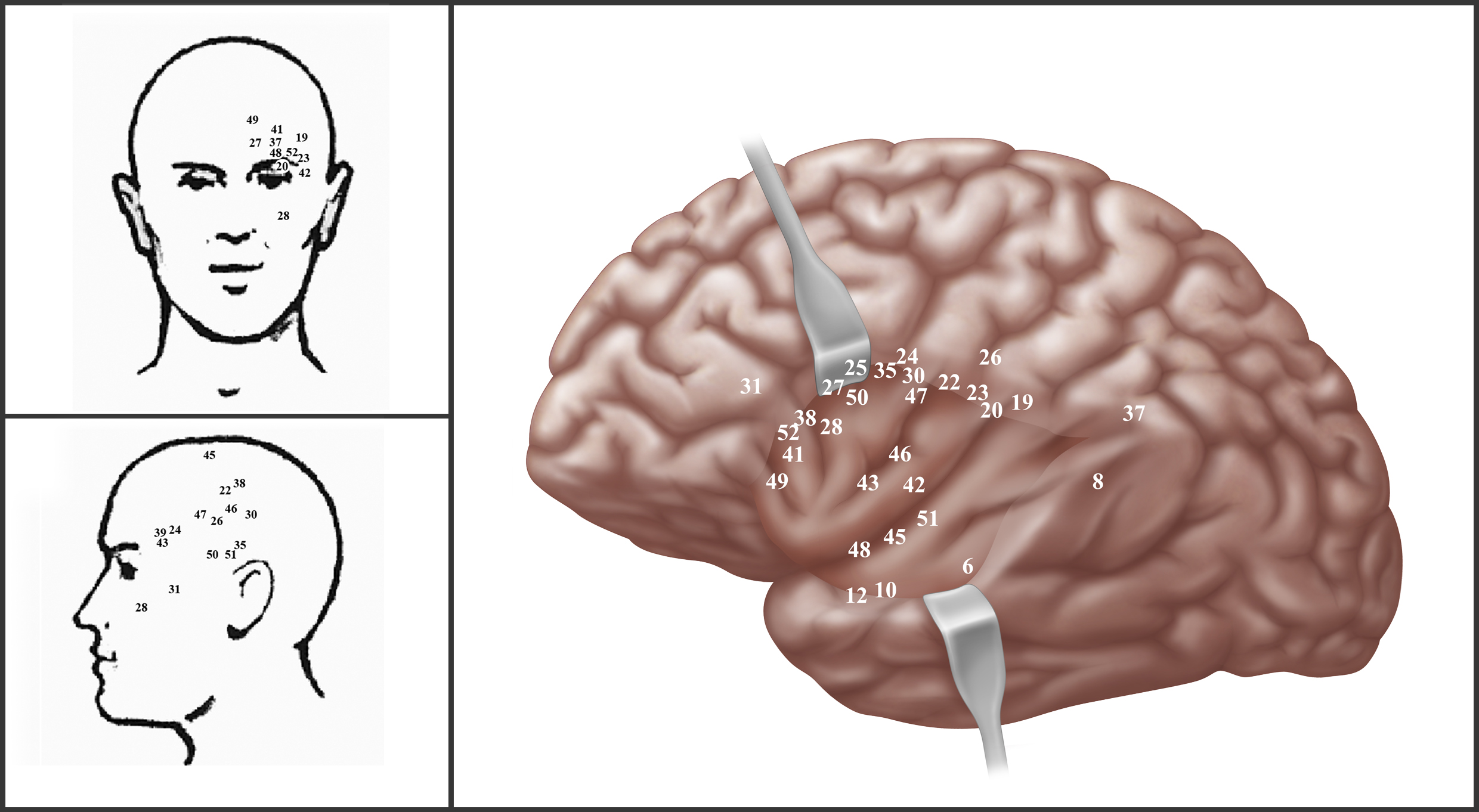
A team coordinated by Paolo Giacobini, Inserm Research Director (Jean-Pierre Aubert Research Center – Neurosciences and cancer, Inserm U1172/Université de Lille/Lille University Hospital), is challenging the hypothesis according to which PCOS only alters the ovaries, by showing that it also modifies the activities of brain neurons in the hypothalamus, which are responsible for controlling reproduction. Implicated is a hormone produced by the ovaries and involved in their functioning: anti-Müllerian hormone (AMH). Patients with PCOS present blood levels of this hormone which are two to three times higher, and directly linked to the severity of the disease.

The team based its research on two observations made in pregnant women with PCOS. The first is its already-known correlation with hyperandrogenism (excessive production of androgens). The second, a new observation, is its correlation with a heightened production of AMH during pregnancy. The researchers have shown that mice treated with AMH during gestation give birth to females that develop the symptoms characteristic of PCOS in adulthood. The production of abnormally high levels of AMH during the prenatal period could therefore be responsible for gestational hyperandrogenism and the abnormal hormonal impregnation of the fetus.

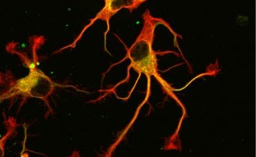
The team also observed that, in mice mimicking PCOS, in utero exposure to abnormally high levels of AMH was responsible for increased activity of the GnRH protein-secreting hypothalamic neurons in adulthood.  This intense GnRH production stimulates the heightened production of another hormone, luteinizing hormone (LH), which itself stimulates the production of androgens.  Paolo Giacobini and his coworkers, including Brooke Tata and Nour El Houda Mimouni, joint first authors of the article, demonstrate here that prenatal exposure to AMH triggers a genuine chain reaction in offspring:  the hypothalamic neurons start secreting more GnRH, which increases LH production by the pituitary gland, in the end triggering this characteristic increase of androgens in the ovaries, which is responsible for the disrupted ovulation observed in PCOS.

Armed with these findings, the researchers applied to mice mimicking PCOS a specific treatment that “normalizes” the increased action of GnRH on LH production, thereby restoring their fertility. These findings observed in the mouse model offer unheard of therapeutic perspectives which remain to be confirmed at human level.

How do we detect danger?

Living beings are able to integrate and identify relevant sensory information, such as smells, sounds or light, in order to regulate how they behave in the presence of potential danger. This is called context discrimination. Inserm researchers based at Neurocentre Magendie in Bordeaux have recently discovered which neurons are implicated in this phenomenon and where they are located. Good news for sufferers of post-traumatic stress disorder in whom context discrimination is disrupted.

This research has been published in Neuron.

 Traumatic experiences, such as natural disasters, terrorist attacks or military combat, can lead to the development of psychiatric disorders, such as post-traumatic stress disorder (PTSD). When sufferers find themselves faced with a similar environment to that in which the traumatic event occurred, they relive the stress of the original trauma with the same intensity. In these patients, anxiety disorders are associated with context generalization.  They have indeed become incapable of integrating and identifying the relevant sensory information produced by their five senses – which is captured in the environment – in order to regulate behavioral responses. The neuronal circuits involved in this phenomenon are unknown.

A team of researchers led by Dr Cyril Herry has for the first time recently identified in mice a population of neurons implicated in context discrimination. These neurons are located in the medial prefrontal cortex.

They did this using optogenetic approaches (see boxed text), in which the activity of neuron populations is activated or inhibited so as to determine their involvement in a particular behavior. In order to evaluate the neuronal circuits implicated in context discrimination, the researchers exposed the mice to an environment comprising various sensory elements (light, smell, sound) in which they received one or more mild electric shocks in order to render them averse to that environment.

The mice were then exposed to the same environment but without the relevant sensory elements (smell, sound, light), in order to have them believe that it was non-aversive. Thanks to real-time recordings of the activity of neurons in the medial prefrontal cortex and their optogenetic manipulation, the researchers were able to identify a population of neurons specifically activated during the context discrimination.

This research shows that neuronal activity in the medial prefrontal cortex is a key element of context discrimination.

In addition, the researchers have demonstrated that this group of neurons projects specifically to the brain stem, a region directly implicated in the motor regulation of emotional behaviors.

“This research, which deepens our understanding of the neuronal activity leading to context discrimination, could contribute to the development of treatments and therapies for people with anxiety disorders” considers Dr Cyril Herry, Inserm research director and investigator of this study.

Optogenetics consists of introducing into neurons natural light-sensitive proteins, such as channelrhodopsin, an extract of algae which is sensitive to blue light, or archaerhodopsin, which is sensitive to green and yellow light. When blue light is introduced into the mouse brain by an optical fiber, activation of the channelrhodopsin generates a depolarizing current: this activates the neurons. However, if the archaerhodopsin is activated by a green or yellow light, it generates a hyperpolarizing current and the neurons are inhibited. These light-sensitive proteins expressed at neuronal membrane level are therefore capable of freely activating or inhibiting nerve impulses. This enables the researchers to identify the neuronal networks implicated in a specific task and to determine the causal role.

A new solution for chronic pain

Neuropathic pain is a chronic illness affecting 7-10% of the population in France and for which there is no effective treatment. Researchers at the Institute for Neurosciences of Montpellier (Inserm/University of Montpellier) and the Laboratory for Therapeutic Innovation (CNRS/University of Strasbourg)[1] have uncovered the mechanism behind the onset and continuation of pain. Thanks to their discovery, they have developed an innovative treatment prototype which produces, in animal models, an immediate and long-lasting effect on pain symptoms. This study was published on March 12, 2018 in Nature Communications.

French researchers have recently revealed the unexpected role played by the molecule FLT3 in chronic pain – a molecule known for its role in various blood functions and produced by the hematopoietic stem cells which generate all blood cells. Neuropathic pain is caused by peripheral nerve lesions caused by diseases such as diabetes, cancer, or shingles, or to accident-related trauma or surgery. In this study, the researchers showed that immune cells in the blood which flood the nerve at the lesion site synthesize and release another molecule, FL, which binds with and activates FLT3, triggering a chain reaction in the sensory system, causing pain. They revealed that FLT3 induces and maintains pain by acting far upstream on other components in the sensory system which are known for making pain chronic (a phenomenon known as “chronicization”).

After discovering the role of FLT3, researchers created an anti-FLT3 molecule (BDT001) to target the FL binding site, using detailed computer analysis of three million possible configurations. This molecule blocks the connection between FL and FLT3, thereby preventing the chain of events which leads to chronic pain.  When administered to animal models, BDT001, after three hours, reduced typical neuropathic pain symptoms such as hyperalgesia, a heightened sensibility to pain, as well as allodynia, pain caused by stimuli which normally do not provoke pain, with effects lasting 48 hours after a single dose.

Neuropathic pain, which affects approximately 4 million people in France, is a debilitating disease with significant social costs. Current forms of treatment, essentially based on off-label uses of medication such as anti-depressants and anti-epileptics, are ineffective: less than 50% of patients obtain a significant reduction in their pain. Furthermore, such treatments can cause substantial side effects. The innovative therapy[2] based on this research is being developed by Biodol Therapeutics, a start-up firm which may, as a result, finalize the very first specific therapy against neuropathic pain, and, in the long term, provide relief to many people.


[1] Researchers from the Institute of Functional Genomics (CNRS/Inserm/University of Montpellier) also took part in the research.

[2] This project was financed by the French National Research Agency (ANR) and the SATT AxLR in Montpellier. INSERM licensed the patent rights resulting from this discovery (WO2011/083124 and WO 2016/016370) to Biodol Therapeutics, a new start-up operating in Montpellier and Strasbourg and supported by BPI and the Region of Occitanie.

Susceptibility to addiction: poor production of new neurons implicated

©AdobeStock

Drug addiction behaviors and vulnerability to relapse are linked to our brain’s ability to produce new neurons. This is the finding of Inserm researchers from Neurocentre Magendie at the University of Bordeaux, after observing the behavior of mice taught to self-administer cocaine. Their results, to be published in Molecular Psychiatry, show a link between the deficient production of new neurons in the hippocampus and addiction to drugs.

In the brain, the hippocampus is one of the centers of memory. It includes the dentate gyrus, which has the particularity of producing new neurons (a phenomenon known as neurogenesis) in adults. Abnormal neurogenesis is correlated with a number of neuropsychiatric disorders, such as memory or mood disorders.

Although a link between erratic neurogenesis and drug addiction has already been suspected, there has been no scientific proof to back up this hypothesis until now. The Inserm research teams of Nora Abrous and Pier-Vicenzo Piazza from Neurocentre Magendie (Unit 1215) of the University of Bordeaux, studied the role of neurogenesis in cocaine addiction.

Two groups of mice were compared: one healthy and one genetically engineered to have reduced hippocampal neurogenesis. The mice were trained to self-administer cocaine by introducing their noses into a hole to trigger the intravenous diffusion of cocaine in their bloodstream. The number of actions that they needed to perform to obtain a similar quantity of drug was then gradually increased. The researchers observed that the transgenic mice showed greater motivation (measured in terms of number of actions in the holes) to “work” to obtain cocaine.

After several weeks of withdrawal, the mice were once again exposed to the environment in which they had learned to self-administer cocaine. It was then that the transgenic mice showed greater susceptibility to relapse by seeking once more to trigger administration of the drug.

The transition to addiction is a process combining repeated exposure to drugs and a vulnerability specific to each individual: by demonstrating that neurogenesis is a key factor in vulnerability to addiction, this research offers new perspectives in understanding individual fragility in the face of drug dependence.

This research also opens new avenues towards understanding addictive behaviors in adolescents. “Adolescence, a period of initiation into drug use, is also a time of major brain maturation, characterized in particular by extremely intense neuronal production in the dentate gyrus” specifies Nora Abrous, Inserm researcher, who had already demonstrated in 2002 the negative impact of drug use on the production and survival of new neurons in the hippocampus. She adds that “drug use, by reducing the production of these neurons, increases addiction in adolescents and renders them more vulnerable to relapse during withdrawal attempts”. In its subsequent research, her team “will try to manipulate new neurons with the help of pharmacogenetic techniques, in order to reduce the motivation of the mice for the drug and block relapse during withdrawal”.

Compensation mechanisms in subjects with Alzheimer’s disease lesions to preserve their intellectual and memory performance

©Tiago Muraro – Unsplash

The INSIGHT-preAD study, directed by Prof. Bruno Dubois, is being carried out by teams from Inserm, CNRS and Université Sorbonne at the Brain and Spine Institute (ICM) and the Memory and Alzheimer’s Disease Institute (IM2A) at AP-HP Pitié-Salpêtrière Hospital in Paris, in collaboration with the MEMENTO cohort. It aims at identifying factors underlying Alzheimer’s disease development in healthy subjects over 70 with no existing cognitive disorders. Sponsored by Inserm, this study shows that after 30 months of follow-up, amyloid lesions (also called Alzheimer’s lesions) did not impact cognition or behavior in affected subjects.Its results, published on February 27, 2018, in The Lancet Neurology, point to the existence of compensatory mechanisms in subjects with these lesions.

Drugs currently under development to treat Alzheimer’s disease demonstrate significant efficacy on brain lesions, albeit without a joint decrease in symptoms. It would therefore appear that these trials are undertaken too late, with patients whosedisease istoo advanced. This led to the idea of testing treatment efficacy at an earlier stage, i.e. at the beginning or even before the onset of symptoms in patients with Alzheimer’s disease lesions. Such an approach demands a thorough understanding of the progression markers of the disease at its pre-clinical stage.

The INSIGHT-preAD study, short for “INveStIGation of AlzHeimer’s PredicTors in subjective memory complainers – Pre Alzheimer’s disease”, led by Prof. Bruno Dubois, Director of the Cognitive and Behavioral Disease Center at AP-HP Pitié-Salpêtrière Hospital and Professor of Neurology at Université Sorbonne, aims to identify these progression factors.

The study is based on longitudinal (over time) monitoring of an active cohort launched in May 2013 at AP-HP Pitié-Salpêtrière of 318 volunteer patients over 70 years of age, with perceived memory problems despite normal cognitive and memory performance during testing.

At the beginning, participants agreed to imaging examinations to determine whether or not their brain presented Alzheimer’s disease lesions (also called “amyloid” lesions).  28% of them were found to have lesions although no factors were identified at this stage.

Amyloid-positive and amyloid-negative patients did not present any differences in cognitive (memory, language, orientation), functional, and behavioral testing when they entered the cohort. Additionally, no difference was observed between the subgroups in terms of severity of perceived memory problems, structural neuro-imaging (MRI) or metabolic neuro-imaging (FDG-PET).

As part of the INSIGHT-preAD study, patients underwent a neuropsychological assessment, an electroencephalogram and actigraphy testing on a yearly basis as well as blood testing (to test for biomarkers) and neuroimaging exams (MRI, FDG-PET and amyloid-PET) every two years. 

The teams involved in the study analyzed the data collected at its launch and two years later, as well as a 30-month follow-up clinical assessment of volunteer subjects. 

They did not find any significant change between amyloid-positive and amyloid-negative patients either for the full set of markers monitored (behavioral, cognitive, functional) or in neuro-imaging testing.  Electroencephalogram data, however, highlighted that patients with lesions displayed modified electrical activity in anterior regions of the brain, especially frontal areas, but maintained intellectual and memory performance.

At a 2.5-year follow-up, only four patients have developed Alzheimer’s disease. When they entered the study, they displayed predictive factors including older age, higher concentration of amyloid lesions, and decreased hippocampal volume.

These results therefore show that the presence of amyloid lesions in the brain does not translate into cognitive, morphological, metabolic or functional modifications in affected patients. Results of the study pertaining to electroencephalographic variations also suggest that compensation mechanisms may exist.

Progression towards Alzheimer’s disease in these patients, with an average age of 76, is therefore low, highlighting the existence of significant cognitive reserve in this type of population. Ongoing monitoring is necessary to determine whether these findings can be verified over a longer time span. 

The next INSIGHT-preAD study update will take place in 2022. 

This research has received financial support from the French Ministry of Research (Investments for the Future), the Plan-Alzheimer Foundation, and Pfizer.

The Biological Clock Sets a Different Rhythm for Each Organ

©Adobe Stock

A team of Inserm researchers led by Howard Cooper (Inserm Unit 1208 “Stem Cell and Brain Institute”) in collaboration with their colleagues in the U.S. have for the first time established a reference map of gene expression, by organ and time of day. A mammoth task that began a decade ago and has required two years of analysis. These results, published in Science, show just how important it is to consider the biological clock in order to administer medication at the right time for optimized efficacy and minimal side effects. The researchers are now preparing an atlas which will be available for use by the entire scientific community.

 Roughly two thirds of protein-coding genes are expressed in a cyclical manner over the 24-hour period with peaks occurring in the morning and late afternoon. However, this expression varies greatly from one tissue to another, confirming that, in addition to the internal central clock, each organ expresses its own clock. An Inserm team has been the first to prove it in a diurnal species, providing a spatiotemporal map of the genetic circadian expression for the various organs. This work marks a major step forward in the field of chronobiology.

Prior to that, studies exploring the circadian rhythm in various organs were generally conducted in animal models such as the fruit fly (research which received last year’s Nobel Prize) and nocturnal species, particularly mice. Since the body clock is primarily synchronized by the light-dark cycle, it would have been tempting to reverse the cycle to obtain data in diurnal animals. However, rodents are not just phase-shifted in relation to humans, their way of life is also very different. Their sleep is fragmented across the whole 24-hour period, unlike diurnal species, which get most of their sleep at night. They also feed continuously over the nocturnal waking phase, whereas humans take their meals at regular times. All of these factors also help to synchronize the biological clock. The time had come therefore to work with species closer to our own in order to deepen knowledge of ourselves.

This involved the researchers analyzing the RNA of over 25,000 genes from 64 organs and tissues, every two hours for 24 hours, in non-human primates. The major organs underwent detailed analysis as well as the various regions of the brain. All in all, the researchers analyzed 768 samples. A mammoth task that began a decade ago and which has required two years of analysis! For each sample, they looked for, quantified and identified the RNA present in the cells. This RNA then either goes on to become proteins or it remains as RNA with regulatory properties on other molecules. This is what we call the transcriptome.

80% of the genes regulated by the biological clock ensure essential cell functions

The authors observed that 80% of the cyclically expressed genes code for proteins that ensure functions essential to cell life, such as waste elimination, DNA replication and repair, metabolism, etc. However, there is a very broad diversity of transcriptomes, i.e. all RNA present in the cells of the various samples over the 24 hours.

The cyclically expressed genes vary in terms of number (with roughly 3,000 in the thyroid or prefrontal cortex versus only 200 in the bone marrow) and type (less than 1% of the “rhythmic” genes in one tissue are also present in the other tissues). Even the 13 known genes of the biological clock, which the authors expected to encounter cyclically in all of the samples, were not all present, neither in the same quantities nor at the same time. What these 64 tissues do have in common are the well-defined peaks of gene expression in the late morning and late afternoon. The first – and biggest – occurs between 6 and 8 hours after waking with more than 11,000 genes expressed at that point in the body. And the second less intense peak sees approximately 5,000 genes in action in the tissues. The cells are then virtually at rest during the night, particularly the first part of the night.

The authors were surprised by the degree of rhythmicity of the organs of the non-human primate and the potential applications. “Two thirds of coding genes are highly rhythmic, that’s a lot more than we were expecting,” clarifies Howard Cooper, Inserm Research Director from the “Chronobiology & Affective Disorders” team of Inserm Unit 1208. “But above all, 82% of these code for proteins that are targeted by medication or which are therapeutic targets for future treatments. This proves just how important it is to consider the biological clock in order to administer medication at the right time for optimized efficacy and minimal side effects. Some experts are working on these questions, particularly in the field of cancer, but in my opinion we need to go much further. That’s why we’re preparing a veritable atlas, in the form of a searchable database, to provide scientists worldwide with the expression profile of each gene in the various organs over the 24-hour period,” explains the researcher.

Migraine: Regions of the Brain We Thought Felt No Pain

©Carson Arias – Unsplash

Could we have been wrong over the past 70 years in thinking that certain regions of the brain are insensitive to pain? This is what the findings of a team of researchers from Inserm, Nice University Hospital, Université Côte d’Azur and St Anne Hospital in Paris would suggest. By collecting observations of brief painful events occurring during brain surgery in awake patients, they found that certain structures – hitherto considered not to feel pain – were at the origin of pain sensations when stimulated mechanically. These findings, to be published shortly in Brain, open up new avenues for research into the treatment of headache and, in particular, migraine.

For more than 70 years it has been commonly accepted that intracranial pain sensitivity is limited to the dura mater – the outermost meningeal layer which lines the vault and base of the cranium – and its nutrient vessels. The pia mater – the delicate innermost layer, which adheres closely to the surface of the brain – and its nutrient vessels are considered insensitive to pain. This assumption enables neurosurgeons to perform painless intracranial surgery (craniotomies) on awake patients. Until now, this principle also influenced research into the treatment of headaches, particularly migraine.

For a deeper understanding of the origin of headaches, researchers from Inserm, Nice University Hospital and Université Côte d’Azur studied this supposed insensitivity of the pia mater and its nutrient vessels. Between 2010 and 2017, 3 neurosurgeons and 53 of their patients with brain tumors requiring removal by awake craniotomy participated. During the surgery, the patients subjected to the mechanical stimulations which form an integral part of the procedure had to indicate when and where they felt pain. The surgeon noted the cranial structures whose stimulation had triggered pain.

On average, almost two pain sensations were reported per patient, which were always on the same side as the stimulus. The pain, which was brief and intense, ceased immediately following stimulation. The researchers observed that stimulation of the pia mater and its nutrient vessels led to pain, localized mainly in the sensitive V1 territory. A territory which innervates the forehead, eye sockets, cornea, superior and anterior temporal regions, nasal bridge and nasal mucosa.


©Denys Fontaine – Correspondence between (on the right) the areas of the pia mater stimulated during surgery and (on the left) the areas where the patients indicated pain.


These observations contradict the existing accepted theory and argue in favor of the pia mater and its nutrient vessels being sensitive to pain. They would also suggest that these structures could be involved in headache, in the same way as the other sensitive cranial structures.

For ethical and practical reasons, it was not possible during this study to systematically explore the cranial structures that appeared sensitive. However, the identification of the receptors implicated in the detection of pain messages could constitute a novel research avenue for the treatment of headache and, in particular, migraine.

Identification of Early Markers of Neurodegenerative Diseases in At-risk People

©Fotolia

A study sponsored by the AP-HP has shown for the first time that asymptomatic individuals who carry the c9orf72 mutation, which means they are at risk of developing frontotemporal degeneration (FTD) or amyotrophic lateral sclerosis (ALS), experience cognitive, anatomical, and structural changes very early on, before age 40.

The ability to identify these markers before disease symptoms appear is a major discovery, as such markers are crucial in developing therapeutic trials and monitoring their efficacy.

This study conducted by the Brain & Spinal Cord Institute – INSERM / CNRS / UPMC – at the Pitié-Salpêtrière AP-HP Hospital by Dr. Isabelle Le Ber, Anne Bertrand, and Olivier Colliot (CNRS researcher) was funded by the Translational Health Research Program (PRT-S).

The results of the study were published on December 2, 2017 in JAMA Neurology.


Frontotemporal degeneration (FTD) and amyotrophic lateral sclerosis (ALS) are neurodegenerative diseases that can have a common genetic cause, the most frequent of which is a mutation of the c9orf72 gene. Certain pre-clinical developments targeting this gene offer encouraging therapeutic directions. Identifying markers to detect the appearance of lesions at an early stage and to monitor the evolution of the disease is crucial in order to be able to test the efficacy of these potential therapies.

Indeed, it has now been established that these neurodegenerative diseases cause biological and morphological changes several years before the first symptoms of the disease appear. These pre-symptomatic stages are probably the best window of therapeutic intervention to stop the neurodegenerative process before it causes irreversible damage to the brain. The objective is therefore to identify markers of the beginning of the lesion-forming process or clinical conversion, meaning the appearance of the first clinical symptoms of disease progression.

This multimodal study was conducted at the Pitié-Salpêtrière AP-HP Hospital on a large cohort of 80 asymptomatic subjects carrying the c9orf72 mutation, meaning they were at risk of developing FTD or ALS within a few years. The subjects were monitored for 36 months using neuropsychological, structural, and micro-structural analyses of cerebral white matter and cerebral metabolism. Clinical examinations and lab work were also carried out, all with the aim of identifying clinical, biological, neuro-imaging, and cerebral metabolism markers.

The results of this study showed for the first time that very early cognitive and structural changes in subjects under 40 years of age are detectable, on average, 25 years before the onset of symptoms. Problems with praxis (difficulties carrying out certain movements) appear early on. These symptoms are not typical of FTD, and one of the hypotheses is that they could be caused by an early change in the development of certain regions of the brain, which could be related to the mutation. Interestingly, changes in cerebral white matter, detected early by MRI, predominate in the frontal and temporal regions, the disease’s target regions, and could be one of the best biomarkers of the disease. As a whole, this study provides a better understanding of the spectrum of the disease caused by changes to c9orf72.

Revealing biomarkers at very early stages is a first step towards developing the necessary tools for evaluating new treatments. Indeed, to prevent the onset of the disease, medications must be administered at the presymptomatic stages. This requires the development of tools that allow us to know when to start treatment and that can be used to measure its efficacy.

What if meditation allowed us to age better?

© vege/fotolia

And what if meditation enhanced the aging process? This is suggested by the results of a pilot study, conducted by Inserm researchers based in Caen and Lyon. 73 individuals, with an average age of 65 years, underwent brain imaging tests. Among these individuals, “meditation experts” (with 15,000 to 30,000 hours of meditation to their name) showed significant differences in certain regions of the brain. By reducing stress, anxiety, negative emotions and sleep problems, which tend to become more pronounced with age, meditation could reduce the harmful effects arising from these factors and have a positive effect on brain aging.

These results have been published in the journal Scientific Reports.

With age, brain volume and glucose metabolism gradually decrease, resulting in declining cognitive function. These physiological changes may be exacerbated by stress and poor sleep quality. The latter two parameters are considered to be risk factors for Alzheimer’s disease. Acting on stress and sleep could therefore be one of many helpful approaches in order to delay the onset of the disease as far as possible. One line of research, notably conducted at Inserm, focuses on the role of meditation to achieve this goal.

Hence, a pilot study conducted by Inserm researchers from Caen and Lyon explored the possibility that meditation is able to delay the age at which brain changes conducive to the development of Alzheimer’s disease appeared, by a few years. They thus studied brain function in 6 individuals who practiced meditation. “The ‘experts’ taking part in the study had an average age of 65 years and had accumulated between 15,000 and 30,000 hours of meditation. We selected these subjects because they practice meditation based on different Buddhist traditions, which allowed us to create a representative panel,” explains Gaël Chételat, Inserm researcher and lead author for these studies. The researchers then compared their brain function with 67 control subjects, who did not practice meditation, also with an average age of 65 years. A broader group comprising 186 individuals aged 20 to 87 years was also included, to evaluate the classic effects of aging on the brain, and to shed light on the specific effects of meditation.

All individuals taking part in this study underwent neurological MRI and PET examinations at the Cyceron biomedical imaging platform in Caen. Significant differences were evidenced in the volume of gray matter and glucose metabolism. The detailed results of the tests show that the frontal and cingulate cortex and insula in the individuals who practiced meditation were larger in volume and/or had a stronger metabolism than the control subjects, even when differences in terms of level of education or lifestyle were taken into account. “The brain regions detected with a larger volume or stronger metabolism in the individuals who practiced meditation are specifically those which decline the most with age,” explains Gaël Chételat. The effects of age evaluated in this study among individuals who do not meditate, aged 20 to 87 years, were effectively concentrated in certain highly specific regions – the same as those which were preserved among elderly individuals who practiced meditation.

These initial results suggest that meditation could reduce the harmful effects of these factors on the brain, and have a positive effect on brain aging, possibly by reducing stress, anxiety, negative emotions, and sleep problems which tend to become more pronounced with age.

Evidently, this is a pilot study, hence these findings will need to be repeated on larger population samples in order to obtain more robust results. Furthermore, the researchers are also endeavoring to shed light on the mechanisms allowing mediation to have this positive impact on brain aging.

The researchers behind this study were awarded EUR 6 million in funding by the European Commission to complete a larger scale study on aging well, known as the Silver Santé Study (https://silversantestudy.fr/). This project will shed light on the lifestyle factors for aging well, and will test the benefits of brain training in meditation or learning English with regard to mental health and wellbeing among seniors. It is coordinated by Inserm (Gaël Chételat, U1237, Caen) and brings together ten partners in 6 European countries (France, Switzerland, the United Kingdom, Germany, Belgium, and Spain). The initial results should come to light in 2019.

The Conscious and Unconscious Work in Unison to Sort Images in Our Brains

©CEA

Our brains are constantly bombarded with sensory information. Far from being overloaded, the brain is an expert in managing this stream of information. Researchers from Neurospin (CEA/Inserm) have discovered how the brain incorporates and filters information. By combining high temporal resolution brain imaging techniques and machine learning algorithms, the neurobiologists were able to determine the sequence of neuron operations which allows the brain to specifically select the relevant information. The key part of this information is processed and filtered unconsciously by our brains. The relevant information within this stream is selected by a three-step operation, and sent to the association areas of the brain to be memorized. These findings were described in Nature Communications on December 5, 2017.

The researchers measured the brain activity of 15 participants, while they attempted to locate a “target” image in a stream of 10 images per second1. The neurobiologists were thus able to observe three successive operations allowing the participants to process and sort the image stream:

► While ten or so images are shown each second, each image is analyzed by the sensory regions of the brain for approximately half a second. This represents the first phase of automatic processing, unconscious and effortless.

► When the participants are asked to pay attention and memorize a specific image, it is not only the ‘target’ image that is selected, but all images still being processed in the sensory regions. The subject’s concentration will have the effect of amplifying the neuronal responses induced by these images.

► The third phase of processing corresponds to the subject’s conscious relationship. Only one of the selected images induces a prolonged cerebral response involving the parietal and frontal regions. This is the image which the subject will claim to have perceived.

In this study, we demonstrate that the human brain is capable of processing several images simultaneously, unconsciously,” explains researcher Sébastien Marti, the author of this study with Stanislas Dehaene, Director of Neurospin (CEA/Inserm). “Concentration boosts neuronal activity and enables a specific image to be selected, which is relevant to the task the subject is in the process of performing. Only this image will be consciously perceived by the subject,” the researcher continues.

Bombarded with an ever-increasing amounts of information, our brains, in spite of everything, are thus able to manage surplus data by effortless automatic filtering, and a three-phase selection process.

Technological advances in brain imaging and information science have led to a spectacular acceleration in neuroscience research, and this study is a fine example of that.

To further explore the way in which the brain processes images

► Several studies have shown that the brain was potentially capable of analyzing up to 70-80 images per second.

► The faster the image stream, the less capable our brains are at distinguishing these images.

► Visit the CEA website to discover the brain, its function, and the major research challenges explored in the laboratory: https://www.cea.fr/comprendre/Pages/sante-sciences-du-vivant/Essentiel-sur- le-cerveau.aspx

A crucial enzyme finally revealed

© L. Peris /GIN

After 40 years of research, researchers at the CEA, the CNRS, the University of Grenoble-Alps, the University of Montpellier and the Inserm have finally identified the enzyme responsible for the tubulin cycle. Surprisingly, it is not one enzyme but two which control the cycle of this essential component of the cytoskeletal structure. This work opens up new prospects for the improved understanding of the role of tubulin, changes in the cycle of which are associated with cancers, cardiac diseases and neural disorders. These results were published on 16th November 2017 in the review Science.

A collaborative international project involving researchers from the CEA (French Atomic Energy Commission), the CNRS (National Centre for Scientific Research), the Inserm (French National Institute of Health and Medical Research), the University of Grenoble-Alps, the University of Montpellier and the University of Stanford[1] has identified an enzyme, Tubulin CarboxyPeptidase (TCP), which is responsible for the biochemical transformation of cellular microtubules, or detyrosination. Detyrosination is a biological reaction for the removal of the terminal amino acid tyrosine[2] from tubulin α, a constituent of microtubules. After four decades of research, biologists have succeeded in isolating this protein by purification, and have gone on to provide evidence of its cellular activity.

Microtubules contribute to essential cellular functions

Microtubules are dynamic fibres which are present in all cells. Formed by the combination of two proteins (tubulin α et tubulin β), microtubules assume numerous functions. They separate the chromosomes which are to be contained in the two daughter cells resulting from cell division, they contribute to the polarity of cells, morphology and cellular migration. They form “rails” upon which cellular constituents, such as proteins or RNA strands, are transported.

These cellular functions are regulated by the existence of “signals” which are present on the surface of microtubules. These signals are biochemical modifications to amino acids (described as post-translational modifications, as they take place after protein synthesis), executed by various enzymes which, in this case, modify the tubulins.

 The enzyme TCP, identified after 40 years of mystery

The activity of one of these enzymes was identified for the first time in 1977 by Argentine researchers, who named it “TCP” (Tubulin CarboxyPeptidase). The function of this enzyme, which had never been identified previously (its size and sequence were unknown) is the removal of the terminal amino acid, a tyrosine, from the end of tubulin α. This is the detyrosination reaction. A reverse enzyme, ligase TTL, is responsible for resetting this tyrosine in its place. This is tyrosination. This detyrosination/tyrosination cycle is vital for the cell and the organism. Massive (abnormal) detyrosination is observed in a number of severe cancers and cardiac diseases.

The identification and characteristic definition of TCP was therefore a major objective for understanding the physiological function of the detyrosination of tubulin α and evaluating the consequences of its inhibition.

In order to isolate TCP, researchers have monitored its activity, employing conventional biochemical techniques, and have involved chemists from the University of Stanford, who have developed a small inhibitor molecule for its activity. This molecule has been used as bait to “reel in” the desired enzyme.

Tubulin detyrosination/tyrosination cycle

Microtubules are fibres which are present in all cells, comprised of a stack of α/β tubulins. Tubulin carries a tyrosine (Y) at its end, which is alternately removed and replaced by two enzymes, thereby modifying the surface of microtubules. TCP (which is represented by a saw comprised of two elements, VASH/SVBP) is responsible for detyrosination. TTL (represented by a tube of glue) resets tyrosine on the tubulin. This cycle is essential to the various functions of microtubules in cells (division, migration, etc.) and is vital for the organism. © C. Bosc, GIN


SVBPSVBP
VASH1,2VASH1,2
scie TCPTCP saw
detyrosinationdetyrosination
tubuline tyrosinéetyrosinated tubulin
tubuline détyrosinéedetyrosinated tubulin
tyrosinationtyrosination
colle TTLTTL glue


Ultimately, not one, but two enzymes have been discovered. The latter, named VASH1 and VASH2, were already known to scientists, but it was not known that these were enzymes associated with the cytoskeleton. Researchers have demonstrated that, provided they are associated with a partner protein called SVBP, VASH1 and VASH2 are capable of the detyrosination of tubulin α. To demonstrate this, researchers have inhibited the expression of the former (or that of their partner SVBP) in neurons. They then observed a very strong decline in the rate of detyrosination of tubulin α, together with anomalies in the morphology of neurons (see Figure). Researchers went further, demonstrating that these enzymes are also involved in the development of the cerebral cortex.

Prospects for the fight against cancer

Thus, forty years after the conduct of the first work on the detyrosination of tubulin α, the enzymes responsible have been revealed. Scientists are now hoping that, by modulating the effectiveness of TCP and improving their knowledge of the detyrosination/tyrosination cycle, they can advance the fight against certain cancers, and achieve progress in the understanding of cerebral and cardiac functions.

ContrôleControl
VASH1 et VASH2 réduitesVASH1 and VASH2 reduced
SVBP réduiteSVBP reduced
Tubuline deTyrosinée / Tubuline TyrosinéeDetyrosinated tubulin / Tyrosinated tubulin

Photographs of the alteration of neurons associated with a reduction in the expression of TCP enzymes (VASH/SVBP). From left to right: control neuron, neurons in which the expression of VASH1 and VASH2 is reduced, neurons in which the expression of SVBP is reduced. Neurons with a reduced enzyme show a delay in development, together with morphological anomalies.

[1] The following institutes are involved: Grenoble Institute of Neurosciences, GIN (Inserm/Univ. Grenoble-Alps); Institute of Biosciences and Biotechnologies of Grenoble, BIG (Inserm/CEA/Univ. Grenoble-Alps); Institute of Advanced Biosciences, IAB (Inserm/CNRS/Univ. Grenoble-Alps), Department of Pathology, Stanford University School of Medicine (Stanford, USA), Institute of Human Genetics, IGH (CNRS/Univ. of Montpellier), Montpellier Centre of Cell Biology Research, CRBM (CNRS/Univ. of Montpellier).

[2] Tyrosine is one of the 22 constituent amino acids in proteins.

fermer