Menu

Discovery of novel mechanisms that cause migraines

©Photo by Anh Nguyen on Unsplash

Researchers at CNRS, Université Côte d’Azur and Inserm have demonstrated a new mechanism related to the onset of migraine. In fact, they found how a mutation, causes dysfunction in a protein which inhibits neuronal electrical activity, induces migraines. These results, published in Neuron on December 17, 2018, open a new path for the development of anti-migraine medicines.

Even though 15% of the adult population worldwide suffers from migraines, no long-term, effective, curative treatment has been marketed to date. Migraine episodes are related, among other factors, to electric hyperexcitability in sensory neurons. Their electrical activity is controlled by proteins that generate current called ion channels, specifically by the TRESK channel, which inhibits electrical activity. The researchers have shown that a mutation in the gene encoding for this protein causes a split between two dysfunctional proteins: one is inactive and the other targets other ion channels (K2P2.1) inducing a great stimulation of the neuronal electrical activity causing migraines.

Though researchers had already shown the hereditary nature of migraines, they did not know the mechanism underlying migraine. By demonstrating that the TRESK split induces hyperexcitability in sensory neurons leading to migraine, this work, carried out at the Institut de Biologie Valrose (CNRS/Inserm/Université Côte d’Azur), opens new research path for the development of anti-migraine medicines. A patent application has been filed1: the scope is targeting K2P2.1 channels to reduce the electrical activity of neurons and prevent migraines from being triggered.

What is more, the researchers propose that this new genetical mechanism, causing the formation of two proteins instead of just one, has now to be considered for the study of other genetic diseases and for diagnosing them.


1 Patent PCT/EP2018/067581 “Methods and compositions for treating migraine”

Autism Spectrum Disorder: a new brain imaging study appears to challenge the prevailing theoretical model.

©Inserm/Arribarat, Germain

As part of the InFoR-Autism* scientific program, supported by Institut Roche, an MRI neuroimaging study investigated the links between local anatomical connectivity and social cognition in individuals with autism spectrum disorder (ASD). Born out of the partnership between Fondation FondaMental, Inserm researchers, NeuroSpin (CEA Paris-Saclay), and Henri Mondor University Hospital-AP-HP, the results appear to challenge the prevailing theoretical model which suggests that ASD is caused by diminished “long-distance” connections between neurons located throughout the brain, associated with increased “short-distance” neural connectivity between adjacent regions of the brain. Published in Brain, these studies could, if confirmed on a larger scale, pave the way for exploring new therapeutic approaches.

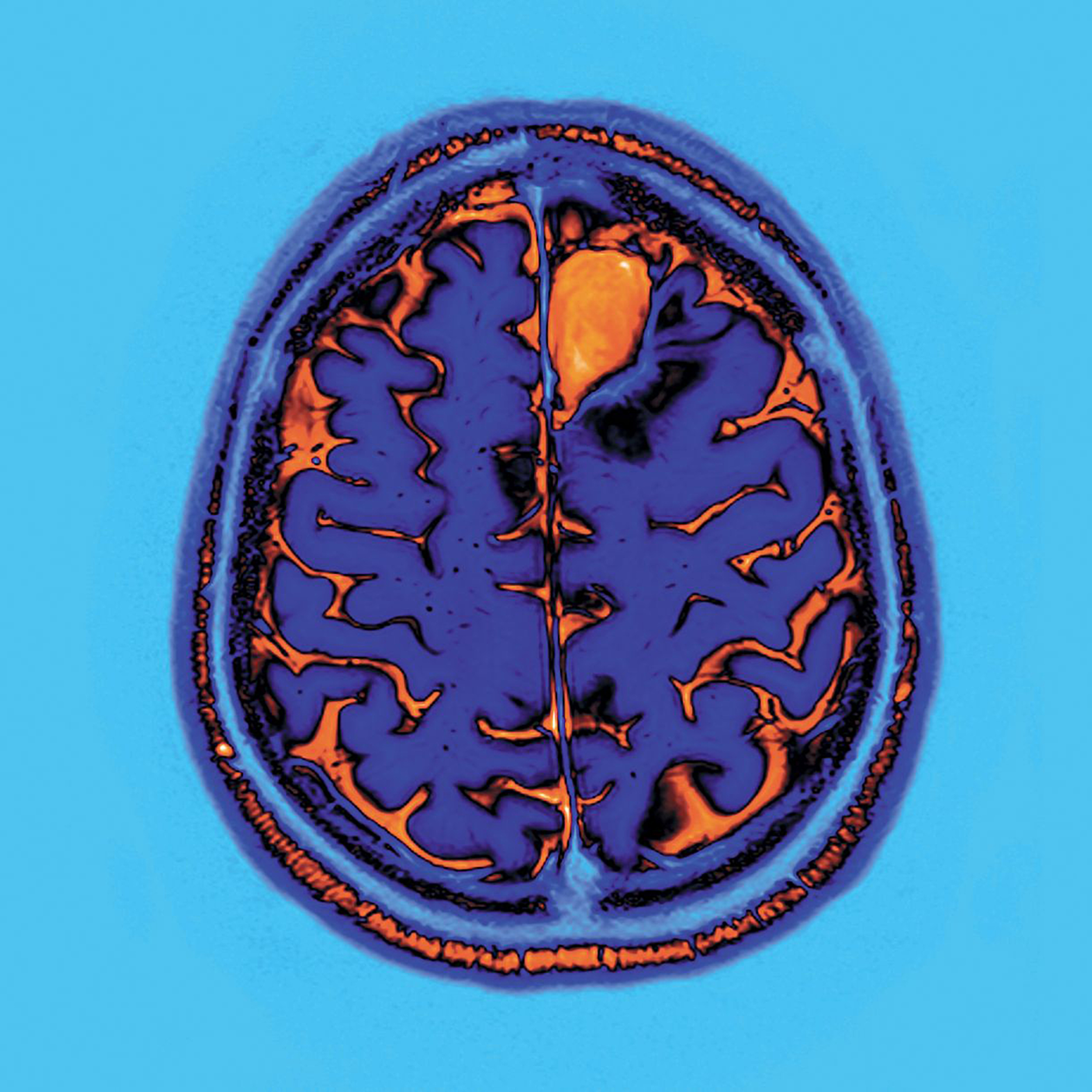
Autism spectrum disorder (ASD) is a neurodevelopmental disorder characterized by communication disorders, diminished social interaction skills, together with sensory and behavioral abnormalities. Genetic and brain imaging studies suggest that abnormal brain development, notably concerning the formation of neural networks and synapse function, could contribute to the onset of ASD.

In recent years, neuroimaging studies have shown that individuals with ASD have functional abnormalities affecting certain areas of the brain known to be responsible for processing emotions, language, and social skills. Studies on brain connectivity in individuals with ASD have notably evidenced diminished “long-distance” connections, in contrast to increased “short-distance” connectivity. These results formed the basis for the development of a theoretical model aiming to shed light on ASD, according to which the deficit in terms of social attention and information processing observed (difficulty in understanding a situation as a whole, attention to certain details) is explained by a saturation of information processed by the brain, linked to the increased neural connectivity between adjacent regions of the brain.

Nonetheless, Professor Josselin Houenou, Professor of Psychiatry at UPEC, Inserm researcher, practitioner at Henri Mondor University Hospital-AP-HP, and last author of the study published in Brain, explains: “This model is based on the study of heterogeneous pediatric populations, comprising autistic children of varying ages and with highly diverse symptoms, and on relatively unspecific neuroimaging methods which do not allow reliable measurement of “short-distance” connectivity.”

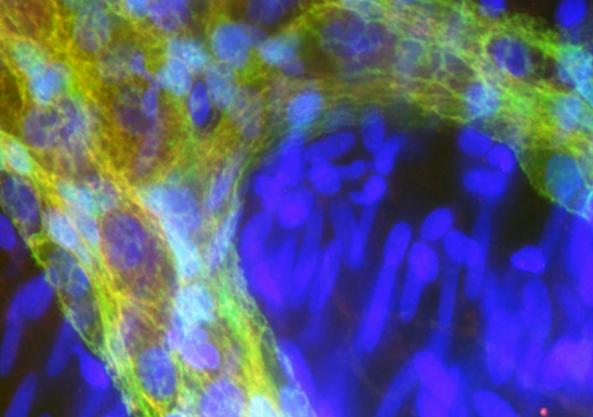
In order to test the current model, the authors of this study used an innovation designed by Miguel Gevara, Jean-François Mangin, and Cyril Poupon at NeuroSpin, namely an atlas specifically focusing on the tractography analysis of 63 “short-distance” connections using images obtained by diffusion MRI (dMRI). dMRI evidences white matter bundles in the brain in vivo, by measuring the diffusion of water molecules, notably along the axons. Tractography can then be used to reconstruct, step by step, the nerve fiber bundle paths, represented in tractogram form.

The authors were thus able to study the links between “short-distance” connectivity and social cognition in a homogeneous adult population of individuals with ASD, originating from the InFoR-Autism cohort* (27 patients with ASD, without intellectual deficit, and 31 control subjects), with one of the most extensive databases per patient and per control.

Discovery of diminished “short-distance” brain connectivity associated with diminished social interaction skills and empathy

The strength of the InFoR-Autism cohort* lies in the wealth of data collected for each subject included. We were thus able to establish a link between the neuroimaging results obtained and the social cognition scores, measuring social skills, empathy, social motivation, etc.,” explains Dr. Marc-Antoine d’Albis, Hôpital Henri Mondor, Inserm U955, lead study author.

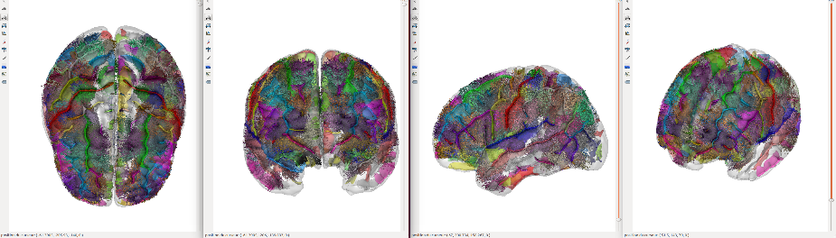
The results show that subjects suffering from ASD display diminished connectivity in 13 “short-distance” bundles, compared to control subjects. Furthermore, this abnormal “short-distance” bundle connectivity is correlated with a two-dimensional deficit in social cognition (namely social interactions and empathy) in subjects with ASD.

Key: 3D image of “short-distance” bundles, © Miguel Guevara

These preliminary results are well and truly at variance with the current theoretical model according to which diminished social attention and information processing in individuals with ASD is explained by increased neural connectivity between adjacent regions of the brain. These now need to be confirmed by studies in children with ASD, as explained by Professor Josselin Houenou.

According to Professor Josselin Houenou, “These preliminary results nonetheless suggest that these “short-distance” connectivity abnormalities could contribute to certain social cognitive deficits observed in autistic subjects. Similar studies now need to be conducted in children in order to confirm the results obtained in adults. Pediatric cohorts allow children of varying ages – and therefore brain maturation – to be studied, which entails taking into account a much larger population of subjects.

If these initial conclusions were confirmed, this would allow the development of new therapeutic approaches for social cognition deficits to be envisaged. For instance, transcranial magnetic stimulation could be explored since brain connectivity between adjacent regions is located on the surface of the brain.”

*InFoR-Autism

Since late 2012, Fondation FondaMental, Inserm, Inserm Transfert, and Institut Roche have been partners in the InFoR Autism scientific program which aims to monitor clinical, biological and brain imaging variables with a view to studying the stability and progression of ASD. 117 patients and 57 healthy volunteers aged 6 to 56 years in total have been included in the study. This cohort has one of the largest databases (clinical, biological, eye tracking, and imaging) per patient and per control.

“Nested sequences”: an indispensable mechanism for forming memories

©Photo by Annie Spratt on Unsplash

A research team from CNRS, Université PSL, the Collège de France and Inserm has just lifted part of the veil surrounding brain activity during sleep.  Though we know that some neurons are reactivated then to consolidate our memories, we did not know how these cells could “remember” which order to turn on in. The researchers have discovered that reactivating neurons during sleep relies on activation that occurs during the day: “nested” theta sequences. These results were published on November 9, 2018 in Science.

Repetition is the best method for memorization, for neurons themselves. This is the principle behind what neurobiologists call sequence reactivations: during sleep, neurons in the hippocampus related to a task activate very quickly in turn in a precise order, which consolidates the memory of this task. Sequence reactivations are fundamental for long-term memorization and for exchanges between the hippocampus and the rest of the brain. These are only present at rest so they appear after initial neuron activity, which implies that they “memorize” the order they should turn on in. But by which mechanism?

A team of researchers from the Centre interdisciplinaire de recherche en biologie (CNRS/Inserm/Collège de France)1 has answered this question by studying activity sequences in rats’ place cells. These are hippocampal neurons that turn on by following the animal’s position in the environment when it moves. First slowly, while it moves, then very quickly during reactivations of sequences during sleep. But neurobiologists know another type of sequence, called theta sequences, which quickly repeat the activation of the same place cells when the animal moves, in parallel with slow sequences. These theta sequences are therefore called “nested”.

Which of these sequences, slow or nested, is necessary for the appearance of sequence reactivations, and therefore causes the consolidation of memories during sleep? Using an ingenious system, the researchers discovered what deactivates nested sequences, without affecting slow sequences: the animals are transported on an electric train, in a car with a treadmill (see image). When the treadmill is stopped, the nested sequences disappear; they return when the treadmill starts again.

The researchers then observed that after several circuits in the train with the treadmill stopped, place cells in the rats’ hippocampi did not reactivate during sleep in the same order as when awake. On the contrary, after one train circuit with the treadmill on, the sequence reactivations are indeed present. So it is these nested theta sequences during movement that are indispensable for the consolidation of memory during sleep.

The researchers are continuing their work, looking now at the integration of non-spatial information such as objects or textures in nested sequences, and their reactivation during sleep.


  1. Associated member of the Université PSL, since 2009 the Collège de France has been conducting a voluntaristic policy for welcoming independent teams that benefit from pooled technical or scientific services and an exceptional multidisciplinary environment. Twenty-two teams are currently housed in the Centre interdisciplinaire de recherche en biologie and in the Instituts de chimie et de physique du Collège de France. Supported by the CNRS in particular, this is available to both French and foreign researchers. It contributes to making Paris a major player as an attractive place for research.

An Unbalanced Maternal Diet Affects the Digestive System of Offspring

©Inserm/Naveilhan, Philippe/U913/IMAD

Inadequate protein intake in a gestating female is linked to lasting digestive abnormalities in her offspring. When studying the link between perinatal malnutrition and digestive system in rats, a team of researchers from Inserm in conjunction with Inra, Université de Nantes and University Hospital Nantes discovered functional digestive abnormalities in young rats and an inappropriate response to stress. Although conducted in animals, this research once again demonstrates the incidence of perinatal stress on health in adulthood and draws our attention to the impacts of restrictive diet-related or involuntary deficiencies during pregnancy. This research has been published in The FASEB Journal.

Gestation is an extremely vulnerable time when it comes to normal fetal development and the maintenance of good health throughout life. Several studies have already revealed effects of maternal malnutrition on the cardiovascular and cognitive development of offspring, with repercussions in adulthood. This time, the focus was on the digestive system of young rats, just before they reached adulthood. This involved halving the protein intake of the mothers throughout the gestation and lactation periods and then reintroducing a normal diet once the offspring were weaned.

The researchers began by studying digestive system functioning in the young rats, particularly intestinal motility and permeability. Motility corresponds to the frequency and speed of stool passage. Permeability represents the capacity of nutrients and other molecules to pass through the intestinal wall into the bloodstream. What they observed was an increase in these two parameters in the animals whose mothers had had an inadequate protein intake. They also detected high levels of stress hormone.

To understand these phenomena, the animals were subjected to a model of psychological stress. In the control rats, the stress situation (mimicked by isolating them on a platform with no way out, placed in the middle of a basin of water) triggered accelerated motility and increased permeability. However, this response was altered in the young rats whose mothers had consumed insufficient protein. Their basic digestive activity was greater but did not increase during the stressful event. “Their stress response appeared inappropriate, as if perinatal stress had desensitized the response to acute stress during future life”, suggests Hélène Boudin, Inserm researcher and co-director of this research.

The researchers then examined whether the digestive nervous system had been modified. They observed that the stress hormone induced an excess of neurons stimulating intestinal motility and permeability. These neurons, in addition to their large number, present the disadvantage of being unable to eliminate waste and toxins naturally. And this is “indicative of a poor capacity to respond to stress” clarifies Boudin.

These various observations enabled the researchers to hypothesize that: perinatal nutritional deficiency leads to an increase in the stress hormone in offspring, itself inducing long-term remodeling of the digestive nervous system. This would be responsible for digestive disorders that can weaken the intestine and impact on wellbeing and quality of life.

This research also reinforces the hypothesis of the prenatal origin of certain digestive diseases and disorders. In addition, the mechanisms brought into play by the perinatal stress studied could be shared with those present in other diseases of malnutrition (overeating and undernourishment) and possibly also in neurodevelopmental diseases including some psychiatric diseases” clarifies Boudin. For her, this research once again shows the incidence of perinatal stress on health in adulthood and draws our attention to the impacts of restrictive diet-related or involuntary deficiencies during pregnancy.

Tools: Sensory Organs in Their Own Right ?

©Photo by Adi Goldstein on Unsplash

What if by holding a tool we could perceive our environment through touch – using the whole tool, and not just the tip? A study by Inserm researchers at the Lyon Neuroscience Research Center (Inserm/Université Jean Monnet Saint-Etienne/Université Claude Bernard Lyon 1/CNRS) has shown just that – the capacity of the human brain to incorporate a tool as an actual sensory organ. This research, published in Nature, raises the question of a new paradigm concerning the sense of touch, its interpretation when developing our use of tools, and in its medical applications – particularly prosthetics.

The sense of touch is essential to the control we have over our hands and, by extension, over the tools we use to perceive our environment through touch.

Inserm researchers at the Lyon Neuroscience Research Center (Inserm/Université Jean Monnet Saint-Etienne/Université Claude Bernard Lyon 1/CNRS) examined the mechanisms which enable the brain to locate touch through tools. To do this, they used three complementary approaches which involved tapping a wooden rod held in the hand.

The first approach involved tapping different locations of a rod held by a volunteer whose vision was obstructed and then asking him or her to locate the impact. Irrespective of where the rod was tapped, the volunteer was able to sense the location of the impact with the same accuracy as when it was his or her own arm which was tapped.

These results demonstrate the human capacity to “incorporate” the entirety of a tool held in the hand as if it was part of the body, with the brain integrating it as a sensory organ in its own right.

The second approach involved recording the vibrations of the rod perceived at the base of the wrist and on the skin of the hand holding it. The researchers observed that the characteristics of the rod’s vibrations transmitted to the hand predictably depended on the location of the impact.

Finally, in the third approach, the characteristics of the vibrations recorded in the second approach were processed by a computerized simulator of skin responses, thereby modeling the responses, to the vibrations, of the mechanoreceptors (sensory neurons of the skin) in contact with the rod. The research team observed that the mechanoreceptors were able to very precisely decode the vibratory motifs of the rod. Since these motifs strictly depend on the location of the impact, the brain is able to interpret their “profile” sent by the mechanoreceptors and, as a result, locate the area of impact.

This study shows that the human brain treats the tools as sensory extensions to the body, a mechanism which the research team suggests calling “sensing with tools“. The phenomenon newly-described here represents a new paradigm which could improve knowledge of tool-incorporation phenomena in humans and the sensory perception of the visually-impaired, as well as the understanding of prosthesis use in amputees.

An Improved Diet Could Protect Against Depression

©Brooke Lark on Unsplash

Researchers from Inserm and Université de Montpellier have produced a meta-analysis of the links between a simple-to-use score measuring adherence to dietary guidelines and the development of depressive disorders. The researchers were able to show that adopting a Mediterranean diet (high in fruit, vegetables, fish and whole grains) was linked to a 33% reduction in the risk of depression. These results were published in Molecular Psychiatry.

Globally, more than 300 million people suffer from depression, representing a frequency of 7% for women and 4% for men. It is the costliest brain disorder in Europe. Using data from 36,556 adults, researchers from Inserm and Université de Montpellier were able to show that adopting a Mediterranean diet (high in fruit and vegetables, fish and whole grains) was linked to a 33% reduction in the risk of depression.

Their study also shows that a pro-inflammatory diet (high in saturated fatty acids, sugar, and processed foods) was associated with a higher risk of depression. The chronic inflammation potentially induced by this type of diet could be directly implicated in the onset of depression. According to Tasnime Akbaraly, Inserm researcher responsible for the study: “These findings support the hypothesis that avoiding pro-inflammatory foods (in favor of an anti-inflammatory diet) helps prevent depressive symptoms and depression.

Other studies have also shown the importance of diet in the functioning and composition of the gut microbiota, directly impacting the link between the gut and the brain – with this relationship playing a key role in depressive disorders.

For Tasnime Akbaraly: “The results of our study show the importance of our dietary habits in the development of depressive disorders and encourage the widespread provision of dietary advice at medical consultations”.

Additional clinical trials are necessary to evaluate the efficacy of diets such as the Mediterranean diet in reducing the risk, severity and recurrence of depressive episodes.

Center-based and Other Forms of Childcare: An Impact on the Behavioral and Emotional Development of Children.

Crédits: AdobeStock

Researchers from Inserm, Sorbonne Université and Université de Bordeaux have published a study based on data from 1,428 children showing that access to a center-based form of childcare between the ages of 0 and 3 years is linked to fewer emotional and peer relationship problems in later life compared with other forms of childcare. These findings were published on October 1, 2018 in Journal of Epidemiology and Community Health.

Researchers from Inserm, Sorbonne Université and Université de Bordeaux (Bordeaux Population Health for Unit 1219) studied in France the impact of the form of childcare used during the three first years of life on the behavioral and emotional development of children. This study is based on 1,428 children from the EDEN cohort (study of the pre- and early postnatal determinants of child health and psychomotor development), based in Nancy and Poitiers which followed up mothers during pregnancy as well as their children up to the age of 8 years.

The mothers reported the main childcare method used for their child at the ages of 4 months, 8 months, 1 year, 2 years and 3 years: informal care (mainly the parents themselves or sometimes the grandparents, neighbors, etc.), home daycare (childminder), or center-based childcare (childcare center, daycare center). Then at 3 years, 5.5 years, and 8 years, they completed the “Strengths and Difficulties Questionnaire” which measures behavioral and emotional symptoms using five scales (emotional symptoms, peer relationship problems, hyperactivity/inattention, conduct problems, and prosocial behavior).

Following adjustment for various sociodemographic characteristics, the study shows that compared with children who stay at home before going to preschool, those attending center-based childcare were around three times less likely to experience emotional problems or peer relationship difficulties at a later stage (between the ages of 3 and 8 years).

Their behavior was also more prosocial, i.e. more empathic (for example, sharing, kindness towards younger children).

“Access to a center-based form of childcare between the ages of 0 and 3 years represents an opportunity for the children concerned because it is linked to better psychological and emotional development down the line.” explains Inserm researcher, Maria Melchior. These findings taken from data from two cities must now be confirmed on a larger scale.

Prenatal exposure to cannabis impacts sociability of male rats

Crédits:  Matteo Paganelli – Unsplash

In a study performed in rats, researchers from Inserm and Aix-Marseille University reveal that prenatal exposure to cannabinoids has sex-specific effects on adult offspring. According to this study published in eLife, consuming cannabis during pregnancy can lead to behavioral and neuronal deficits in male descendants. The findings also point towards a potential pharmacological strategy to help reverse these effects in humans.

A study performed in rats by researchers from Inserm and Aix-Marseille University at the Mediterranean Institute of Neurobiology suggests that using cannabis during pregnancy can lead to less sociability and increased neuronal excitability in males.

According to Olivier Manzoni, Inserm Research Director in charge of the study at the Mediterranean Institute of Neurobiology and Director of the Inserm-Indiana University International Associated Laboratory CannaLab, says: “As cannabinoids can cross the placenta, they may interfere with fetal endocannabinoid signaling during neurodevelopment, which is involved in regulating a variety of processes (synaptic plasticity, appetite, pain sensation), and mediating the pharmacological effects of cannabis. This could in turn lead to some serious long-term deficits. But despite increasing reports of cannabis consumption during pregnancy, the long-term consequences of prenatal cannabinoid exposure remain incompletely understood.”

To fill this knowledge gap, the researchers in Marseille together with their counterparts from the University of Rome (Italy) and Indiana University (USA) examined how prenatal cannabinoid exposure influences the synaptic and behavioral functions of the medial prefrontal cortex – a brain region often implicated in neuropsychiatric disorders – in adult male and female rats.

Their results revealed that males exposed to cannabinoids while in the uterus were less sociable than normal animals, and spent less time interacting with others. Their social behaviors (interactions and play) were impaired, while the number of attacks among males remained unchanged.

Additionally, the researchers saw that the exposed males had a heightened excitability of pyramidal neurons in the prefrontal cortex and a loss of the synaptic plasticity normally mediated by the endocannabinoid system. None of these effects were seen in females.

The deleterious effects of prenatal exposure to cannabinoids on social behavior were specific to male offspring only,” explains co-first author and doctoral student Anissa Bara. “But while social interaction was specifically impaired in males, locomotion, anxiety and cognition remained unaffected in both sexes, suggesting sex-specific behavioral consequences.”

The results also revealed that expression of the mGlu5 gene – an effector of the endocannabinoid system in the prefrontal cortex – was reduced in the males exposed to cannabinoids in utero. The team discovered that amplifying mGlu5 signaling could normalize the synaptic and behavioral deficits induced by prenatal exposure to cannabinoids partly by activating the cannabinoid type 1 receptor (CB1R). Similarly, later tests also revealed that enhancing levels of anandamide (a type of endocannabinoid) in exposed males helped to restore normal social behaviors via the CB1R receptor.

However, prenatal exposure to cannabis does not leave females entirely unscathed. The researchers observed major modifications in the expression of synaptic protein genes in females exposed to cannabinoids in utero. The functional and behavioral consequences of these modifications remain to be identified.

“Altogether, these results provide compelling evidence for sex-specific effects of prenatal cannabinoid exposure,” concludes co-first author Antonia Manduca, also an Inserm Postdoctoral Researcher at the Mediterranean Institute of Neurobiology. “The fact that increasing mGlu5 signaling and enhancing anandamide levels helped to reverse the negative effects of early exposure in rats also hints at a new pharmacological strategy that could one day be trialled in humans.”

When Infection Strikes, Our Brain and Immune System Join Forces

©Adobestock

When infection strikes, what if our immune system was not alone in the fight? What if its major ally was in fact the brain? Researchers from Inserm, CNRS and Aix-Marseille University (AMU) have observed mechanisms of cooperation between the nervous system and the immune system in the response to pathogenic aggressions. This research, published in Nature Immunology, reveals the role of the brain in regulating the inflammatory reaction induced by the immune system in the event of infection and its protective effect against a potential self-destructive exacerbation of that inflammation.

In the event of infection with a virus or other pathogenic organism, the immune system springs into action to eliminate the infectious agent. The immune cells release inflammatory molecules called cytokines, which are responsible for the inflammation process necessary to fight the dissemination of pathogens in the body. There are times, however, when the inflammatory reaction is excessive and toxic to the body. It can provoke lesions in the infected organs which, when too severe, can lead to death.

Previous studies have shown that, in the event of infection, the brain is mobilized to regulate the inflammatory reaction. When it detects the cytokines produced by the immune cells, the brain induces the blood secretion of hormones known to be negative regulators of inflammation: glucocorticoids. The properties of these hormones are widely used in medicine in many pathological conditions, but little is known about their specific mode of action.

In this context, researchers from Inserm, CNRS and Aix Marseille University (AMU) at the Center of Immunology Marseille-Luminy (CNRS/Inserm/AMU) studied, in mice, the mechanism of action of the glucocorticoids produced following activation of the brain in controlling the intensity of the inflammatory reaction caused by viral infection.

Their findings show that glucocorticoids regulate the activity of a population of immune cells which produce inflammatory cytokines and exert a major antiviral and anti-tumor action: natural killer (NK) cells.

These cells possess a receptor which is activated by the glucocorticoids produced after the infection. This activation leads to the expression on the surface of the NK cells of a molecule called PD-1, which is attracting considerable interest within the medical community and is targeted in many cancer treatments due to its inhibitory action on the activity of the immune cells that express it.

The researchers observed that mutant mice which do not express the glucocorticoid receptor in their NK cells were more likely to develop a severe hyper-inflammation reaction and die in the event of an infection. This research demonstrates that expression of the glucocorticoid receptor by the NK cells is necessary to regulate the intensity of the inflammation so that the response against the virus does not become toxic to the body. In addition, the study shows that this regulation is governed thanks to the inhibitory effect of PD-1 which, in the infectious setting, limits the production of inflammatory cytokines by the NK cells.

According to Sophie Ugolini, Inserm researcher and study director: “The most unexpected part of our discovery was that this regulation prevents the immune system from going into overdrive and destroying healthy tissues while fully maintaining its antiviral properties necessary for the effective elimination of the virus. “

This study could enable the development of new therapeutic strategies which would target this regulation pathway. Aside from infections, the researchers especially hope to explore the potential role of this regulation pathway in some cancers.

Myositis: A New Classification Representing a Decisive Step Towards Improved Diagnosis and Personalized Treatment

Coupe transversale de muscle humain, régénération de fibres musculaires après un traitement de la myopathie de Duchenne. Crédits: Inserm/Fardeau, Michel

Prof. Olivier Benveniste’s Inflammatory myopathies and innovative targeted therapies team at the Institute of Myology has produced a new classification of the different forms of myositis (rare inflammatory muscle diseases). Four new types of myositis taking into account the various clinical criteria of patients have now been defined. This research, involving teams from the Institute of Myology, Inserm, the Paris public hospitals system (AP-HP) and Sorbonne Université, was published in September in JAMA and paves the way for reliable diagnosis and personalized treatments.

Myositis (rare inflammatory muscle diseases) is a group of rare autoimmune muscle diseases in which the immune system, in charge of protecting the body from external aggressions (bacteria, viruses…), dysfunctions and attacks the body (in this case, the muscle). These diseases affect between 3,000 and 5,000 adults and children in France.

While the different forms of myositis all have an autoimmune component, each has its own specific triggering mechanisms. Until now, three types of myositis (polymyositis, dermatomyositis, inclusion body myositis) had been identified according to a classification system established in 1975 and updated in 2017 (ACR/EULAR rheumatologist criteria) based essentially on clinical and histological criteria. Prof. Olivier Benveniste, head of the Inflammatory myopathies and innovative targeted therapies team at the Institute of Myology who has been monitoring patients daily at Pitié-Salpêtrière Hospital AP-HP over the past 20 years, had identified major diagnostic errors related to this incomplete and, as a consequence, non-homogeneous, classification which would sometimes even lead to errors in patient treatment. Some patients mistakenly diagnosed with inclusion body myositis were given high-dose steroids which made their condition worse.

That is why, together with his team and the Center of Reference for Neuromuscular Diseases of the Institute of Myology, Prof. Benveniste launched a study on 260 patients in whom he collected and analyzed the various clinical characteristics –and particularly the presence of autoantibodies, which are sometimes causes or consequences of the disease. Using innovative statistical methods, without a priori, in which the mathematical algorithm works unsupervised to aggregate similar patients into subgroups (cluster analysis), the researchers revealed a new classification with four major types of myositis: inclusion body myositis, dermatomyositis, immune-mediated necrotizing myopathy, anti-synthetase syndrome (with polymyositis no longer forming a type of myositis as such).

Characteristics of the four forms of myositis:

Inclusion body myositis: This form of myositis more often affects men over 60 years of age. It progresses slowly but ultimately leads to a highly disabling motor deficit. It particularly affects the quadriceps (thigh muscles used to climb stairs, get up from a chair, maintain stability when walking…), the muscles used to close and shake hands and the muscles used for swallowing. This disease is resistant to standard immunosuppressive treatments such as steroids. It is due to the presence of an inflammatory reaction (myositis) in the muscle and a neurodegenerative process related to Alzheimer’s disease (presence of inclusions).

Dermatomyositis: This form more often affects women. Children can also be affected. There is an associated cancer risk in the most elderly subjects (usually after 60 years). In addition to myositis, which causes predominant muscle weakness in the shoulders, this disease is characterized by the presence of typical dermatological lesions. Dermatomyositis is due to an imbalance of the immune system involving type 1 interferon that helps protect against viruses. New therapies specifically targeting this interferon pathway are under development. Dermatomyositis-specific antibodies are anti-Mi2, anti-SAE, anti-NXP2, and anti-TIF1 gamma.

Immune-mediated necrotizing myopathy: This is characterized by muscular weakness affecting patients of all ages. In the absence of treatment, this type of myositis leads to the most severe and disabling muscle atrophy. This disease is related to the presence of two specific anti-SRP or anti-HMGCR antibodies which directly attack and destroy the muscles. Anti-HMGCR may appear after taking statins. Treatment aims to remove these antibodies.

Anti-synthetase syndrome: This disease affects muscles but also joints (leading to rheumatism), and the lungs (leading to shortness of breath that is sometimes severe). Here too, certain antibodies appear to be responsible: anti-Jo1, anti-PL7 and anti-PL12.

This new classification is decisive in establishing a diagnosis and offering personalized treatment to patients.

“We realized that the current myositis classification was unsuitable and could often lead to the failure of a potential treatment due to non-homogeneous patient groups within a given trial. So our aim was to define a classification based on phenotypic, biological and immunological criteria in order to better diagnose the different types of myositis and ultimately find suitable treatments for patients. This new classification is becoming a reference because even the FDA, which up until then was using the US classification, recommends using our research as a basis. ” explains Prof. Benveniste.

Using Light to Switch Off Nicotine Addiction?

Crédits: AdobeStock

Researchers from Inserm, CNRS and Sorbonne Université at the Paris-Seine Neuroscience laboratory1 in collaboration with Institut Pasteur2, New York University (NYU) and University of California Berkeley (UC Berkeley) have succeeded in controlling the activity of nicotine receptors in the brains of mice. To do this, they developed an optogenetic pharmacology strategy in which light is used to block the nicotine receptors, with the result being the possibility to control the addictive effects of nicotine. This study was published in eLIFE on September 4, 2018.

Every year, more than 7 million people die from smoking worldwide. Nicotine, the main addictive substance in tobacco, acts on the brain by binding to the nicotine receptors. At present, standard pharmacology techniques do not enable precise and reversible action on these receptors. That is why researchers had the idea of producing molecular tools able to interrupt the functioning of these receptors in the brain through the use of light.

In this study, the researchers modified the nicotine receptor in mice in order to attach a chemical nanoswitch which reacts to light. Under the effect of violet light, the switch folds away, preventing the nicotine from binding: the receptor is “off”. Under the effect of green light, or in darkness, the switch unfolds and allows the nicotine to act: the receptor is “on”.

For this study, the researchers focused on a specific nicotine receptor –type β2– and on a key area of the reward circuit, which delivers dopamine. When nicotine is injected intravenously, the dopamine neurons respond with an increase in their electrical activity, with the resulting dopamine release being key to acquiring the addiction. In this research, this effect of nicotine was found to be greatly reduced when the nanoswitch was triggered by the violet light but was rapidly restored under green light.

The researchers then demonstrated that it was possible to inhibit the attraction for nicotine by triggering this switch.

To do this, they compared the time spent by mice in two compartments, with and without nicotine. Under green light, when nicotine could exert its effect, they observed that the animals preferred the compartment with nicotine. However, under violet light, the mice spent equal amounts of time in each compartment, proving that they were no longer attracted by the nicotine.

Crédits : A.Mourot/S.Mondoloni/R.Durand de Cuttoli

This study proves that it is possible to manipulate the attraction for nicotine in mice, both rapidly and reversibly. Alexandre Mourot, Inserm researcher in charge of the study, states: “This innovative technology provides us with a better understanding of the role of the various nicotine receptors and neuron pathways in acquiring and maintaining nicotine addiction, and also in the processes of withdrawal and relapse. This step is particularly important when it comes to identifying suitable new therapeutic targets for fighting nicotine addiction“.

1 Laboratory located on the Pierre et Marie Curie campus of Sorbonne Université, shared with CNRS and Inserm

2 At the Genes, synapses and cognition laboratory (Institut Pasteur/CNRS).

fermer