Menu

New forms of expression of the variant of Creutzfeldt-Jakob disease observed within the scope of experimental transmissions

A team of French researchers from the CEA, AP-HP, CNRS, Inserm, Inra, ENVT, Institut Pasteur and MacoPharma have demonstrated that multiple variants of prions can coexist and manifest themselves in different clinical forms depending on the conditions of transmission. The results of this study are published in Nature Communications, dated 2 November 2017.

Fewer than 250 cases of variant Creutzfeldt-Jakob disease (vCJD) have been identified in the world, mainly in the United Kingdom, following the exposure of human populations to Bovine Spongiform Encephalopathy (BSE, or ‘mad cow disease’). However, over 99% of the human infections by these prions of bovine origin could remain silent according to British studies carried out on samples of appendices, which suggests a risk of inter-human transmission linked to such apparently healthy carriers, in particular through blood transfusions.

Assessment of the transfusional risk

Within the scope of the assessment of the transfusional risk linked to vCJD, researchers from the CEA, La Pitié-Salpêtrière university hospital, AP-HP, CNRS, Inserm, Inra, ENVT, Institut Pasteur and MacoPharma[1] exposed mice and macaques to blood products from vCJD-infected donors.

Through techniques traditionally used for the diagnosis of prion diseases (histology, biochemistry), the researchers found few cases of transmission of vCJD in its classic form (less than 3% of the animals exposed). These cases were mainly observed when the donors were strongly infected, which corresponds to the situation observed in humans[2].

Observation of myelopathies

On the other hand Emmanuel Comoy, CEA researcher and principal author, specifies, “in a significantly higher proportion (two to five times more according to the models studied, i.e. almost 10% of the animals exposed), some recipient animals developed fatal neurological syndromes different from vCJD”.

These syndromes do not present all of the classic characteristics of prion diseases (vacuole formation in the brain and accumulation of an abnormal form of the prion protein[3] resistant to deterioration by enzymes). They are referred to as myelopathies in affected primates because they are mainly centred on the spinal cord. These syndromes show themselves to belong to prion diseases, since their retransmission to the mice leads to the appearance of the classic characteristics of prion diseases.

Infection by healthy carriers

The myelopathic form of the disease was observed in animals transfused with blood from apparently healthy donors, i.e. donors exposed to prions and without an identifiable pathology, as well as from blood from patients presenting the clinical signs of vCJD.

The study thereby reveals a diversity of syndromes, from the healthy carriers to the typical form of vCJD: multiple variants of prions could therefore coexist and emerge under different clinical forms depending on the conditions of transmission (route of exposure, dose, nature of the contaminating product, aggregated or dispersed state of the prions).

In a context where human exposure through food intake to the BSE agent could have resulted in the emergence of numerous, apparently healthy, carriers, the researchers note that their study – which challenges the unicity of the vCJD/BSE origin – underlines the difficulty in detecting and preventing the transmission of all forms of prion infections. These questions are currently the subject of additiona.

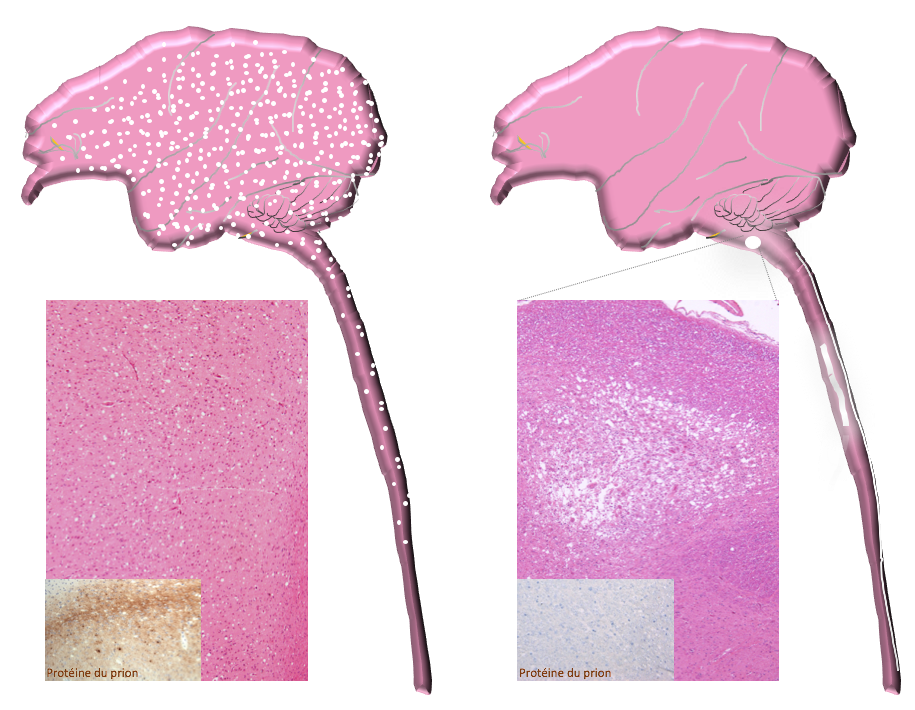
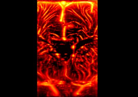
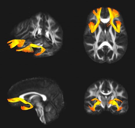
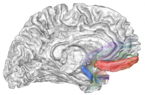
Comparison of the lesions observed: The macaques developing a vCJD present a vacuolation and an accumulation of the abnormal prion protein in the entire nervous system (figure on left), whereas the lesions in the myelopathic macaques are concentrated in the lower cervical spinal cord and the brain stem, with no accumulation of abnormal prion protein detectable using classic techniques (figure on right). © CEA/E. Comoy.


[1] Institut François Jacob (CEA), Brain and spinal cord institute (CNRS/Inserm/UPMC/La Pitié-Salpêtrière university hospital, AP-HP), Neuromyogenics institute (CNRS/Inserm/Univ. Cl. Bernard Lyon 1/Lyon Civil Hospices), Host-pathogen Interactions laboratory (Inra/ENVT), Macopharma, Biology of Infections unit (Institut Pasteur), laboratory of Neurodegenerative diseases: mechanisms, therapies, imaging (CNRS/CEA/Univ. Paris Sud).

[2] Over the last ten years, four cases of contamination by the vCJD agent in the United Kingdom have been attributed to the transfusion of non-deleukocyted blood products (so-called ‘whole’ blood, whose white cells have not been removed) coming from donors who themselves developed a vCJD shortly after their blood donation. At present, blood products are systematically deleukocyted.

[3] A pathological prion is a pathogenic agent made up of a protein whose conformation or withdrawal is abnormal and which, unlike infectious agents such as viruses or bacteria, or even parasites, has no nucleic acid (DNA or RNA) as a medium for infectious information.

Interaction between brain and heart may be new indicator of state of consciousness

©Fotolia

How do we know whether a patient is conscious when he or she is unable to communicate? According to an Inserm study conducted in 127 patients aged 17 to 80, changes in heartbeat in response to sound stimulation is a good indicator of state of consciousness. This is what Inserm researcher Jacobo Sitt and his team, based at the Brain & Spine Institute (ICM) at Pitié-Salpêtrière Hospital, AP-HP, demonstrate in an article published in Annals of neurology. This easy-to-perform examination complements the existing tests and enables finer predictive diagnosis, useful for both doctors and the patient’s loved ones.

The study of consciousness disorders distinguishes the vegetative state, in which the patient is awake but not conscious, from the minimal state of consciousness, which involves a certain degree of consciousness. Distinguishing between these states is very important in order to establish a prognosis on the patient’s neurological outcome, inform the patient’s loved ones, and implement suitable treatment. The tools developed up until now to determine state of consciousness, such as the electroencephalogram (EEG), functional MRI or PET scan, all concentrate on the brain. These methods require either heavy equipment or complex analysis.

Researchers from Inserm have used a novel approach, which consists of exploring the interaction between the heart and the brain.

Previous studies revealed that the “unconscious” processes of the autonomic nervous system, such as breathing or heartbeat, could be modulated by conscious cognitive processes. The perception of external stimulation, auditory stimuli, for example, could therefore be expressed by an effect on cardiac activity, and all the more easily if the subject is conscious.

By studying the data of 127 patients in vegetative or minimally conscious states, researchers observed that cardiac cycles were indeed modulated by auditory stimulation only in conscious or minimally conscious patients. They also showed that these results complemented those obtained with an EEG. Combining these tests (cardiac and EEG) markedly improves performance when predicting a patient’s state of consciousness.

These findings open up new perspectives for an overall approach in evaluating patient state of consciousness. The researchers now wish to extend the scope to include other physiological signals modulated by conscious processes, such as breathing or pupil dilation, in order to develop a comprehensive tool for better bedside evaluation of patient state of consciousness.

What does the auditory test involve?

The test involves playing sound sequences to the patient, which are repetitive to begin with and then, at random and infrequent intervals, present variations. During these variations, the researchers are able to determine whether there is a change in heartbeat, which would mean that the patient is aware of ambient sounds.

Is There a Long-term Link Between Childhood Environmental Harshness and Health?

©Fotolia

 A study, conducted by Coralie Chevallier, Inserm researcher at Unit 960 “Cognitive Neuroscience Laboratory” (Inserm/ENS), suggests that an unfavorable childhood environment leads to earlier reproduction and a decreased effort in looking after health during the course of life. These findings have been published in Evolution and Human Behavior.

Current models derived from evolutionary biology show that it can be advantageous for an organism living in a harsh environment (with low levels of resources or high levels of stress or violence) to adopt behaviors favoring benefits in the short term. While these benefits are more modest than those obtained over the long term, they are at least more certain. As such, the receipt of signals by a juvenile organism indicating that the environment is dangerous leads it to adapt its strategy in favor of reproducing sooner – to the detriment of longer-term investments in body maintenance and repair.

On the basis of this observation in animals, Coralie Chevallier, Inserm researcher at the “Cognitive Neuroscience Laboratory” of the École Normale Supérieure (ENS) in Paris, conducted a study to determine whether this model could be applied to humans. To do this, she and her team selected a panel of 1,000 men and women, between the ages of 19 and 87, and representative of the French population.

The panel members answered a series of questions concerning, for example, their age, gender, household composition, socioeconomic status and level of education. They then received a second questionnaire addressing three main themes:

  • childhood environment: parental involvement, upbringing, personal experiences, family difficulties, etc.;
  • reproductive strategy: number of children, age at first pregnancy, age of first sexual intercourse, and number of short-term partners;
  • state of health: body mass index (BMI), how they feel about their health, their desire to remain in good health, and tobacco consumption.

Analysis of these data shows a link between childhood environmental harshness and health and reproductive strategy, with sexual activity starting earlier, the first child being born at a younger maternal age, and poorer health in adulthood (overweight, smoking, etc.). In other words, reduced investment in health is indeed associated with a reproductive strategy that is advantageous in the short term, and these behaviors are associated with an unfavorable childhood environment.

These results therefore suggest that behaviors we could have a priori considered unrelated are actually part of more general strategies that adapt according to the environment. According to Coralie Chevallier, this research is important for the public authorities: “the exposure to harsh environments during childhood has major consequences throughout life and should be the subject of targeted public policies.”

A Novel Animal Model to Speed Up the Fight Against Alzheimer’s Disease

For the first time, an animal model can express the two biological characteristics of Alzheimer’s disease. Researchers from CEA, Inserm, Paris-Sud and Paris-Descartes Universities, and CNRS have developed an animal model that reproduces the progression of the human disease. These results offer new possibilities for testing drugs and developing a diagnostic method involving a simple blood test. Their findings were published in Cerebral Cortex on October 18, 2017.

Alzheimer’s disease remains incurable. The medicines currently available alleviate certain symptoms only partially and the scientific community is experiencing major difficulties in developing effective therapies.

One of the obstacles encountered is the impossibility of diagnosing the disease before it reaches an advanced stage. Yet the biological characteristics of Alzheimer’s appear at least 20 years before the onset of the symptoms (memory loss, etc.). That is why it is necessary to understand this silent phase if we are to hope to treat patients while the brain effects can still be reversed. However, until now, in vitro or animal models making it possible to study this long period preceding the apparition of cognitive symptoms did not exist.

What is more, Alzheimer’s disease, at an advanced stage, is characterized by two types of brain degeneration: the aggregation of Tau proteins in the neurons and the apparition of Aβ42 peptide plaques on the outside of the neurons. Current animal models only express one or the other of these two forms of degeneration.


The challenges facing Alzheimer’s research

► Worldwide, around 45 million people suffer from Alzheimer’s disease. A figure that could reach 52 million in 2020, 81 million in 2040 and 115.4 million in 2050.

► The therapeutic strategies currently available are considered insufficiently effective.

► Clinical trials in Alzheimer’s fail in 99.6% of cases (according to Jeffrey L Cummings, Travis Morstorf and Kate Zhong (2014) Alzheimer’s disease drug-development pipeline: few candidates, frequent failures; Alzheimer’s Research & Therapy20146:37).

► Patients are treated too late to be cured and the current animal models are not representative of the human disease.


A collaboration (1) which began in 2013, involving teams from CEA, Inserm, Paris-Sud and Paris-Descartes Universities and CNRS, led to the development of rodent models (2) which present very early stages of the disease as well as the two types of degeneration. Their findings have been published in the journal Cerebral Cortex (REF). For the first time, an animal model presents disease characteristics comparable to those of human patients.

The researchers now intend to use this animal model, called AgenT, in order to:

► Test potential drugs to determine their efficacy and effects on the two types of degeneration;

► Study the early phase of the disease, during which its development could be reversed;

► Search for blood markers of the earliest stage of the disease, making its diagnosis in living patients (ante-mortem diagnosis) possible from 45 or 50 years of age.


A patent was filed for this model by Inserm Transfert in Europe (WO/2015/067668) in November 2013 which was then extended internationally (WO/2015/067668). The granting of this patent is currently being examined in Europe, the United States and Japan.

Jérôme Braudeau (Doctor of Neuroscience, inventor of the model and last author of this paper) and Baptiste Billoir (a graduate of HEC Paris) are currently creating a start-up that will implement this model.

It will conduct preclinical testing for third parties, research laboratories and pharmaceutical industries, as well as its own research to develop the first early diagnostic blood test in humans.

The objective is to be able to screen for Alzheimer’s at least 10 years earlier than we can right now, thereby having a window for the early treatment of the disease before it progresses to an incurable stage.


InfracliniqueSubclinical
CliniqueClinical


Comparison between the progression of Alzheimer’s disease: 1/ in patients, 2/ in AgenT models (the progression of cognitive disorders and formation of senile plaques is very similar), and 3/ with the transgenic animal models available up to present. © AgenT


First clinical proof of concept for functional ultrasound imaging of brain activity in newborns

Coronal image of the brain vasculature of a premature neonate, obtained non-invasively using ultrafast Doppler ultrasound imaging.  Photo credit: Inserm U979 “Wave Physics for Medicine”, Langevin Institute – Waves and Imaging.

Physicians from Inserm Unit 979 “Wave Physics for Medicine” at the ESPCI Paris together with clinician researchers from the neonatal intensive care unit of Robert-Debré AP-HP pediatric hospital and Inserm Unit 1141 have just made a scientific and medical breakthrough: the non-invasive imaging of brain activity in newborns using ultrasound. This will open up new avenues for bedside neurological diagnosis in full-term and premature babies. The details of their research have been published in the October 11, 2017 issue of Science Translational Medicine.

 The technique used, called functional ultrasound imaging of brain activity, was invented in 2009 at the ESPCI Paris in Inserm Unit 979 “Wave Physics for Medicine”, led by Mickael Tanter, Inserm Research Director. Its originality lies in the use of ultrasound technology which, unlike other methods of brain imaging, is simple and portable. Physicians generally use magnetic resonance imaging (MRI) or positron-emission tomography (PET) to image brain activity. Although major technical progress has been made with these methods, they remain restrictive and costly, with long-waiting times for patients.


Similar in appearance to the ultrasound scanners used in obstetrics or echocardiography, the research prototype used has the particularity of being able to acquire images at very high speed. When combined with cutting-edge data processing algorithms, it is possible to map, with very high sensitivity, the subtle variations in blood flow in the small vessels of the brain, variations that are linked to neuronal activity. This new method combines ultrafast image acquisition with very high spatial resolution and a great depth of image. Until now, this had been applied only in pre-clinical studies, using animal models.


Therefore, the research published today establishes the first proof of concept of non-invasive functional ultrasound brain imaging in humans, performed in the neonatology and neonatal intensive care unit of Prof. Olivier Baud at Robert Debré Hospital, AP-HP, currently directed by Prof. Valérie Biran. The brain activity of premature neonates has been recorded in large regions of the brain, at rest and during seizures, at 1,000 images/sec and with a spatial resolution of 150 µm. These hitherto unheard-of data show propagation of cerebral blood flow between and during seizures, and make it possible to locate where they are coming from. Thanks to an ultrafast ultrasound prototype used at the patient’s bedside, images are acquired non-invasively by placing an ultrasound scanner on the baby’s head, above the fontanelle.


For Mickael Tanter and his colleague Charlie Demené, “this first proof of concept of a non-invasive form of neuroimaging that makes it possible to record neuronal activity across an extensive area of the brain, marks the entry of ultrasound into the world of clinical neurosciences with a method that is highly sensitive, portable and can be used directly at the patient’s bedside”.

This study demonstrates the potential of functional ultrasound imaging for the monitoring of premature neonates, who are tricky to examine and in whom it is difficult to diagnose neurological disorders. This technology is not heavy to handle and no patient transportation, contrast agents or ionizing emissions are needed. For Olivier Baud, “functional ultrasound brain imaging could represent a genuine revolution in the field of medicine by bringing new knowledge of neurovascular dynamics, brain development and neuroprotection mechanisms, as well as more early diagnosis of brain functional connectivity alterations”.


This study is part of the FUSIMAGINE project funded by the European Research Council (ERC) for the development of functional ultrasound brain imaging (https://fultrasound.eu)

Voies EEGEEG channels
DroiteRight
GaucheLeft


Identification of a molecular mechanism associated with schizophrenia and bipolar disorder

©Fotolia

A research team in psychiatry at CEA-Neurospin, together with the Mondor Institute of Biomedical Research (INSERM) and the Henri-Mondor (AP-HP) university hospitals, has shown that a genetic variant associated with several psychiatric conditions alters a prefrontal-limbic network, which may increase the risk of developing schizophrenia or bipolar disorder. The results of this study were published online on October 2, 2017 in Journal of Neuroscience.

The authors studied an allelic variation of the SNAP25 gene, which is involved in neurotransmission and associated with schizophrenia, bipolar disorder and attention-deficit/hyperactivity disorder.

The researchers combined a genetic association study in 461 patients with schizophrenia, an in vitro genetic construction, and an approach known as “genetics imaging[1]” involving two cohorts: one with 71 subjects (including 25 bipolar patients) and the other with 121 healthy subjects. They also interpreted the post mortem genetic expression of SNAP25 using the brain tissue of schizophrenic patients.

The results reveal that the variation of the SNAP25 gene changes the expression of the associated protein in the brain, which may impact the processing of information between the brain regions involved in regulating emotions. In line with this mechanism, the genetics imaging study, combining structural and resting state functional magnetic resonance imaging, shows that in both cohorts the at-risk variant is associated with a larger amygdala (a region of the brain) and altered prefrontal-limbic connectivity.

This study confirms the existence of a risk factor shared by schizophrenia and bipolar disorder: variation of the SNAP25 gene. These very common and debilitating diseases each affect 1% of the adult population. In addition to elucidating their mechanism, the results of this study point to the existence of symptoms potentially present in patients with various diseases in which the gene is implicated.


Frontal-limbic connections associated with the SNAP25 variation visualized by diffusion MRI tractography

(Credit: Stéphane Jamain (data from diffusion-imaging.com)


Amygdala-prefrontal network associated with the SNAP25 variation visualized by diffusion MRI tractography

(Credit: Josselin Houenou /BrainVisa/Connectomist 2.0)

[1]Genetics imaging uses imaging (here, magnetic resonance imaging (MRI)) to compare two populations of subjects which differ only by their genetic predisposition (here, SNAP25 variation).

Identification of a gene linked to brain lesions in preterm infants

© Fotolia

Researchers from Inserm, the Université Paris Diderot, King’s College London, and the Duke-NUS Medical School in Singapore have identified a gene that may be associated with brain lesions that can be caused by preterm birth. This study is published today in Nature Communications.

In the context of deliveries, preterm labor is associated with inflammation in the mother and/or baby,  often linked to an infection. This inflammation can cause brain injury, with potentially life-long consequences such as cerebral palsy, autism, or behavioral problems in 30% of preterm babies.

A new study, published today in Nature Communications, has investigated the role of microglial cells, which control immune response in the brain, in responding to this inflammation. The researchers have demonstrated expression of a gene, known as DLG4, in these cells which control the inflammatory pathway.

DLG4 is found in various forms among all human beings. Previously, it was thought to play a role only in neuron function. This new discovery suggests that it is also involved in the process leading to brain lesions in some preterm babies, and may open up new directions for research into the most effective treatments for such lesions.

The collaborative study used an approach that included both mice models of inflammation and a genomic analysis of over 500 brain examinations of infants born preterm. This approach identified differences in the way DLG4 is expressed in mice microglia, and demonstrated an association between DLG4 and cerebral anomalies in preterm infants.

This discovery suggests a previously unknown mechanism involved in causing brain lesions in preterm infants. In the view of Pierre Gressens: “We have shown that the DLG4 gene is expressed differently in the microglia when the brain is damaged by inflammation. We hope that our work will offer a new avenue for studying and understanding the cause of this inflammation and resulting brain damage, so that scientists can develop more effective treatments for diseases such as autism and cerebral palsy, by stopping or even preventing the inflammation associated with preterm birth.”

Schizophrenia: the link between personality disorders and the perception of time

© Fotolia

A recent study conducted by Anne Giersch and her team of researchers (Inserm Unit 1114 /University of Strasbourg) showed that some people with schizophrenia are unable to perceive and anticipate the passage of time. These results, published in the Scientific Reports journal, also reveal a link between fragile temporal prediction and minimal self disorders (self-perception, “I am here, now”).    

Schizophrenia is a psychiatric disease that affects about 0.7% of the world population, which includes 600,000 people in France. The disease, which most often appears in adolescents between the ages of 15 and 25, can be diagnosed in the following two ways: clinical symptoms (hallucinations, delusions, disorganization, etc.), and cognitive and neurobiological disorders.

A recent study conducted by Anne Giersch of the “Cognitive Neuropsychology and Physiopathology of Schizophrenia” Inserm Unit, located in Strasbourg, tested 28 patients with schizophrenia and 24 healthy subjects. Scientists have sought to demonstrate a link between the perception of “self” and “time”. Together, these two perceptions help us understand our experience as it is lived in time.

The Strasbourg group first ran a cognitive test to study each subject’s temporal prediction.  This type of prediction, for example, makes one ready to press the accelerator before a light turns green. More broadly, it allows us to bridge isolated events, and to create a feeling of temporal continuity, which is crucial for stability and the continuity of subjective life. In some people suffering from schizophrenia, a fragile temporal prediction was observed.  During cognitive tests, the passage of time allowed one to prepare a response, but some patients with schizophrenia cannot do so.

The patients were subsequently evaluated on a phenomenological scale (study of experiences reported by the patients), in order to detect minimal self disorders. Personality disorders and the dissolution of the self awareness are frequent symptoms of schizophrenia.  The latter, for example, attribute thoughts or actions to people other than themselves, which leads to a confusion between “self” and “others”.  Researchers observed that the patients who suffered the most from minimal self disorders were also those who had the most trouble anticipating the passage of time.  These results therefore reinforce the hypothesis of a link between fragile prediction (cognitive disorders) and minimal self disorders (clinical symptoms).

“The purpose is to determine the neurological foundations of temporal prediction. By studying the source of the problem, we will come to better understand the origin of the clinical symptoms of schizophrenia”, concluded Anne Giersch.

A Tool to Predict Cognitive Decline Within 10 Years of Parkinson’S Disease Onset

©Fotolia

An international study with the participation of physicians from the AP-HP Paris public hospitals network and researchers from Inserm, UPMC and CNRS within the Brain & Spine Institute (ICM) has identified a clinical-genetic score to predict cognitive impairment in Parkinson’s disease sufferers. Cognitive impairment is one of the most debilitating characteristics to manifest in certain patients with the disease. The ability to predict its emergence within ten years of the onset of Parkinson’s is of major importance for their treatment and for the set-up of targeted clinical trials.

This study, published in The Lancet Neurology and funded notably by the National Institutes of Health (NIH), brings together U.S. teams from Harvard Medical School and Brigham and Women’s Hospital (Boston).


After several years of living with Parkinson’s, patients can suffer cognitive impairment in addition to the movement disorders characteristic of the disease. In their study, the researchers built an algorithm to identify those patients most subject to this impairment. It was developed using clinical and genetic data from 9 cohorts of patients with Parkinson’s from Europe and North America, i.e. roughly 3,200 patients who were followed over a 30-year period, from 1986 to 2016. 

In France, the DIG-PD cohort, sponsored by AP-HP and coordinated by Prof. Jean-Christophe Corvol from the Department of Neurology and Head of the Clinical Investigation Center at Pitié Salpêtrière Hospital, is part of the project, which is called “Drug Interaction With Genes in Parkinson’s Disease – DIG-PD”. In the French cohort, over 400 patients were followed annually for 6 years.

A number of factors were taken into account and analyzed. Age at disease onset, motor and cognitive severity, education level, sex, depression, as well as β-glucocerebrosidase gene mutation status turned out to be the biggest predictors of cognitive impairment and were included in the prediction model developed by the researchers. The study also revealed that education could play a role in the onset of impairment and that this factor could be associated with potential patient “cognitive reserve”.

Based on this data, the clinical score developed by the researchers precisely and reproducibly predicts the onset of cognitive disorders within 10 years of that of Parkinson’s disease. It was developed thanks to the clinical and genetic analysis of the 9 cohorts, i.e. the analysis of over 25,000 associated elements of data.

This tool is of considerable importance for the prognosis of cognitive impairment in patients with Parkinson’s disease. It could also be used to identify more precisely those at high risk of developing such disorders to enable them to anticipate treatment or participate in targeted clinical trials.

Omega-3 Essential for the Brain from Adolescence into Adulthood

© Fotolia

Omega-3 fatty acids are crucial for the brain. Deficiency in these acids can lead to depressive mood. A new study led by Inserm and Inra researchers from Unit 901, “Mediterranean Institute of Neurobiology” (Inserm/Université d’Aix-Marseille) and UMR 1256, “Nutrition and Integrated Neurobiology” (Inra/Université de Bordeaux) reveals the mechanisms of disease that develop in adult mice on a low omega-3 diet since adolescence. It also demonstrates therapeutic approaches. The results have been published in The Journal of Neuroscience.

The rapid growth of western societies has been associated with significant dietary changes. The western diet is low in omega-3 essential fatty acids, which are found in large quantities in oily fish such as salmon as well as in chia seeds, nuts, and soy. This type of diet is a risk factor for mental health disorders such as depression and stress. It is important therefore to gain a better understanding of the mechanisms linking imbalanced diet to mental health disorders.

An Inserm team based at Marseille’s Mediterranean Institute of Neurobiology, in collaboration with an Inra team from Bordeaux, has developed a mouse model of omega-3 deficiency lasting from adolescence into adulthood. The researchers observed that introducing this low omega-3 diet from adolescence onward reduces the level of fatty acids in both the prefrontal cortex (which is involved in complex cognitive functions such as decision-making, executive control, and reasoning) and in the nucleus accumbens (which is involved in the reward system and the regulation of emotions), resulting in anxious behaviors and reduced cognitive function in adulthood.

The researchers then investigated the mechanisms underlying these results, and discovered that two primary forms of neuronal learning (in the synapses, the communication zones between neurons) are altered in the prefrontal cortex and the nucleus accumbens in omega-3-deficientmice.

With a view to developing innovative therapeutic solutions, the scientists showed that two methods were effective in completely restoring both the brain function and the emotional and cognitive behavior of omega-3-deficient adult mice. “We simply had to enhance the capacity of mGlu5 (the receptor for glutamate, the most important neurotransmitter in the central nervous system) in the neurons in order to re-establish communication, or inhibit the degradation of the main cannabinoid naturally secreted by the brain, which controls synaptic memory,” explained Olivier Manzoni and Sophie Layé, the researchers who led the study.
These results suggest that nutrition is a key environmental factor that influences brain function and behavior until adulthood, long after the end of the perinatal period. This study identifies nutritional risk factors for neuropsychiatric disorders, and points toward new therapeutic options for behavioral disorders associated with omega-3 deficiency.

Blood test developed to detect rare neurological disease

Groupe sanguin

Fotolia

Teams from the AP-HP Paris public hospitals network, in collaboration with researchers from the Brain & Spine Institute (ICM) (Inserm/CNRS/UPMC), and Metafora biosystems, a start-up with links to the CNRS, have recently developed a diagnostic blood test for a rare but treatable condition called De Vivo disease.

It was tested on 30 patients with the disease, which causes neurological deficits such as epilepsy and movement disorders.

Compared with current diagnostic tests that use an invasive procedure (lumbar puncture) or complex DNA analysis, this new test[1], the results of which have been published in Annals of Neurology, will be able to screen for the condition rapidly (within 48h) and with ease in both adults and children.

De Vivo disease or glucose transporter type 1 deficiency syndrome (GLUT1-DS) is most often characterized by developmental delay, epilepsy and/or movement disorders in children. Formes frustres[2] have been described in children (abnormal movement episodes) but also in adults. On the basis of an estimated prevalence of 1/83,000 in the Danish population, the number of those affected in the French population[3] is thought to be 800, a little over one hundred of whom will be diagnosed. Once diagnosed, metabolic therapies are available to alleviate the symptoms.

Dr Fanny Mochel from AP-HP Pitié-Salpêtrière University Hospital, along with teams from other hospitals of the AP-HP network (Bichat, Raymond-Poincaré and Robert-Debré) and the Brain & Spine Institute (Inserm/CNRS/UPMC), have developed a simple and rapid (within 48h) diagnostic blood test for De Vivo in collaboration with Metafora biosystems. Current diagnosis is restrictive because it involves an invasive procedure (lumbar puncture) and complex genetic analysis.

In this study, blood samples from 30 De Vivo patients, with profiles differing according to age and symptoms, were analyzed. Following comparison with samples from 346 healthy controls, the results show that the test is significantly conclusive in that it identified 78% of the De Vivo patients, including those for whom the genetic analysis was inconclusive.

Backed by these results, the researchers recommend the use of this new test in routine clinical practice in all neuropediatric and neurology departments. They suggest that its simplicity should enable more patients to be diagnosed in France.

Thanks to this innovative novel blood test, it will be possible to screen for the disease in all patients presenting with cognitive impairment, movement disorders, epilepsy or a combination of the three. The therapies at our disposal can considerably improve symptoms, including, for example, the control of epileptic seizures. However, since these are more effective when started early, timely diagnosis of the condition is crucial.


[1] Protected by patent CNRS WO2004/096841.

[2] When patients do not present all the characteristic symptoms of a disease or when such symptoms are mild

[3] [1] Larsen J, et al. The role of SLC2A1 mutations in myoclonic astatic epilepsy and absence epilepsy, and the estimated frequency of GLUT1 deficiency syndrome. Epilepsia. 2015 Dec;56(12):e203-8.

fermer