Menu

Cough syrup effective in stroke

cerveau avc apprentissage

© Fotolia

N-acetylcysteine (Mucomyst®, Exomuc®, Fluimucyl®) is commonly used as a treatment for clearing mucus from the airways, but researchers from Inserm Unit 1237, “Physiopathology and Imaging of Neurological Disorders” (Inserm/ UNICAEN), led by Denis Vivien, have shown that it is also capable of unblocking arteries obstructed by a blood clot. This study, coordinated by Maxime Gauberti and Sara Martinez de Lizarrondo, opens the way to a new treatment strategy in stroke. The work was published in May in Circulation.

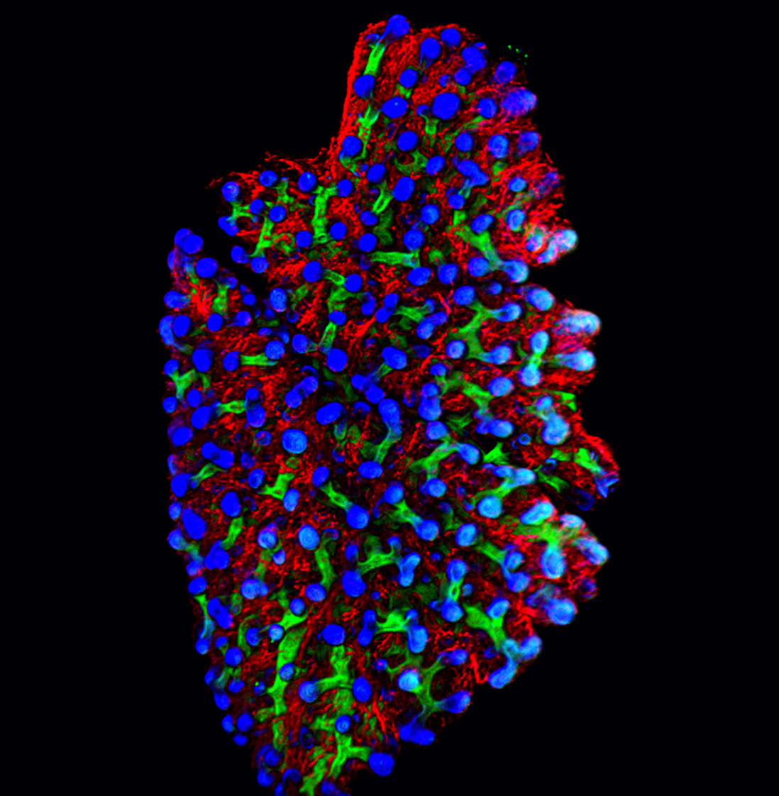
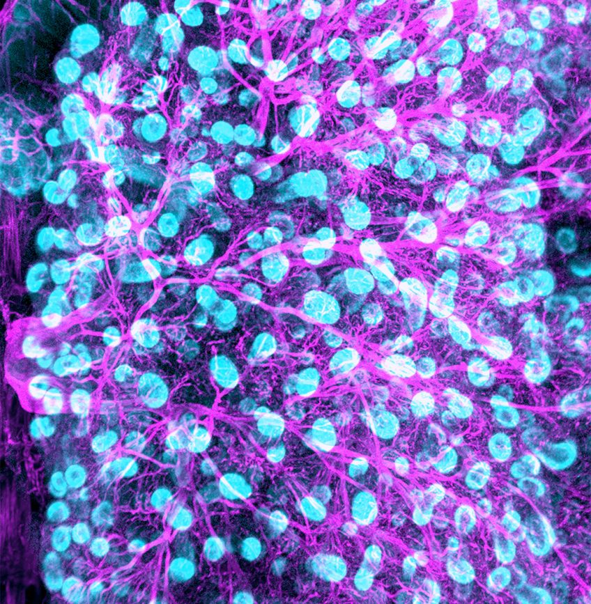
Obstruction of an artery by a blood clot is the leading cause of death worldwide. Known as a thrombosis, it leads to ischemic strokes (deprivation of oxygen and other nutrients), myocardial infarction, and limb ischemia. When a thrombosis forms, it requires emergency care: the clot must be destroyed as quickly as possible in order to restore blood flow and prevent irreversible damage.

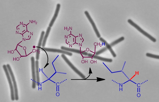
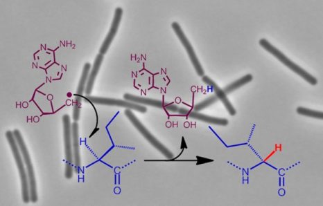
N-acetylcysteine (sold under the brand names Mucomyst®, Exomuc®, and Fluimucil®) is very widely used as a mucolytic drug: it helps thin out bronchial secretions, and assists with expectoration. Its mechanism of action is very simple: N-acetylcysteine breaks the molecular bonds between mucin proteins (the main component of mucus). In doing so, it breaks down the mucin macromolecules into smaller fragments, making the mucus more fluid and easier to expectorate.

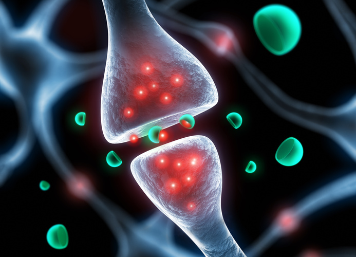
Interestingly, the mucin in the pulmonary mucus is not the only protein in the human body that forms such molecular bonds. The same type of bonds can be found in blood vessels, resulting in the formation of thrombi. Rather than mucin, here it is Von Willebrand factor that is involved: the protein able to cause the accumulation of platelets and the formation of blood clots.

Researchers from Inserm Unit 1237 (in collaboration with researchers from Inserm Unit 1176 and the University of Pennsylvania) have now shown that intravenous injection of N-acetylcysteine enables the breakdown of blood clots and thus unblocks the arteries. In several ischemic stroke models, N-acetylcysteine was even more effective than currently available treatments.

According to the authors of the paper, “N-acetylcysteine is a low-cost treatment that is already used around the world as a cough medicine, and the demonstration of its thrombolytic effects could have very wide application in the management of stroke and myocardial infarction patients. We hope to build on this work and to have a clinical trial underway as soon as possible.”.

This work was supported by the Fondation pour la Recherche sur les AVC (the French Stroke Research Foundation).

From context to cortex: Discovering social neurons

Crowd

© Fotolia

The existence of new “social” neurons has just been demonstrated by scientists from the Institut de neurosciences des systèmes (Aix-Marseille University / INSERM), the Laboratoire de psychologie sociale et cognitive (Université Clermont Auvergne / CNRS), and the Institut de neurosciences de la Timone (Aix-Marseille University / CNRS). Their research on monkeys has shown that when these animals are made to perform a task, the presence or absence of a conspecific—that is, another monkey—determines which neurons are activated. Published in Social Cognitive and Affective Neuroscience, these findings broaden our knowledge of the social brain and help us better grasp the phenomenon of social facilitation.[1]

Understanding how the brain functions within a social context is a major challenge facing neuroscientists.  Through their unique multidisciplinary collaboration, a primate neurophysiologist and an experimental social psychologist have now discovered two new classes of neurons in the prefrontal cortex: social and asocial neurons.

Most areas of the brain are associated with specific tasks. Some are specialized in the processing of information related to life in society: they make up the so-called social brain. In connection with thesis research conducted by Marie Demolliens,[2] CNRS researchers Driss Boussaoud and Pascal Huguet assigned monkeys the task of matching a picture shown on a touch screen with one of four different items displayed at the corners of the same screen. Executing such a task requires use of the prefrontal cortex but not the “social” areas of the brain. The researchers made daily recordings of neuronal electrical activity in this region of the brain while monkeys performed the task in the presence or in the absence of a conspecific.

Though the monitored neurons of the prefrontal cortex are primarily involved in execution of the visuomotor task, the study showed most of them also reacted strongly to either the presence or absence of another monkey. During the experiment, some of these neurons were only strongly activated in the presence of a conspecific. They have thus been dubbed social neurons. On the other hand, the activity levels of other, asocial neurons only spiked in the absence of a fellow monkey. Even more surprisingly, the greater the intensity of social neuron activity in the presence of a conspecific, the better the subject performed the task. Social neurons are hence at the root of social facilitation. Likewise, the greater the activity of asocial neurons in the absence of conspecifics, the better the subject performed the task—though not as well as in the presence of another, when social neurons are stimulated. The researchers also demonstrated that in the other, rare permutations—activation of social neurons in the absence of conspecifics, or of asocial neurons in their presence—the monkeys did not perform as well.

This work reveals the important connection between social context and neuronal activity, and the consequences on behavior: which neurons the brain uses depends on whether a conspecific is present, even if the task is the same. Thus, rather than being limited to the areas of the brain principally associated with social activity, social neurons might actually be dispersed throughout the brain and play a role in various tasks—whether or not the latter are social in nature.  These findings cast new light on the nature of the social brain as well as certain behavioral disorders characteristic of autism and schizophrenia.

neurones_sociaux_web

Figure 1: Differential activation of social and asocial neurons depending on whether the monkey performing the touch screen task is in the presence or absence of a conspecific.

© M. Demolliens

 [1]. Social facilitation refers to enhanced performance of an activity due to the presence of a conspecific. It is observed among all species whose members live in groups (i.e., social species).

[2]. Under the joint supervision of Driss Boussaoud and Pascal Huguet.

Does our childhood shape our political choices?

portrait of beautiful girl reading a newspaper at home/in the news/modern children

©Fotolia

Do our childhood experiences shape our political attitudes? A team of Inserm researchers from Unit 960 “Cognitive Neuroscience Laboratory” (Inserm/ENS) have discovered the answer to this question, the results of which have recently been published in the journal Evolution and Human Behavior. Childhood poverty is associated with stronger adherence to authoritarian political attitudes in adulthood, not only in the French population, but also in a sample of 46 European countries.

Understanding the origins behind the success of authoritarianism is an important key to preserving current democracies. Since the early 2000s, most Western countries have seen a historic rise in authoritarian parties. At the same time, authoritarian attitudes are becoming the norm in many political parties. Analysis of these political phenomena is usually based on contextual factors, such as the economic crisis or terrorist threat, which tend to favor authoritarian attitudes. However, recent studies in biology and psychology have shown that a person’s childhood environment can also shape their behavior in adulthood. Inserm researchers, in collaboration with SciencesPo, wished to determine whether these processes came into play in shaping political attitudes. The researchers focused more specifically on the effect of childhood poverty on authoritarian attitudes.

The researchers used tests in which the participants described their first impressions of faces, to ascertain their political preferences. Previous psychology studies have, in fact, shown that political attitudes influenced preferences for certain types of faces, and that simple judgments based on candidates’ faces could predict election outcomes. Inspired by these studies, the researchers from the Cognitive Neuroscience Laboratory determined the preference for fictitious male politicians, represented by computer-modeled faces and calibrated to represent variable degrees of dominance and trustworthiness.

The “trustworthiness” and “dominance” dimensions are orthogonal to each other. All combinations are possible: a face can be very dominant and less trustworthy, very dominant and very trustworthy, less dominant and less trustworthy, or less dominant and very trustworthy.

im1


The researchers developed two tests: a simplified version for children and another for adults.

Forty-one 7-year-olds were asked to choose their team captain to lead a mountain expedition, from among faces showing varying degrees of dominance and trustworthiness.

im2

This initial test showed that children exposed to negative socioeconomic conditions preferred more dominant and less trustworthy captains, compared with their classmates living in more comfortable surroundings.

Based on this early effect of poverty, the researchers then examined its impact on shaping subsequent political preferences. In partnership with the IPSOS polling institute, they determined the preferences of a representative sample of the French population (1,000 participants, quota sampling) for male politicians displaying varying degrees of dominance and trustworthiness. In this part of the study, faces displaying varying degrees of dominance and trustworthiness were randomly shown to pairs of participants, with the following question: “Who would you vote for?”

im3

This study revealed that childhood poverty increased the preference for dominant and less trustworthy male politicians in adulthood, irrespective of the participants’ current socioeconomic status and level of education.

Lastly, the research team focused more specifically on explicitly authoritarian attitudes, by asking the study participants to describe the extent to which they agreed with the following phrase: “I think having a strong man who does not care about parliament or elections as the leader of a country is a good thing“. Analysis of these responses showed that childhood poverty increased adherence to explicitly authoritarian attitudes, not only in the French population sample interviewed, but also in a panel of 46 European countries.

Based on three different tests, these studies highlight the importance of early factors in determining political attitudes, and thus provide a more in-depth understanding of the dynamics of democracies.

Communication between neurons implicated in autism spectrum disorders and intellectual disabilities

vignettecp-web

© Fotolia

An international collaborative study coordinated by Frédéric Laumonnier (Unit 930 “Imaging and Brain” Inserm/University of Tours) and Yann Hérault of the Institute of Genetics and Molecular and Cellular Biology (Inserm/ CNRS/ University of Strasbourg) provides new and original findings on the pathophysiological role of the contact areas between neurons in certain brain disorders. The study reveals that mutation of one of the genes involved in intellectual disability and autism spectrum disorder leads to dysfunction of the synapses, which are essential for neuronal communication. The research was published on April 18, 2017, in Molecular Psychiatry.

Autism spectrum disorder (ASD) and intellectual disability (ID) are neurodevelopmental disorders that generally emerge when a child’s brain is developing and often persist into adulthood. Behavioral disorders and inabilities to communicate and establish social interactions are observed in people with ASD. In addition, those with ID present difficulties with comprehension, memory, and learning. While the origins of these disorders remain poorly understood, we now know that a significant proportion are associated with genetic mutations.

During the brain development process, synapse formation is essential for brain functions such as memory and learning. Synapses are the points of contact between neurons which enable neurons to connect with each other and propagate information.  Some synapses are inhibitory and others excitatory, to enable the establishment of functional neuronal networks. However, mutations of the so-called PTCHD1 (Patched Domain containing 1) gene, which is located on the X chromosome and enables the expression of a protein potentially involved in synaptic functioning, have recently been identified in boys with the aforementioned disorders. These mutations stop the gene from expressing itself.

In order to validate the involvement of PTCHD1 gene mutations in ASD and ID, Hérault and his co-workers created a mouse model that was deficient for the PTCHD1 gene. In these animals, they observed major memory deficits and significant symptoms of hyperactivity, thus confirming the gene’s involvement in ASD and ID. Parallel studies by Laumonnier’s team showed a presence of the PTCHD1 protein in the excitatory synapses and also detected changes in the same mice’s synapses.

These changes to synaptic structure and activity in the excitatory neuronal networks were found to be particularly significant in a central brain region known as the hippocampus. This region plays a major role in cognitive processes, particularly those involving memory and the formation of new memories.

Genetic abnormalities impacting the structure or functioning of these synapses constitute a pathophysiological target in ASD and ID. In this context, this research defines a new “synaptic disease” caused by a PTCHD1 gene mutation. This dysfunction emerges during the development of the central nervous system and is associated with ID and ASD. Understanding of the pathophysiological mechanisms that underlie these neurodevelopmental disorders, particularly through the study of model organisms, is essential to improve therapeutic strategies.

In utero exposure to endocrine disruptors : learning from diethylstilbestrol

Equipe Inserm-CNRS ATIP - Avenir de Céline Colnot, U781, Hôpital Necker - Enfants Malades, Paris

©Inserm/Latron, Patrice

A research team from Université Paris Descartes, Inserm and Centre Hospitalier Sainte‐Anne, led by Prof. Marie‐Odile Krebs, has shown that patients suffering from psychotic disorders, and exposed in utero to diethylstilbestrol, presented specific epigenetic changes. These changes correspond to genomic regions notably comprising gene ZFP57, known to play a role in neurodevelopment. Owing to this new project, the researchers examine the broader issue regarding the impact of in utero exposure to endocrine disruptors (including diethylstilbestrol) on neurodevelopment and the emergence of psychiatric disorders.

This study was published in PlosOne on April 13, 2017.

Diethylstilbestrol (DES), a synthetic estrogen and endocrine disruptor, was prescribed for dozens of years to pregnant women worldwide, to reduce the risk of miscarriage. Use of this substance, limited over time, at high doses and during a particularly vulnerable period, may be thought to represent the typical pattern of exposure to endocrine disruptors in humans. It is associated with numerous medical conditions, particularly gynecological cancers and urogenital malformations among individuals exposed in utero. It has also been suggested to affect several generations.

The precise mechanism explaining these various syndromes has not been fully elucidated. Altered epigenetic homeostasis1 has been put forward as one hypothesis. Animal studies identified epigenetic changes (notably DNA methylation) after exposure to diethylstilbestrol. The study investigators examined the possibility of a correlation between prenatal exposure to diethylstilbestrol, associated with DNA methylation, and a possible increase in the risk of onset of psychotic disorders.

The consumer group Hhorages (Halte aux HORmones Artificielles pour les GrossessES) [which means “stop artificial hormones in pregnancy”] helped the team to recruit 247 individuals whose mothers were prescribed diethylstilbestrol during pregnancy, with the aim of creating a cohort. The analyses focused on 69 participants attending face-to-face interviews, which enable psychiatric diagnoses to be made via standard questionnaires. A blood sample was then taken with a view to performing the molecular analyses.

Exposed subjects were compared to their non-exposed siblings. This within-family comparison allowed shared genetic and environmental inheritance to be taken into consideration. Researchers did not observe any significant difference between exposed and non-exposed individuals in terms of DNA methylation.

However, they identified a differentially methylated region comprising gene ZPF57 in a sample of individuals exposed to diethylstilbestrol and suffering from a psychotic disorder (versus exposed subjects not presenting such disorders). This result suggests that altered expression of this gene, which is moreover known to play a role in neurodevelopment, could be linked to the emergence of psychiatric disorders in subjects exposed to diethylstilbestrol.

In conclusion, this study encourages further research efforts on in utero exposure to endocrine disruptors and the possible effects on neurodevelopment.

1 Epigenetic mechanisms are a group of mechanisms, which modulate gene expression without changing the underlying DNA sequence.

Decision-making, a contagious process

Risk, safety - wooden signpost

© Fotolia

Individuals learn to assess the level of prudence, patience or laziness shown by others from observing their behaviour, but most importantly, this influences their own decisions, without their even noticing it. A discovery that could have implications for neuroscience. Do our neighbours’ decisions affect our own? Such is what we are led to believe by the work of Jean Daunizeau and Marie Devaine, two Inserm researchers at the Brain and Spine Institute (ICM) in Paris (Inserm/ CNRS/ UPMC). They studied the behaviour of people making choices requiring prudence, patience or effort, and showed that when these people observe the behaviour of other individuals, they then start imitating them, without even knowing it !

 Underlying this work is a question fundamental to understanding how we make decisions in everyday life: is it a matter of personality, embedded in our DNA, or a process acquired through our education and social interactions? To find out, the researchers studied three characteristics that guide most of our decisions: prudence, patience and effort (or, depending on one’s point of view: risk-taking, impatience and laziness). To do this, they combined mathematics with cognitive psychology. “Social psychology is often criticised for being an over-empirical science, with results that are difficult to reproduce. To circumvent this problem, we use mathematical modelling,” explains Jean Daunizeau, who supervised this work.

In practice, the researchers recruited volunteers and subjected them to decision-making tests. A computer offered them choices requiring various degrees of patience, effort and prudence. For example, they had to choose between winning €2 immediately or €10 a few days later, pressing a soft handle for a small reward or a very hard one for a higher sum, or opting for a lottery that offered a strong chance of winning a small amount or a smaller chance of a big win. The volunteers responded to a series of forty choices of this type, thus allowing the authors to create an algorithm representative of their personality.

 In a second phase, the volunteers had to predict the choices of a fictional character invented using the algorithm, and more (or less) prudent, lazy or patient than the subject himself/herself. When left to their own devices, all the volunteers imagined that this character would make the same choices as themselves, regardless of how they behaved. However, after several errors and a period of adaptation, they ultimately made increasingly better predictions of the algorithm’s responses. Everything happens as though people presume that others think and act like themselves; this is what is known as the false consensus bias. “This phenomenon has already been described in other contexts,” explains Jean Daunizeau, “for aesthetic or moral choices, for example. It stipulates that people believe that their own judgement is shared by most other people. We find that here for choices requiring patience, effort or prudence.” But this bias is progressively offset by learning: having observed the behaviour of the fictional character, the volunteers correctly predict 85% of its choices. “On average, people are therefore able to closely interpret other people’s attitudes,” explain the researchers.

 Finally, the authors subjected the volunteers to a third series of tests, and found that the volunteers’ choices had become more similar to those of the fictional character. “This type of mimicry is relatively unconscious: when they are asked the question, the volunteers are unaware that the nature of their choices has changed, and that they are showing more patience or prudence. This phenomenon is known as the social influence bias, and means that our attitude tends to align with that of others. It was known in relation to certain behaviours, but here we reveal it in decision-making.”

 An improved understanding of how peoples’ decision-making is influenced by the way other people make decisions could have medical implications. Strong mimicry is observed in healthy subjects. What about individuals with psychiatric illnesses that affect social relationships, such as autism or schizophrenia? Here is what the researchers want to confirm: “If there are differences at that level; absence of mimicry could perhaps become a diagnostic element. There would then be a clinical aspect,” concludes the team.

The human embryo as you have never seen it

att00001

© Institut de la vision

A team coordinated by Alain Chédotal, Inserm research director at the Institut de la Vision (Inserm/UPMC/CNRS) and Paolo Giacobini at the Jean Pierre Aubert research centre (Inserm/Lille University) have just made a series of new observations of the anatomy of human embryos from 6 to 14 weeks old. A feat made possible by combining two recent techniques in immunomarking and 3D microscopy and a technique making the tissues transparent. These discoveries are published in the journal Cell dated 23 March 2017.

Plunging into the heart of a living being. From drawing to reality. This is what Alain Chédotal and his colleagues have done using their new technique to explore the anatomy or embryos. In the journal Cell, they reveal new photos and three-dimensional films of several tissues and organs of human embryos and foetuses from 6 to 14 weeks old, inaccessible until now.

Medical textbooks contain plenty of pictures of embryos but we always see the same thing: to represent their organs, we rely only on drawings. And the moulded embryos kept in medical faculties are made of wax. It should be said that the models, until now, were based on thin sections observed under the microscope, requiring illustrators to assemble and interpret all the information to represent an entire organ. This process belongs to the past with the advent of new three-dimensional imaging methods. In practice, the authors of this work have succeeded in combining three techniques, immunofluorescence, tissue clarification and microscopic observation, to be able to publish the first real 3D images of embryo tissues and organs.

They first used immunofluorescence to mark the intact organs. This technique consists of using fluorescent antibodies that bind specifically to the proteins expressed by certain cells, making it possible to locate them. Next, to view the fluorescent signal, they made the embryo tissues transparent using a technique perfected in mice in 2011. To do this, they immersed the tissues in several solvents, enabling them to strip the membrane-bound lipids from the cells, so as to retain only their protein architecture/skeleton and allow light to pass through. Finally, once this work was completed, they used a special microscope with a thin light source. A laser beam two millimetres thick scans the transparent samples, making it possible to photograph each plane and then the 3D image of the organ is built by computer.

Imaris Snapshot

© Institut de la vision


3D image of embryonic human lung. Future bronchi and bronchioles (blue and green) are visible, as are also the blood vessels (red).

New observations available to everyone

Marking embryos using antibodies made it possible to reveal the presence of specific cells and to obtain images of the peripheral nervous system, vascular system, lungs, muscles and the urogenital system. “What we observed has confirmed existing data in embryology, but this is the first time that we have obtained real images of the organisation of tissues in so much detail. In particular, we have discovered things that it was not possible to see without specific marking. For example, we succeeded in distinguishing sensory nerves (that transmit sensory signals to the brain) from motor nerves (that are connected to muscles), which was previously impossible“, explains Alain Chédotal. Another discovery: the variability of the nerve pathways in the hands. The development of the principal nerves is conserved in all hands, but the small peripheral nerve pathways develop much more randomly between the left and right hands and between individuals. Finally, the last advantage emphasised by the researchers: “we are able to have an idea of the rate of cell growth for each organ by counting the fluorescent cells at different embryo ages“.

To make these data available to the widest audience, the team has created a dedicated internet site subsidised by the French Voir & Entendre [See & Hear] Foundation*. “Here we offer free access to our films and we will continue to add new ones as we produce them. We would also like other laboratories to be able to add to it from their own work. The aim is to make it an initial image bank to provide a complete 3D atlas of the human embryo during the first development trimester, with organ-by-organ searches possible. There is both an education objective but also a clinical purpose, particularly for surgeons that operate in utero and who in this way will have precise images of embryo tissues or their nervous and vascular system“, the researchers conclude.

* https://transparent-human-embryo.com/

Gender discrimination also exists in science

At university lecture

© Fotolia


Gender discrimination can be found in quite unexpected places. An international team that includes Demian Battaglia, a CNRS Researcher at the Neurosciences Systems Institute (INS) (Inserm/Aix-Marseille University), and researchers from Yale and the Max Planck Institute (Germany) has just demonstrated that women are under-represented in the review process for scientific publications. This research is published in the 21 March 2017 issue of the journal eLife.

Gender discrimination is a well-known phenomenon. Scientific research is not spared by these issues, including academic publication, one of the cornerstones of scientific endeavour. In order to be validated, every article must be approved by independent researchers. Obviously, these reviewers are meant to be selected according to their competences and not their sex. An international team has focused on the question of the gender of these reviewers. The results are shocking: women scientists, already a minority in their field, are under-selected for reviewing papers. The blame falls on the natural and unconscious tendency of editors, mainly male, to choose someone of their own sex.

Demian Battaglia, a CNRS researcher at the Neurosciences Systems Institute (INS) (Inserm/Aix-Marseille University), and Markus Helmer and his colleagues worked on journals from Frontiers, the only publishing house to publicly disclose the names of reviewers. After studying 41,000 publications in different areas (science, health, engineering and social science), published between 2007 and 2015, and a database of 43,000 reviewers, they found that women are under-represented in various scientific areas. But they are also asked to provide reviews less often than might be statistically expected.

The reason is simple: editors, be they men or women, have a tendency, known as homophily, to select reviewers of their own sex. The practice is common in friendships and the professional network. But it manifests itself in different ways for the different sexes. This behaviour is widespread in men (over 50% of individuals), and limited, but practised in an extreme manner, in women (about 10% of female editors are strongly homophilic).

The result of this process is that even where there is a policy of parity, with equal representation of women and men, the authors believe that this homophilic bias may persist. The real challenge is therefore to change these behaviours. The authors of the study thus propose tools, such as tables that would be posted when proofs are published online, recalling the figures on sexism.

The team now hopes to repeat its analyses in a few years in order to see if its recommendations have been adopted.

What if optimism were learnt?

How does our brain learn from our mistakes? Does it prefer good news to bad news? These are the questions answered by a team of researchers led by Stefano Palminteri (Inserm-ENS), laureate of the ATIP-Avenir programme, from the Laboratoire de Neurosciences Cognitives. The results will be published in Nature Human Behaviour.

Generally speaking, humans tend to overestimate the likelihood of a positive event in the near future, whereas they underestimate that of a negative event. In cognitive psychology, this is known as the optimism bias. This and other cognitive biases influence our rational logic, our judgements and our decisions, and hence our behaviours. Optimism bias is a tendency to take “positive” information (good news) into account more than “negative” information (bad news). This basic asymmetry is assumed to generate and reinforce this bias, and leads us to believe that our future outlook is, on average, better than that of others. This has particularly been shown in heavy smokers who underestimate their risk of premature death, and in certain women who underestimate their risk of getting skin cancer.

A research team from the Laboratoire de Neurosciences Cognitives (LNC) wished to know more about this phenomenon and understand its origin. Is this phenomenon linked only to our beliefs regarding possible future events, or more generally, is it also present in any type of learning, including the most basic: learning by trial and error? To do this, the researchers studied behaviour in a group of people involved in a process of learning by trial and error, which consisted of choosing between two symbols associated with a monetary reward. Depending on the choice of the participant, the latter could win €0.50 (“good news”), win nothing, or lose €0.50 (“bad news”). Results demonstrated that the participants attributed 50% more importance, on average, to the “good news” than to the “bad news.” This general tendency of our brain to learn in an asymmetrical manner, preferring the good news and ignoring the bad news, may be the basis for the optimism bias.

One question remained unanswered, that of the relationship between this deeply rooted learning bias and the brain reward circuits. To answer this question, the LNC researchers studied the brain activity of subjects carrying out the learning task just described, using functional magnetic resonance imaging (fMRI). According to Stefano Palminteri, who headed this study: “The brain activity recorded in the main structures of the brain reward circuit is almost twice as high in an optimistic subject compared to a more realistic subject, for the same monetary reward. This activity shows the existence of distinct profiles, more or less optimistic or realistic.”

Apart from providing a neuropsychological explanation for optimism, this work provides additional evidence for the existence of a deeply rooted learning bias in human cognition. The optimism bias could therefore be involved in psychopathologies such as depression (absence of the bias) or certain addictions (overexpression of the bias). “In order to better understand the cause and persistence of these behaviours, with their high social and human cost, it is therefore essential to study of these basic biases in the learning,” the authors of the study believe.

Toward the synthesis of antibiotics by a new bacterial enzyme

cp-enzymebact_synthantib

©Inra

Researchers at the French National Institute for Agricultural Research (Inra) and Inserm have discovered a new family of bacterial enzymes that can produce hitherto unidentified peptides with antibiotic activity. Published in Nature Chemistry, this work holds promise for the synthesis of molecules of pharmaceutical interest and the design of new antibiotics.

As part of research involving study of the enzymes of the gut microbiota, researchers from Inra and Inserm studied the model bacterium Bacillus subtilis. Genetic analysis of the organism showed the presence of genes conserved by common bacteria from the gut microbiota, such as enterococci. *

The scientists were especially interested in two Bacillus subtilis genes potentially coding for a peptide and an enzyme from the superfamily known as “radical SAM enzymes.” Their work has made it possible to describe a new enzyme mechanism that can transform a peptide into a bioactive molecule. Known as epimerisation, this enzymic transformation brings about a change in the configuration of certain amino acid residues from the L- configuration (normally found in peptides) to the D-configuration. The researchers discovered how this enzyme works: it removes a hydrogen atom from the alpha-carbon atom of the amino acid residues and donates another, bringing about their epimerisation. It is a mechanism not previously described in nature.

It is the first time that researchers have demonstrated the ability of “radical SAM” enzymes to catalyse epimerisation within a peptide in vitro. Surprisingly, the peptide thus modified and called an “epipeptide” is able to inhibit growth of Bacillus subtilis very effectively. These epipeptides thus represent a new class of natural products that might be used to develop new antibiotics against Gram positive bacteria (such as staphylococci, enterococci or streptococci), which are becoming increasingly antibiotic resistant, constituting a major public health problem.

Voluntary eye movements, a new indicator of postural control in Parkinson disease ?

Oeil vision yeux

© Fotolia

Postural instability is the main factor associated with falls in patients with Parkinson disease, but there are other symptoms, such eye disorders. Teams from Pitié-Salpêtrière Hospital, AP-HP, and the Brain and Spine Institute (Inserm, CNRS, UPMC) have examined the link between these eye disorders observed in some patients and postural instability. Their results demonstrate a potential new marker of posture control in Parkinson disease. These results are published in the journal Neurology.

Some patients with Parkinson disease show impairment of the voluntary eye movements, the “antisaccades.” Dr Claire Ewenczyk, from the team led by Prof Marie Vidailhet, and Prof. Stéphane Lehéricy, from the Brain and Spine Institute, (Inserm, CNRS, UPMC) at Pitié-Salpêtrière Hospital, AP-HP, conducted a study in 30 patients with Parkinson’s disease with or without postural control disorders, and 25 healthy subjects.

The researchers examined the patients’ walk and balance, and recorded the initiation of walking and eye movements. At the same time, they studied the interactions between two areas of the brain: the frontal oculomotor area, which plays an important role in visual attention and eye movements, and the mesencephalic locomotor area, which is strongly involved in the control of posture and eye movements.

Their results indicate that patients with posture disorders also show abnormal reaction time (latency) in their voluntary eye movements. This abnormality is correlated with an alteration in the parameters during initiation of a step, particularly the duration of anticipatory postural adjustments, mechanisms used by the central nervous system to maintain balance in a standing position while performing voluntary movements.

Studying the interactions between the frontal oculomotor and mesencephalic locomotor areas of the brain reveals, in healthy subjects, a correlation between the frontal connections of these areas and the latency of voluntary eye movements. In patients with Parkinson disease, this association disappears. This suggests extensive involvement of the mesencephalic area in the disease, which affects both posture control and eye movements.

The prolongation of the reaction time of eye movements or “antisaccades,” a simple and reliable parameter, might constitute a prognostic marker of postural control in Parkinson disease, and be used for patient assessment in future longitudinal studies.

fermer