Menu

An antibody-based drug for multiple sclerosis

Inserm Unit U919, directed by Prof. Denis Vivien (“Serine Proteases and Physiopathology of the Neurovascular Unit”) has developed an antibody with potential therapeutic effects against multiple sclerosis. The study, directed by Fabian Docagne and published in Brain, paves the way for a new strategy to control the disease.

Multiple sclerosis is a disease that affects the central nervous system, particularly the brain and spinal cord. It is the most common cause of neurological disability in young adults.

The disease is considered autoimmune since the immune system, which is there to protect the body from external assault, attacks its own constituents. The cells of the immune system, particularly the lymphocytes, bring about the destruction of the myelin sheath that surrounds and protects the extensions (axons) of the neurons. This demyelination, which marks the beginning of axon degeneration, disrupts the transmission of nerve impulses. Lesions in the form of “plaques” are dispersed over the brain and spinal cord. They cause symptoms that vary greatly from one individual to another.

Usually, the disease is characterised by exacerbations, with the appearance of motor, sensory and cognitive disorders, followed by remission a few weeks later. But with the passage of years, these symptoms can progress to irreversible disability. Current treatments reduce the exacerbations and improve the quality of life of patients, but do not control the progression of the disease.

sclérose en plaq

(C) Inserm/Fumat, Carole

In order for the cells of the immune system circulating in the bloodstream to reach the central nervous system, they must penetrate the blood-brain barrier (haematoencephalic barrier) and blood-spinal cord barrier (haematomedullary barrier).

During previous work on a mouse model of stroke, the team from Inserm Unit 919 studied a factor involved in opening the blood-brain barrier, the NMDA receptor. In particular, they observed that blocking the interaction of this receptor with tPA (a member of the serine protease family) has beneficial effects associated with maintaining the integrity of the barrier.

In this study, the researchers developed a strategy for blocking the interaction of tPA with the receptor, in multiple sclerosis. In the laboratory, they developed a monoclonal antibody (Glunomab®) directed against the specific site on the NMDA receptor to which tPA binds.

schéma docagne english

(C) Fabian Docagne, Inserm/Servier Medical Art

In cellular models of the human blood-brain and blood-spinal cord barriers, the use of this antibody prevented opening of the barrier under inflammatory conditions, limiting the entry of lymphocytes. The team then tested the therapeutic effects of the antibody in an experimental mouse model of multiple sclerosis. After intravenous injection of Glunomab, the progression of motor disorders (partial or total paralysis of the limbs), as assessed by a clinical score, was blocked. In these treated mice, this effect was associated with reduced infiltration of lymphocytes into the nervous tissue, and reduced demyelination.

By thus preventing myelin destruction by the cells of the immune system, this strategy might represent a promising therapy for the control of multiple sclerosis.

A patent application has been filed on this work.

Can we suppress the antipsychotic drug side-effects?

Since their development in the 1950s, antipsychotic drugs have been widely used to treat psychoses and neuropsychiatric disorders like schizophrenia. A debilitating side-effect of these drugs called parkinsonism limits their efficacy.Irvine scientists led by Emiliana Borrelli, Inserm research director at University of California and colleagues have discovered the key cellular mechanism that underlies the antipsychotic-induced parkinsonism – which includes involuntary movements, tremors and other severe physical conditions. These studies present evidence that will stimulate a targeted approach for the design of novel antipsychotics without side-effects. The results have been published in Neuron on July, 6th.

borrelli_101115_01_daa

© Daniel A. Anderson/UCI

The researchers report that antipsychotics side-effects are due to blockade of the dopamine D2 receptor in a specialized type of neurons in the striatum, called interneurons. Blockade of D2 receptor in these neurons increases neurotransmitter signaling (acetylcholine) above threshold on neighbor neurons leading to motor abnormalities in rodents (catalepsy) which correspond to parkinsonism in humans. Catalepsy is marked by severe muscular rigidity and fixity of posture regardless of external stimuli. Indeed, in mouse studies, the Borrelli team discovered that removing D2 receptors in nerve cells (cholinergic interneurons) did not result in catalepsy in the mice upon antipsychotic treatment.


Borrelli said the importance of this study is twofold.

It clarifies a long-waited mechanism that allows to explain the motor side-effects of antipsychotic drugs and will help future design of drugs deprived of nasty side-effects. It also generates important information for combined therapies (using drugs that block D2 but also acetylcholine receptors) that should be used to improve the life of people treated for debilitating psychiatric disorders.”

Brain tumours: for the first time, ultrasound makes blood vessels permeable to enhance treatment delivery

Teams from the Paris Public Hospitals (AP-HP), Pierre and Marie Curie University, Inserm and the CarThera company (which is hosted by the Brain and Spine Institute [ICM]), coordinated by Prof. Alexandre Carpentier, a neurosurgeon at Pitié-Salpêtrière Hospital, AP-HP, have successfully used ultrasound to temporarily permeabilise blood vessels in the brains of patients affected by recurrent malignant brain tumours. This innovative method allows increased delivery of treatments, including chemotherapeutic agents, to the brain, and represents hope for other brain pathologies. This work was published on 15 June in the international journal Science Translational Medicine.
Treatment of primary malignant brain tumours is currently based on a neurosurgical procedure followed by sessions of chemotherapy and/or radiotherapy. These treatments bring about disease remission for varying periods, depending on the patient. The blood-brain barrier (BBB), this particularly impermeable wall of vessels that limits the exposure of the neurons to toxic agents, restricts the entry and hence the delivery of treatments to the brain.
Given this observation, the respective teams led by Prof. Alexandre Carpentier and Dr Ahmed Idbaih, and the neuro-oncology group from Pitié-Salpêtrière Hospital, AP-HP, launched a phase 1/2a clinical trial in July 2014, sponsored by AP-HP, in patients with recurrent malignant brain tumours.

The objective is to permeabilise the blood-brain barrier, in order to increase the penetration and delivery of chemotherapeutic drugs to the brain, using the “SonoCloud®” ultrasound device developed by the CarThera company.

Implanted in the thickness of the skull bone, this device is activated a few minutes before intravenous injection of the product. Two minutes of sonication is enough to temporarily permeabilise the BBB for 6 hours, thereby allowing a 5-fold greater delivery of the drug to the brain than would normally happen.
To date, and for the first time in the world, several repeated “openings” of the BBB could be observed in the 20 patients treated. Moreover, tolerance is excellent: the technology invented by Prof. Carpentier and developed by the CarThera company, with the help of the Inserm LabTAU physics laboratory, does not damage the neurons, and the BBB spontaneously closes again 6 hours after the intravenous infusion.

According to Prof. Alexandre Carpentier, “this innovative method offers hope for the
treatment of brain cancers, as well as other brain pathologies, such as, potentially, Alzheimer’s disease, where the existing drugs have difficulty in penetrating the brain. This technique must continue to undergo evaluation to allow its entry into routine clinical use in a few years.”

A novel research program on traumatic memories

How will the traumatic events of the terrorist attacks of 13 November 2015 evolve in people’s memories, whether collective or individual? How does individual memory feed on collective memory and vice versa? Is it possible, by studying cerebral markers, to predict which victims will develop post-traumatic stress disorder and which will recover more quickly? These are a few of the questions addressed in the ambitious 13-Novembre program, coordinated by the CNRS, Inserm and héSam Université, with the collaboration of numerous partners. This transdisciplinary research program, codirected by the historian Denis Peschanski and neuropsychologist Francis Eustache, is based on the collection and analysis of the accounts of 1000 volunteers, interviewed four times over ten years. Involving several hundred people, this study is a worldwide first in terms of size, number of disciplines encompassed and protocol used. Results are expected to benefit the socio-historical and biomedical fields, but also have implications for public policy and public health.

Following the appeal launched last November[1] by Alain Fuchs, president of the CNRS, the research community is seeking to elucidate the issues facing society in the wake of the terrorist attacks that hit France in the course of last year. This call for projects gave rise to 13-Novembre, an interdisciplinary program that will run for 12 years. Coordinated by the CNRS and Inserm, in collaboration with héSam Université, it aims to study the construction and evolution of memory after the attacks of November 13th 2015, and also the relationship between individual and collective memory. “The 13-Novembre project illustrates the role of the CNRS, which is to support two scientists set to monitor studies involving 150 researchers from different disciplines in a long-term program of unparalleled scope,“ Alain Fuchs says. “From the very beginning, Inserm has been committed to the project, which combines human and social sciences and the latest advances in the neurosciences. This ambitious interdisciplinary program will answer the questions we are asking ourselves. I believe that this is part of the mission of two organizations like Inserm and the CNRS,” says Yves Lévy, CEO of Inserm.


1000 people monitored over 10 years

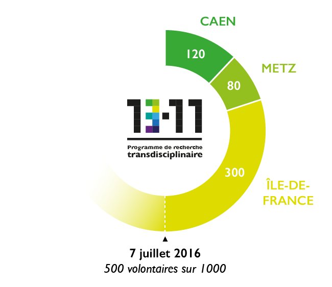
The testimonies of 1000 volunteers will be collected and analyzed. Some of these volunteers experienced the events at close hand: survivors, their family and friends, the police, the military, the fire brigade, the doctors and caregivers involved. Others were affected indirectly, i.e. the residents and users of the affected neighborhoods; Parisians from other areas; and finally, inhabitants of several French cities, including Caen and Metz.


The scale of this study makes it novel: the 1000 participants will be followed for 10 years over four campaigns of filmed interviews (in 2016, 2018, 2021 and 2026), with the contribution of the INA (which will conduct the Paris interviews) and ECPAD[2] (for the interviews outside of Paris). Its design is also unprecedented as the guidelines for the interviews were written jointly by historians, sociologists, psychologists, psychopathologists and neuroscientists, in such a way that the material collected is used by each discipline. This study is a world first.


Individual testimonies will be put in perspective with the collective memory as it is built over the years: television and radio news broadcasts, press articles, reactions on social networks, texts and images of commemorations, etc., are all examples of records held by the INA and analyzed by its research teams, in relation with other laboratories. Additionally, a partnership with the Crédoc[3] will make it possible to gauge public opinion at the dates of the interview campaigns. Eleven specific questions were thus integrated into the Crédoc’s traditional half-yearly questionnaire in June and July 2016.


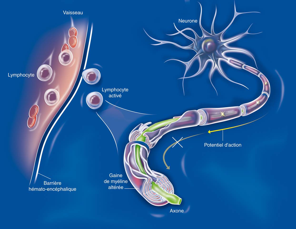
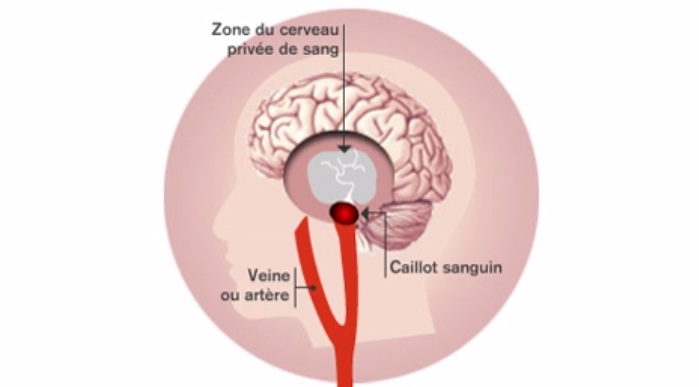
A biomedical study named “Remember” will be performed on 180 of the 1000 participants: 120 people directly affected by the attacks, some of whom are suffering from post-traumatic stress disorder, and 60 who live in Caen. Interviews and brain MRI scans, conducted at the same frequency as the video interviews, will help to shed light on the impact of traumatic stress on memory (including intrusive thoughts and images, characteristic of post-traumatic stress disorder), and to identify markers associated with cerebral resilience to trauma. The participants, of course, will not need to be re-exposed to traumatic thoughts or images.


In parallel, the “ESPA” (post terrorist attack public health study) was initiated by Santé publique France[4] in collaboration with the 13-Novembre program in order to analyze—via an Internet questionnaire—the psychotraumatic impact of attacks on those directly exposed, but also the validity of healthcare channels.


Transdisciplinary study and civic commitment

The program is of crucial interest for all of the scientific disciplines represented. Historians and sociologists will try to understand how individual testimonies and collective memory are co-constructed. The linguist will measure the evolution of vocabulary and syntactic constructions. The neuropsychologist will focus on the consolidation and reconsolidation of memory and its functioning, which depends on whether one has experienced the event itself or is recalling the conditions in which they heard of it. As for the neuroscientists, they will work on the modifications to mental representations, post-traumatic stress disorder and the potential to eliminate painful memories. The psychopathologists will concentrate on the impact of the attacks on self-image, and will look into defense mechanisms or the relationship with destructiveness. In addition, the 13-Novembre program will be useful for criminal law, victim support policies, crisis management and commemorative practices. The filmed interviews will also have a heritage value: they will preserve and transmit the memory of the November 13th attacks. This is a civic commitment by the scientific community and the INA and ECPAD professionals who will be in charge of the recordings and documentary descriptions, as well as of making them available to researchers and archiving them permanently.


This program also takes some of the multidisciplinary concepts and methods developed by Denis Peschanski and Francis Eustache on the collective memory of World War II and September 11th and applies them to the Paris terrorist attacks within the framework of the “Matrice” Equipex (equipment of excellence) project, coordinated by héSam Université and in which the INA is already a partner. For the two researchers, it is impossible to understand collective memory without considering the cerebral dynamics of memory. Likewise, these dynamics cannot be fully grasped without considering the contribution of social determinants. The researchers were also inspired by the questionnaire in print elaborated by US psychologist William Hirst as part of a survey carried out, a week, a few months and a few years after the terrorist attacks of September 11th 2001. A comparative analysis of the two studies is also planned.


Multiple partners and supports

The 13-Novembre study started on May 13th in Caen (northwestern France) and June 2nd in Bry-sur-Marne (Paris region) for the filmed interviews. The biomedical study Remember started on June 7th at the biomedical imaging facility “Cyceron” in Caen in collaboration with Normandie Université. The call for volunteers is ongoing, notably via the French daily Le Parisien-Aujourd’hui en France (and its website). The first findings should be available in autumn 2017 and final results are expected in 2028, two years after the last interviews.


The CNRS and Inserm are in charge of coordinating the scientific aspects of the 13-Novembre program, while the administrative side has been entrusted to héSam Université. 13-Novembre is funded by the French Research Agency (ANR) as part of the French government’s Investments for the Future Program (PIA).


The program involves several research laboratories:

  • Centre de recherche sur les liens sociaux, Cerlis (CNRS/Université Paris Descartes/Université Sorbonne Nouvelle – Paris 3),
  • Laboratoire Neuropsychologie et imagerie de la mémoire humaine (Inserm/EPHE/Université de Caen Normandie),
  • Institut des systèmes complexes – Paris-Île-de-France (CNRS),
  • Laboratoire Neuropsychiatrie: recherche épidémiologique et clinique (Inserm/Université de Montpellier),
  • Centre de recherche sur les médiations (Université de Lorraine),
  • Laboratoire Bases, corpus, langage (CNRS/Université Nice Sophia Antipolis).


It has a large number of associated partners:

  • INA,
  • Santé publique France,
  • ECPAD,
  • EPHE,
  • Archives nationales,
  • Archives de France,
  • Université Paris 1 – Panthéon Sorbonne,
  • Université de Caen Normandie,
  • Cyceron biomedical imaging facility
  • Caen University hospital,
  • Le Parisien- Aujourd’hui en France national daily,
  • Universcience,
  • Crédoc.


It also benefits from the support of several ministries, local authorities and associations:

  • Ministry of National Education, Higher Education and Research,
  • Ministry of the Interior,
  • Ministry of Culture and Communication,
  • Secretary of State for Defense, chargé des Anciens combattants et de la Mémoire
  • Paris authorities,
  • 10th arrondissement [borough] local authorities in Paris,
  • 11th arrondissement [borough] local authorities in Paris,
  • The town of Saint-Denis,
  • The urban community of Caen la mer,
  • Normandy region,
  • Normandie Université
  • Institut national d’aide aux victimes et de médiation (Inavem),
  • The association “Life for Paris : 13 novembre 2015”,
  • The association “13 novembre : Fraternité et Vérité”,
  • The association “Paris aide aux victims”,
  • B2V joint social protection group
  • Institut mémoires de l’édition contemporaine (IMEC).

[1] See: https://intranet.cnrs.fr/intranet/actus/160225-attentats-recherche.html (in French).

[2] Établissement de communication et de production audiovisuelle de la Défense.

[3] Centre de recherche pour l’étude et l’observation des conditions de vie.

[4] Santé publique France is the new public health agency formed from the amalgamation of the Institut national de prévention et d’éducation pour la santé (Inpes), Institut de veille sanitaire (InVS) and Etablissement de préparation et de réponse aux urgences sanitaires (Eprus), on May 1st 2016.

Anorexia nervosa: pleasure at getting thin more than fear of getting fat

A study from Inserm, Paris Descartes University and Sainte Anne Hospital suggests that anorexia nervosa might not be explained by fear of gaining weight, but by the pleasure of losing it… and that the phenomenon might be genetically influenced. Published in Translational Psychiatry, this study, directed by Prof. Gorwood, head of the Clinic for Mental and Brain Diseases, challenges the notion of fear of weight gain in anorexia patients.

Dish with a tomato and a dial of a bathroom scale

(c) Fotolia 

Often associated with major psychological distress, anorexia nervosa is an eating disorder that mainly affects girls and young women. Diagnosis is based on three international criteria: restriction of food intake leading to weight loss, a distorted perception of weight and body, and an intense fear of becoming fat.

Although there is no pharmacological treatment, Prof. Philip Gorwood’s team has focused on these clinical criteria. As the researcher explains: “When research is going nowhere, it is important to call into question the criteria at the very root of the disorder. We have therefore re-evaluated the last criterion, although it is quite prominent in patient discourse, by assuming that it is a mirror image of what is really involved, i.e. a reward for losing weight. We established the postulate that patients felt pleasure at becoming thin rather than fear of becoming fat.”

So as not to be influenced by patients’ discourse and analysis of their eating disorders, the researchers used a “skin conductance test,” which measures the subject’s sweating rate when exposed to various images. The emotion caused by certain images actually leads to a rapid and automatic increase in sweating.

The researchers showed images of people of normal weight or overweight people to 70 female patients consulting the Clinic for Mental and Brain Diseases (CMME) of Sainte Anne Hospital. For these patients, of varying weight and with different degrees of disease severity, viewing these images caused much the same reaction as in healthy subjects. Conversely, when looking at images of thin bodies, the patients showed positively evaluated emotions, whereas healthy subjects had no particular reaction.

Anorexia nervosa is a highly heritable disorder (70%). One of the genes most often associated with anorexia nervosa codes for BDNF, a factor involved in neuron survival and neuroplasticity. In patients with anorexia nervosa, the study indicates that the increase in sweating experienced when viewing images of thin bodies is explained by the presence of a specific form (allele) of the gene in question. This result was confirmed after examining potential confounding variables such as weight, type of anorexia or duration of the disorder.


The conclusions of this work:

-support the genetic approach as a different way of addressing the key symptoms of anorexia nervosa;

– orient research toward reward systems rather than phobic avoidance;

– finally, they suggest that certain therapeutic approaches, such as cognitive remediation and mindfulness therapy, might have a clear beneficial effect on this illness.

Stroke study prompts call for revised treatment guidelines

Doctors should rethink how they treat patients who have suffered the deadliest form of stroke, a clinical trial suggests.The study recommends a change in guidelines for treating any patients who have had a stroke caused by bleeding into the brain – known as intracerebral haemorrhage (ICH) – while regularly taking aspirin.

AVCAccident vasculaire cérébral ischémique (AVC)

(c) Inserm/Koulikoff Frédérique


Prof. Yvo Roos (Academic Medical Center Amsterdam) who initiated the study said: “Until now, a treatment known as platelet transfusion has been used by some doctors in the hope that it will aid recovery. It had been thought that platelets – blood cell fragments that play a vital role in blood clotting – could block ruptured blood vessels and prevent further bleeding into the brain in patients who are using anti-platelet therapy”.

However, the team from the Netherlands, France, UK has found that giving platelet transfusions to patients who had suffered an ICH while taking aspirin reduced their chances of recovery.


Prof. Charlotte Cordonnier (Inserm Unit 1171, Univ. Lille, CHU Lille) adds :”Each year, about two million adults worldwide suffer stroke caused by ICH, which accounts for half of all stroke deaths. Two out of five people die within one month, and a further two out of five become dependent on carers. One quarter of patients are taking aspirin before they suffer an ICH.”

Stroke patients who participated in the clinical trial received standard stroke care, but they were also randomly assigned either the platelet transfusion or no extra treatment. Researchers found that platelet transfusions increased the risk of death and long-term disability compared with usual care.

It is unclear why patients given platelet transfusions were worse off than those who were not, researchers say. They suggest that the treatment may cause blood clots to form or trigger inflammation in the brain, worsening bleeding.

Prof. Rustam Al-Shahi Salman, of the University of Edinburgh’s School of Clinical Sciences, who co-led the study, said: “Our study shows that platelet transfusion seems harmful, and certainly is not beneficial, for people who take aspirin and have a stroke caused by bleeding into the brain. These findings should change clinical guidelines.”

Today, the study results are presented at the European Stroke Organisation Conference and are published in The Lancet.

Enhanced hippocampal-cortical coupling improves memory

For the first time, scientists in the Center for Interdisciplinary Research in Biology (CNRS/INSERM/Collège de France) have produced direct evidence that the long-term storage of memories involves a dialogue between two brain structures, the hippocampus and cortex, during sleep; by enhancing this dialogue, they succeeded in triggering the consolidation of memories that would otherwise have been forgotten. This work is published in Nature Neuroscience on 16 May 2016.

learing concept

(c) Fotolia

Since the 1950s, the principal theories on memory have posited that such traces are initially formed in the hippocampus and then gradually transferred to the cortex for long-term storage. Although supported by numerous experimental studies, this hypothesis had never yet been directly verified.

In order to prove it, the scientists first recorded the activity of the hippocampus and cortex during sleep. They found a correlation between the oscillations observed in these two structures: when the hippocampus emitted sharp wave-ripples, the cortex in turn emitted delta waves and spindles like a series of questions and answers. To establish a link with memory, the scientists then trained rats to memorize the position of two identical objects in a room. During testing the next day, one of the objects had been moved and the rodents had to determine which one. Those that had spent 20 minutes in the room on the first day passed the test, while those that had only been there for three minutes failed. This difference was also reflected in the hippocampal-cortical coupling during sleep just after the initial exploration: coupling was more visible in rats that passed the test the next day. It was then necessary to prove that this was indeed the cause of memorization.

The scientists then developed a system for real-time detection of hippocampal sharp wave-ripples and immediate triggering of cortical delta waves and spindles, or in other words to generate coupling between these two structures on demand. They applied this system in rats that had been trained for just three minutes the first day, and were therefore not expected to remember the position of the objects the next day: these rodents passed the test perfectly. By contrast, if a variable delay was introduced between the hippocampal and cortical waves, the effect disappeared.
To better understand the mechanisms at play, the scientists also recorded cortical activity during learning, sleep and the test. They observed that selected neurons changed their activity in the context of coupling during sleep, and that the next day the cortex responded to the task by becoming more active in the vicinity of the object that had been moved.

By demonstrating the mechanisms underlying long-term memorization, this work may shed new light on certain memory disorders in humans. It might thus be possible to envisage overcoming certain memory deficits if they result from the same mechanism as that studied here. However, the ethical issues related to these techniques will need to be addressed and methods will have to be refined to enable selective action on the memories that need to be enhanced, before any clinical application can be envisaged. The team is now set to elucidate the dialogue between the hippocampus and cortex, notably when several memories need to be remembered, or not.

Control of fertility: a new player identified

Individual small RNAs are responsible for controlling the expression of gonadoliberin or GnRH (Gonadotropin-Releasing Hormone), a neurohormone that controls sexual maturation, the appearance of puberty, and fertility in adults. This has just been demonstrated by the “Development and Plasticity of the Neuroendocrine Brain” team led by Vincent Prévot, Inserm Research Director (Jean-Pierre Aubert Research Centre, Lille). The involvement of microRNAs, transcribed from DNA, occurs around birth, and marks a key step in postnatal development. Failure of these microRNAs to act leads to the disruption or even total cessation of GnRH production by the hypothalamic neurons that synthesise it, and hence to infertility. In the most serious cases, sterility may result. Details of this work in mice are published in the 2 May 2016 issue of the journal Nature Neuroscience.

Figure images Anglais

(c) Inserm/Prévot Vincent

Images showing the expression of the neurohormone GnRH (green) by the hypothalamic neurons that synthetize it in mice in which GnRH neurons have been genetically tagged by the Tomato gene (gene coding for a red fluorescent protein).


Reproductive function is determined by events that take place in the brain. Gametogenesis (the production of spermatozoa and oocytes) and the secretion of hormones by the ovaries and testes are heavily dependent on the hypophysis, a small gland located below the brain, to which it is connected by a capillary network. The latter is in turn controlled by a glandular “orchestra conductor” located at the base of the brain, the hypothalamus. During postnatal development, activation of a small number of highly specialised neurons (the GnRH neurons) in the hypothalamus leads to the synthesis of a hormone, gonadoliberin or GnRH (Gonadotropin Releasing Hormone), and this process leads to the appearance of puberty.

This step, known as “mini-puberty” constitutes the first activation of the reproductive axis by the brain. It occurs between the first and third months of life of the infant, and is important to the correct course of sexual maturation*. At puberty, GnRH stimulates the synthesis by the hypophysis of other hormones, which in turn enter the bloodstream to promote the growth of the gonads (ovaries and testes), and to subsequently ensure reproductive function.

The appearance of puberty remains one of the greatest scientific enigmas of the 21st century. In the last 30 years, the discovery of mutations in various parts of the genome in patients with disorders of puberty has made it possible to identify some genes involved in this process.

However, physicians and scientists believe that these genes are responsible for only a third of the disorders of puberty encountered in patients. The discovery of the involvement of microRNAs opens up considerable prospects for the medical management of these patients, from both a diagnostic and therapeutic point of view.

MicroRNAs are small non-coding RNAs transcribed from our DNA. In contrast to messenger RNAs (mRNA), they are not translated into proteins. Because of this, microRNAs are not part of the “coding genome,” but constitute what some people call the epigenome. Regulation of gene expression, e.g. expression of the GnRH gene, by microRNAs is therefore considered “epigenetic” regulation.

Research conducted in mice by Vincent Prévot’s team shows that birth induces a radical change in the expression of microRNAs in the hypothalamic GnRH neurons. This modification of the microRNA expression profile is essential to the inhibition of the expression of transcription factors (proteins that activate or inhibit gene expression) that have a repressive effect on GnRH expression. This inhibition of inhibitory factors allows increased production of GnRH, which is indispensable to infantile and juvenile sexual maturation, and the occurrence of puberty. Indeed, in the absence of microRNAs, the expression of transcription factors that inhibit GnRH expression increases, and leads to the extinction of GnRH synthesis in the brain, leading to the arrest of sexual maturation, absence of puberty, and complete sterility in adult individuals. Analysis of the GnRH gene in humans shows that analogous phenomena might occur in our own species. The mechanism elucidated by this team might therefore explain the absence of puberty and the occurrence of infertility in some patients for whom no mutation or polymorphism (variation in DNA sequence) has been identified in the coding genome.

Figure Anglais_fusion (c) Vincent Prévot Inserm

(c) Andrea Messina/Inserm

In terms of diagnosis, the study carried out by Vincent Prévot’s team in Lille shows the interest of analysing DNA segments from which microRNAs are transcribed, as well as the genome segments that encode their binding sites on the target genes.

“The work published today shows the importance of studying the genome sequences that will be transcribed into mRNA molecules, to which microRNAs bind in order to regulate their translation into protein,” add the researchers.

From a therapeutic standpoint, the interaction of microRNAs with the genes they regulate may be prevented or mimicked by the administration of small analogous molecules, for which the study done by Vincent Prévot’s team provides proof of concept.

This research received financial support from the French Medical Research Foundation (FRM).   

* This mini-puberty is seriously compromised in premature infants, who are more likely to develop disorders of puberty and adult infertility than infants born at full term.

How can the onset of psychosis be explained?

A research team from Paris Descartes University, Inserm and Sainte-Anne Hospital, led by Professor Marie-Odile Krebs, has demonstrated that epigenetic modifications accompany the onset of a psychotic episode in a cohort of young at-risk people aged 15–25 years. These modifications compromise systems for responding to oxidative stress and inflammation. Through this new work, the researchers have shed new light on this disease, for which the main biological explanation, before now, was based on disruption of dopamine secretion in the brain.

The study was published in Molecular Psychiatry on 26 April 2016

desperate

© fotolia

Psychotic disorders preferentially affect a young population, and have a major social impact. Several years before the onset of a true psychotic episode, certain changes in behaviour (isolation, aggressiveness), or certain non-specific (anxiety, problems with concentration or sleep) or more specific symptoms (perceptual abnormalities, fixed ideas, etc.) are generally present. Certain assessment tools have make it possible to define “at-risk mental state” criteria. Approximately one third of people with an “at-risk mental state” will develop a psychotic disorder within three years. There is therefore strong clinical relevance in understanding the physiopathological mechanisms that accompany this change, in order to better define monitoring strategies and, especially, therapeutic interventions.

To study the onset of psychosis, the team of researchers adopted an original approach: they studied modifications in the methylation profile[1] (measured using a blood sample) of young at-risk subjects (ICAAR cohort), monitored for a period of one year. They compared the profiles of individuals who had experienced a psychotic episode with profiles of those who had not become ill. Their conclusions indicate that epigenetic modifications are involved in the onset of a psychotic episode. The modifications preferentially occurred in promoters of genes involved in protection against oxidative stress, in axonal guidance and in the inflammatory response.


Dynamic epigenetic changes accompany the onset of psychosis

The study was carried out on 39 young subjects, 14 of whom developed a psychotic transition in the year following their entry into the cohort. Analyses were concerned with over 400,000 methylation sites, distributed throughout the entire genome (also known as the “methylome”). They included the temporal dimension (comparison before and after the onset of psychosis), but also required the constitution of an appropriate control group (made up of young people who sought care and/or psychological support, but who did not meet the criteria of at-risk subjects). From the start of monitoring, people who went on to develop psychosis showed hypermethylation of the GSTM5 gene promoter[2]. During monitoring, hypomethylation of the GSTT1 gene promoter was observed, and hypermethylation of the GSTP1 gene. These three genes protect against oxidative stress. Other significant modifications were found in genes associated with inflammation and axonal guidance of neurons.


Approaches to developing molecular tools for early detection and targeted therapeutic agents

These results will lead to improved understanding of the biological upheavals that accompany the onset of psychosis. Until now, disruptions in dopamine secretion at brain level were the main physiopathological explanation for psychosis. With the help of these new data, its onset may be linked to an inflammatory or oxidative stress that disrupts the balance (homeostasis) that has already been weakened by a genetic, environmental or neurodevelopmental vulnerability. These results pave the way to the development of tests for the early detection of the illness and monitoring of its progress in these at-risk populations, since this disruption of homeostasis can be easily detected via blood samples, on a repeat basis if necessary. They also point to new therapeutic strategies aimed at preventing psychotic conversion.

[1] Epigenetic modifications are represented by biochemical tags present on the DNA. They do not modify the DNA sequence, but do, however, induce changes in gene activity. The best characterised are methyl groups (CH3: one carbon atom and three hydrogen atoms) attached to the DNA.

[2] A member of the glutathione transferase family, this gene encodes key enzymes that protect against oxidative stress.

Recycling an anti-hypertensive agent to fight brain tumors

Treatments available for glioblastoma—malignant brain tumors—have little effect. An international collaboration[1] led by the Laboratoire Neurosciences Paris-Seine (CNRS/ INSERM/UPMC)[2] tested active ingredients from existing medications and eventually identified one compound of interest, prazosin, on these tumors. Not only did it seem to be effective in this type of cancer, but it also acted on a signaling pathway that is common with other cancers. These promising findings are available online (advance publication) in EMBO Molecular Medicine.

arzneimittel

(c) Fotolia

Turning old into new is what recycling is all about—and what is being attempted by an international collaboration of research scientists coordinated by Marie-Pierre Junier and Hervé Chneiweiss at the Laboratoire Neurosciences Paris-Seine (Paris). The researchers chose to study the most common malignant tumors that develop from brain cells, glioblastomas, which represent the fourth most frequent cause of cancer deaths among adults and the second in children. This is due to the inefficacy of current treatments. Indeed, a glioblastoma can resist treatment and reawaken from a very small number of tumor cells called glioblastoma-initiating cells (GIC). It is these cells—whose characteristics and properties resemble those of stem cells—that were targeted in the study.

Rather than trying to discover new compounds, the team opted for repositioning existing drugs. In other words, they tested a collection of substances used for so long to treat other conditions that their patents have now fallen into the public domain[3]. This method makes it possible to develop new active ingredients cheaply and very rapidly. Twelve hundred compounds were thus tested on normal human neural stem cells and on glioblastoma-initiating cells from different aggressive tumors.

Twelve of these compounds showed a toxic effect on GIC—and none on the normal neural stem cells. The most effective was prazosin. Tested in mice carrying glioblastoma-initiating cells, prazosin significantly reduced the size of tumors and prolonged survival of the mice by more than 50%.

This compound, which has been used for many years to treat hypertension, is an alpha-adrenergic receptor antagonist. The researchers nonetheless made a surprising finding: glioblastoma-initiating cells are devoid of these receptors. The compound therefore acts via an “off-target” mechanism, or in other words through another pathway than standard interaction. The scientists thus identified an intracellular signaling molecule, PKCδ, which is over-expressed in GIC when compared with normal neural stem cells. In the presence of prazosin, it is only cleaved in GIC, which leads to their death.


Clinical trials will be initiated this year to confirm these findings. If they are conclusive, the compound could rapidly be introduced to complement existing therapies and improve the management of patients with brain cancer. Already, the scientists have discovered that other cancer cells display altered PKCδ signaling, including those in colorectal, pancreatic and liver cancer. Understanding the mechanism of action of prazosin may therefore pave the way for the development of new potential treatments for other cancers.

[1] Including scientists from the Laboratoire d’Innovation Thérapeutique (CNRS/Université de Strasbourg), the Stanford University Institute for Stem Cell Biology and Regenerative Medicine (USA) and the Instituto Estadual do Cérebro Paulo Niemeyer in Rio de Janeiro (Brazil).

[2] This laboratory forms part of the Institut de Biologie Paris-Seine.

[3] Pharmaceutical compounds are protected by a patent for 20 years after their discovery. Because of the length of the clinical trials that are necessary before a drug can be put on the market, the duration of their patent protection does not normally exceed 10-15 years after a Marketing Authorization (MA) is granted.

An effective drug for myoclonus-dystonia, a rare disease of the nervous system

A team coordinated by Prof. Emmanuel Flamand-Roze from Pitié-Salpêtrière Hospital, AP-HP, has tested, at the clinical investigation centre of the Brain and Spine Institute (Inserm /CNRS/UPMC) , the efficacy of zonisamide, a drug currently used to treat certain forms of epilepsy, in 23 patients with a rare disease of the nervous system, myoclonus-dystonia. The promising results from this trial, which was funded by AP-HP (the Paris Public Hospitals), are the subject of a publication in the journal Neurology on 6 April 2016.

Healthy Brain Pills

(c) Fotolia

Myoclonus-dystonia is a rare disease that results from poor control of movements by the brain, leading to abnormal muscle contractions. It features two types of symptoms: jerky muscular movements (myclonus), and an abnormal posture of some parts of the body (dystonia). The unpredictable muscular jerks that accompany every one of the patient’s movements are the most disabling symptom. They are usually strongest in the upper limb and neck regions. The mobility problems associated with this disease can seriously impede the activities of daily living. These highly visible disorders often lead to stigmatisation, loss of self-esteem, and social withdrawal among patients. There is presently no effective drug for this disease. There is, however, a neurosurgical treatment that gives good results, but it is invasive, and restricted to severe forms of the disease.

The scientific team, made up of physicians and researchers, conducted a randomised, double-blind, placebo-controlled trial, to test the efficacy of zonisamide in 23 patients with myoclonus-dystonia. Zonisamide is a drug that has been used in Europe for the last ten years to treat certain forms of epilepsy. It is well tolerated by most patients who use it for this purpose.

The results of this trial show that zonisamide brings about a highly significant reduction in the myoclonus and associated disability. The patients’ dystonia is also alleviated by this treatment.

“This is the first trial to demonstrate the efficacy of a drug against this disease,” explains Prof. Flamand-Roze. “Zonisamide may therefore be offered to patients with mild and moderate forms of myoclonus-dystonia, and to all patients who choose not to have, or who cannot have neurosurgical treatment.”

fermer