Menu

Glutamate, a new player in addiction

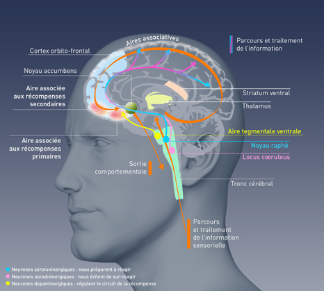
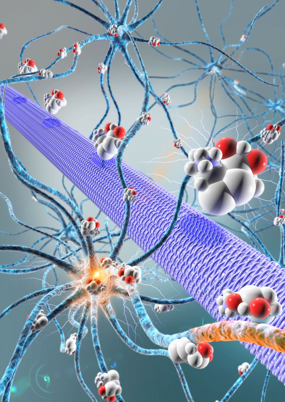
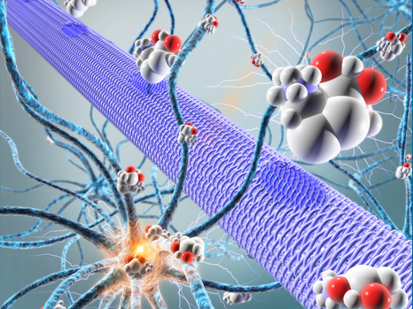
Scientists have just identified in the mouse, and then confirmed in humans, a new factor that regulates addiction. Glutamate, a neurotransmitter[1], contributes to regulating dopamine release in the nucleus accumbens, one of the cerebral structures of the reward system. More precisely, it is its subtle balance with another neurotransmitter – acetylcholine – that prevents up-regulation of the system and entry into addiction. This discovery, which opens up new therapeutic perspectives, was made by neurobiologists in the Neurosciences Paris-Seine laboratory (Institut de Biologie Paris-Seine, CNRS/Inserm/UPMC) and the Douglas Mental Health University Institute (McGill University, Montreal, Canada), working in association with human genetics specialists at Institut Mondor de Recherche Biomédicale (Inserm/UPEC). Their work was published on 4 August 2015 in Molecular Psychiatry.

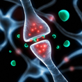
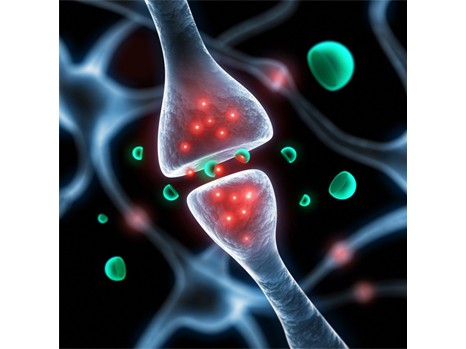
In the context of drug taking, dopamine levels rise in the cerebral structures that form the reward system.  The intensity and rapidity of dopamine release provide a basis for the processes that will lead to the development of addiction. The cholinergic neurons in the nucleus accumbens, one of the centers of reward, are known to regulate this dopamine release. While most neurons only release a single neurotransmitter, a French-Canadian team led by CNRS researcher Salah El Mestikawy showed in 2002 that these acetylcholine-using neurons are also able to utilize glutamate. These neurons, which are to some extent “bilingual”, can thus both activate (via acetylcholine) and inhibit (via glutamate) dopamine secretion.

In this new study, much of it carried out by Diana Yae Sakae for her thesis project supervised by Salah El Mestikawy, the scientists showed that when they inhibited a gene essential to this signaling by glutamate (called VGLUT3) in mice, the animals became more vulnerable to cocaine. They experienced enhanced stimulant effects of the drug, developing “addiction” more easily and being more likely to “relapse” after a period of abstinence. The glutamate from these acetylcholine neurons therefore plays an important regulatory role in limiting cocaine addiction.

The scientists then wanted to determine whether this mechanism also applied to humans. In cocaine and/or opiate-dependent adults, they looked for mutations of the gene that had rendered the mice “addicted”. At the Institut Mondor de Recherche Biomédicale, Stéphane Jamain’s team observed that a mutation of this gene was ten times more common among severely dependent patients than in individuals without psychiatric symptoms. This mutation may explain the greater vulnerability to addiction of these patients[2]. In any case, these observations appear to confirm the role of glutamate in the addictive mechanism.

This work has thus clarified the neuronal mechanisms that underlie the search for hedonic sensations: contrary to what scientists thought until now, these findings show that it is not acetylcholine alone that regulates dopamine release, but a balance between acetylcholine and glutamate.

 At the same time, the scientists identified an unsuspected target for the treatment of drug addiction. Indeed, although acetylcholine has numerous other functions in the brain and muscles, this glutamate signaling is more specific. The next step is to identify the receptor involved so that pharmacological therapies can be developed.

This work was funded in particular by the Fondation pour la Recherche Médicale (FRM) and the Agence Nationale pour la Recherche (ANR).

[1] In order to communicate, neurons use chemical substances called neurotransmitters. Conventional neurotransmitters include dopamine, serotonin, acetylcholine and glutamate, etc.

[2] That said, even within the group of severely dependent patients this mutation was only present in 5% of cases, indicative of the multifactorial nature of addiction and more generally the complexity of psychiatric diseases

An advance in understanding the origins of language

Researchers from the Cognitive Neuroimaging Unit at NeuroSpin have just identified a network of areas of the brain that are organised in a way that could at least partially explain the specificity of the cognitive functions in the human species. These regions are specifically activated in humans, but not in the macaque monkey, in response to specific variations in the auditory sequences played. They coincide with the classic language areas, particularly Broca’s area. The human faculty of language could therefore have its origins in the emergence of a brain circuit capable of combining, in a single region, information coming from the other regions of the brain into a coherent whole. These results, obtained through a collaboration between the French Atomic Energy Commission (CEA), Inserm, Collège de France, Versailles-Saint-Quentin-en-Yvelines University and Paris-Sud University, are published in Current Biology.

In this study, conducted at NeuroSpin, Stanislas Dehaene (a professor at Collège de France, and director of the Inserm/CEA/Paris-Sud University Cognitive Neuroimaging Unit) and Bechir Jarraya (Professor of Neurosurgery at Versailles-Saint-Quentin-en-Yvelines University), together with Liping Wang and Lynn Uhrigh, used a noninvasive functional imaging method, 3 Tesla functional MRI. They exposed three macaque monkeys and twenty volunteers to regular auditory sequences, e.g. three identical sounds followed by a fourth different sound (a sequence notated as AAAB). Occasionally, they presented a sequence that violated this regularity, either because it included a different number of sounds (e.g. AAAAAB), or because the sequence of sounds was abnormal (e.g. AAAA, which does not end in a B sound).

The monkey’s brain reacted to changes in numbers and sequences, which denotes a certain capacity for abstraction. However, it did so in distinct areas, specialised for either number or sequence. In contrast, the human brain combined the two parameters in regions that coincide with the language areas.

Thus, whereas the monkeys detected isolated properties, such as “four sounds” or “the last one is different,” evolution seems to have endowed our species with a specific ability to combine these pieces of information into a coherent whole, a formula such as “three sounds, then another”—the very beginnings of an inner language?


Thus, although the abstract representation of sound sequences is possible in non-human primates, the evolution of a new brain circuit, connected to the auditory areas, might have enabled our species to acquire the unique ability to compose and recognise the complex sequences characteristic of human languages.Dehaene_LangageCerveau

This figure illustrates the unique ability of the human brain to combine pieces of abstract auditory information. Some regions of the brain are associated with the detection of a change in number of sounds by the brain, independently of a concomitant change in the sequence of sounds (red areas in the figure). Conversely, some regions of the brain detect changes in the sequence of sounds, independently of their number (green areas). In the monkey brain, these two sets of regions are unconnected. Places where they intersect (shown in yellow), i.e. regions that combine the two types of information, “change in sequence of sounds” and “change in number of sounds,” are found only in the human brain. All activations detected are projected on a lateral view of the right hemisphere for purposes of representation. © Liping Wang

Unable to cope with uncertainty: the gateway to psychosis?

Imagine being plunged into a world in which events did not always have the same consequences, and with rules that changed without your knowledge. How would you adapt? Uncertainty as a factor in decision making is a fundamental issue in general psychology. Our world turns out to be more or less predictable, and our brain has to adapt to this uncertainty to make the best possible choices in any situation. This is the subject that attracted Fabien Vinckier and Raphaël Gaillard, researchers at St Anne’s Hospital, Inserm and Paris Descartes University, in collaboration with Mathias Pessiglione, an Inserm researcher at the Brain and Spinal Cord Institute at Pitié–Salpêtrière Hospital, AP-HP, and Paul Fletcher, from the University of Cambridge in Great Britain. This study, which has been published in Molecular Psychiatry, reveals that our ability to adapt our decisions to the uncertainty inherent in any choice may be disrupted in the early stages of psychosis.

Participants were invited to play a computer game during which they had to decide whether to bet on symbols. The rules were not always applied, and were reversed from time to time (a symbol that always won money started to lose it, and vice versa). When subjected to these conditions, participants, in order to adapt their choices, had to be able to simultaneously detect changes in the rules of play and times of stability. It was possible to show, with the help of mathematical models, that to be most effective, participants use their confidence in the rules of play to make their choices.

In order to reproduce the conditions for the early stages of psychosis, participants were intravenously administered either a placebo or a very low dose of ketamine. Ketamine is an anaesthetic that is used daily in high doses in operating theatres, and which, at low doses, causes symptoms strongly resembling the early stages of a psychotic episode. Continuous measurement of the participants’ behaviour and brain activity using functional magnetic resonance imaging (fMRI) made it possible to identify the effects of ketamine.

Using this model, the researchers demonstrated that ketamine affects the ability of participants to distinguish times when the rules of play are stable, and optimise their behaviour accordingly.

Thus, they did not did not come to a point where they systematically bet on the winning symbol (i.e. betting 100% of the time, even though the symbol only actually won 80% of the time), as if a persistent doubt unsettled them. This impairment is correlated with a disturbance in the fronto-parietal brain network.

“This study characterises the key role of adaptation to uncertainty in decision-making, and its disruption in the early stages of psychosis. It should enable a better understanding of the onset of psychotic illness, and guide therapeutic innovation,” explains Raphaël Gaillard, Professor of Psychiatry at Paris Descartes University, and Head of the Department of Mental Health and Therapeutics at St Anne’s Hospital.

This study reveals, in a pharmacological model of psychosis, the disruption of a person’s ability to finely adapt his/her behaviour to the uncertain nature of the environment. The brain bases for this impairment have been identified (a fronto-parietal network), and can be linked to the molecular pathway on which ketamine acts, and which is currently the focus of a search for new treatments for schizophrenia.

These findings are a continuation of a publication that appeared in the journal Science (Whitson, Science, 2008) on the onset of apparently psychotic phenomena (superstitions, conspiracy theories) in people who are subjected to strong uncertainty. Some psychotic symptoms, such as the emergence of delusions, could be a type of inappropriate response to the inability to construct and maintain a stable representation of the worldPhotoCP web

©Fotolia

Autism: the value of an integrated approach to diagnosis

Researchers at Inserm (Inserm Unit 930 “Imaging and Brain”) attached to François-Rabelais University and Tours Regional University Hospital have combined three clinical, neurophysiological and genetic approaches in order to better understand the brain mechanisms that cause autism. When tested on two families, this strategy enabled the researchers to identify specific gene combinations in autistic patients that distinguished them from patients with intellectual disabilities.

This study, published in the journal Molecular Psychiatry, offers new prospects for the diagnosis and understanding of the physiological mechanisms of autism.

Autism is a condition characterised by great heterogeneity, both in terms of clinical manifestations and genetics. It is currently estimated that nearly 400 genes may be involved in this disorder. Diagnosis of this condition is all the more complex because it is often associated with other developmental disorders involving the same genes.

To improve diagnosis, the Inserm researchers used an original multimodal approach combining:

  • Clinical assessment
  • High-throughput genomic analysis to sequence all the genes
  • Analyses of the electrical activity of the brain in response to the perception of a change (electroencephalography – EEG)

Two families with members affected by autism and/or intellectual disability were given the benefit of this integrated approach. In these two families, all individuals affected by the condition carried a mutation in the NLGN4X gene, which manifested in the brain as problems in transmitting information by the neurons.

Using EEG, the researchers primarily observed an abnormal brain wave pattern, characteristic of patients with autism. The other family members, including those with intellectual disabilities, did not show this feature.

Thanks to this new approach, a second rare mutation was characterised and linked to atypical brain activity measured by EEG in autistic patients.

For Frédéric Laumonnier and Frédérique Bonnet-Brilhault, the main authors of this work, “This study helps us realise that there is no ‘gene for autism,’ but combinations of genes involved in neurodevelopment that affect the development of the neuronal networks targeted by this condition.”

Identifying these combinations is a key step in understanding the physiopathology, and ultimately in the development of targeted therapeutic drugs.

Autism is a pervasive developmental disorder that appears early in childhood and continues into adulthood. It presents as an altered ability to establish social interactions and communicate, and as behavioural problems. People with autism often seem imprisoned in a kind of inner world.

This work was supported by Fondation de France and the European Union (EU FP7 project Gencodys)

Caring for blindness: a new protein in sight?

Vasoproliferative ocular diseases are responsible for sight loss in millions of people in the industrialised countries. Many patients do not currently respond to the treatment offered, which targets a specific factor, VEGF. A team of Inserm researchers at the Vision Institute (Inserm/CNRS/Pierre and Marie Curie University), in association with a team from the Yale Cardiovascular Research Center, have demonstrated in an animal model that blocking another protein, Slit2, prevents the pathological blood vessel development that causes these diseases. This work is published in Nature Medicine.

Vasoproliferative ocular diseases are the main cause of blindness in the industrialised countries. Age-related macular degeneration (ARMD), diabetic retinopathy and retinopathy of prematurity (in newborns) are characterised by progressive involvement of the retina, the area of the eye that receives visual information and transmits it to the brain. This damage is caused by abnormal growth of the blood vessels in the retina. These weakened vessels allow leakage of serum—which causes a swelling that lifts the retina—and/or blood, which leads to retinal haemorrhage.

This process involves several proteins required for normal or pathological development of the blood vessels. The action of vascular endothelial growth factor (VEGF) is a particularly decisive factor in this ocular disorder. At present, the main treatments are aimed at blocking its action by injecting inhibitors into the eye. Some patients are or become resistant to these anti-VEGF therapies.

For this reason, the team led by Alain Chédotal, in collaboration with a team led by Anne Eichmann[1],

sought to identify new factors involved in the growth of new blood vessels, angiogenesis. They paid particular attention to Slit2.

Slit2 is a protein already known for its role in the development of neural connections. By acting on its receptors, Robo1 and Robo2, it is also involved in the development of many organs and certain cancers. The researchers therefore formulated the hypothesis that this factor might have a role in the abnormal vascularisation observed in vasoproliferative ocular diseases.

To test this postulate, the scientists inactivated Slit2 in a mouse model. They observed that ramification and growth of the retinal blood vessels were severely reduced, without any change in the stability of the pre-existing blood supply. Surprisingly, they discovered that without this protein, VEGF action was also partly reduced. By simultaneously blocking Robo1 and Robo2, they obtained the same results. They thus demonstrated that Slit2 is essential for angiogenesis in the retina.

“The success of these initial experiments led us to hope that controlling Slit2 might block the chaotic development of blood vessels in ocular diseases,” explains Alain Chédotal, Inserm Research Director.


The team therefore repeated these tests in an animal model for retinopathy of prematurity. As they had suspected, the absence of Slit2 protein prevented abnormal vascularisation of the retina in these young mice.

This work suggests that therapies targeting Slit2 protein and its receptors, Robo1 and Robo2, might be beneficial for patients with vasoproliferative ocular disease, especially those who are resistant to conventional anti-VEGF therapies.

Moreover, it would be interesting to set up other studies to obtain a better understanding of the mechanism of action of Slit2 and its relationship with VEGF. This could open up new avenues for the treatment of tumours.inserm

© Alain Chédotal /Inserm. Retinal blood supply of a one-week-old mouse. Growth is upward. The endothelial cells constituting the vessel walls are shown in blue and their nuclei in red. Green staining indicates the nuclei of proliferating cells, which will give rise to new vessels. Proliferating endothelial cells therefore appear yellow (green added to red).


[1]  Yale Cardiovascular Research Center (Yale University) and the Center for Interdisciplinary Research in Biology (CNRS/INSERM/Collège de France)

An electronic micropump to deliver treatments deep within the brain.

Many potentially efficient drugs have been created to treat neurological disorders, but they cannot be used in practice. Typically, for a condition such as epilepsy, it is essential to act at exactly the right time and place in the brain. For this reason, the team of researchers led by Christophe Bernard at Inserm Unit 1106, “Institute of Systems Neuroscience” (INS), with the help of scientists at the École des Mines de Saint-Étienne and Linköping University (Sweden) have developed an organic electronic micropump which, when combined with an anticonvulsant drug, enables localised inhibition of epileptic seizure in brain tissue in vitro. This research is published in the journal Advanced Materials.

Drugs constitute the most widely used approach for treating brain disorders. However, many promising drugs failed during clinical testing for several reasons:

  • they are diluted in potentially toxic solutions,
  • they may themselves be toxic when they reach organs to which they were not initially directed,
  • the blood-brain barrier, which separates the brain from the blood circulation, prevents most drugs from reaching their targets in the brain,
  • drugs that succeed in penetrating the brain will act in a non-specific manner, i.e. on healthy regions of the brain, altering their functions.

Epilepsy is a typical example of a condition for which many drugs could not be commercialised because of their harmful effects, when they might have been effective for treating patients resistant to conventional treatments [1].

During an epileptic seizure, the nerve cells in a specific area of the brain are suddenly activated in an excessive manner. How can this phenomenon be controlled without affecting healthy brain regions? To answer this question, Christophe Bernard’s team, in collaboration with a team led by George Malliaras at the Georges Charpak-Provence Campus of the École des Mines of Saint-Étienne and Swedish scientists led by Magnus Berggren from Linköping University, have developed a biocompatible micropump that makes it possible to deliver therapeutic substances directly to the relevant areas of the brain.

The micropump (20 times thinner than a hair) is composed of a membrane known as “cation exchange,” i.e., it has negative ions attached to its surface. It thus attracts small positively charged molecules, whether these are ions or drugs. When an electrical current is applied to it, the flow of electrons generated projects the molecules of interest toward the target area.

To enable validation of this new technique, the researchers reproduced the hyperexcitability of epileptic neurons in mouse brains in vitro. They then injected GABA, a compound naturally produced in the brain and that inhibits neurons, into this hyperactive region using the micropump. The scientists then observed that the compound not only stopped this abnormal activity in the target region, but, most importantly, did not interfere with the functioning of the neighbouring regions.

This technology may thus resolve all the above-mentioned problems, by allowing very localised action, directly in the brain and without peripheral toxicity.

“By combining electrodes, such as those used to treat Parkinson’s disease, with this micropump, it may be possible to use this technology to treat patients with epilepsy who are resistant to conventional treatments, and those for whom the side-effects are too great,” explains Christophe Bernard, Inserm Research Director.


Based on these initial results, the researchers are now working to move on to an in vivo animal model and the possibility of combining this high-technology system with the microchip they previously developed in 2013. The device could be embedded and autonomous. The chip would be used to detect the imminent occurrence of a seizure, in order to activate the pump to inject the drug at just the right moment. It may therefore be possible to control brain activity where and when it is needed.

In addition to epilepsy, this state-of-the-art technology, combined with existing drugs, offers new opportunities for many brain diseases that remain difficult to treat at this time.


PhotoCP-micropompe

The organic electronic micropump (represented by a purple cylinder) directly releases among the neurons, active molecules (spheres) that will control the activity of these neurons (here they will stop the epileptic activity).

© Adam Williamson, Christophe Bernard, ID Labs, Arab4D (Christophe Bernard: Controlling Epileptiform Activity with Organic Electronic Ion Pumps. DOI: 10.1002/adma.201500482. 2015. Copyright Wiley-VCH Verlag GmbH & Co. KGaA. Reproduced with permission)

[1]Epilepsy in brief

This disease, which affects nearly 50 million people in the world, is the most common neurological disorder after migraine.

The neuronal dysfunctions associated with epilepsy lead to attacks with variable symptoms, from loss of consciousness to disorders of movement, sensation or mood.

Despite advances in medicine, 30% of those affected are resistant to all treatments.

A pharmacological compound for restoring neuromuscular transmission

Researchers from Inserm, CNRS and Paris Descartes University have just demonstrated the beneficial effect of lithium chloride on a group of genetic disorders responsible for muscle dysfunction known as congenital myasthenias. These results, obtained using a new mouse model of the disease, are published in The Journal of Neuroscience, and constitute an important step in the search for treatments for these rare diseases.

Congenital myasthenic syndromes (CMS) usually begin in the neonatal period, but sometimes begin later in childhood, during adolescence, or even in adulthood. They manifest as muscular weakness in the arms and legs, with involvement of the eyes, and facial and bulbar muscles (sucking, swallowing and vocal function [dysphonia]). They are characterised by dysfunction of neuromuscular transmission. This rare disease affects 1-2 individuals in 500,000 (source: Orphanet).

Fotolia_31590778_S

© Fotolia. Neuron Synapses enable the transmission of neuromuscular messages.


Researchers from the “Centre for Neurophysics, Physiology and Pathology” (French National Centre for Scientific Research [CNRS] / Paris Descartes University) in Paris study the physiopathological mechanisms of these diseases of neuromuscular transmission.

Using a transgenic mouse model of the disease, they have just shown the beneficial effect of a pharmacological compound known as lithium chloride (LiCl). Lithium chloride was already known as a treatment for some diseases of the central nervous system, such as depression and bipolar disorder.

“Treating these mice with lithium chloride reduces muscle weakness and fatigability,” explains Laure Strochlic, Inserm researcher.

Specifically, the researchers injected the product once a day into the peritoneum of the mice. The compound substantially restores the altered structures of the synapses, the structures that allow the transmission of information between nerve cells. It acts by inhibiting an enzyme known as “GSK3” in the muscle, making it possible to restore the motor deficit caused by the disease.

The researchers are now working to see if these results can be reproduced in other models of congenital myasthenia, and are adjusting the dose and duration of treatment. They are planning a clinical trial to test the efficacy of LiCl and other inhibitors of the GSK3 enzyme in 2-3 years’ time.

These results are protected by a patent filed by Inserm Transfert.

Repairing the cerebral cortex: it can be done

A team led by Afsaneh Gaillard (Inserm Unit 1084, Experimental and Clinical Neurosciences Laboratory, University of Poitiers), in collaboration with the Institute of Interdisciplinary Research in Human and Molecular Biology (IRIBHM) in Brussels, has just taken an important step in the area of cell therapy: repairing the cerebral cortex of the adult mouse using a graft of cortical neurons derived from embryonic stem cells. These results have just been published in Neuron.

The cerebral cortex is one of the most complex structures in our brain. It is composed of about a hundred types of neurons organised into 6 layers and numerous distinct neuroanatomical and functional areas.

Brain injuries, whether caused by trauma or neurodegeneration, lead to cell death accompanied by considerable functional impairment. In order to overcome the limited ability of the neurons of the adult nervous system to regenerate spontaneously, cell replacement strategies employing embryonic tissue transplantation show attractive potential.

A major challenge in repairing the brain is obtaining cortical neurons from the appropriate layer and area in order to restore the damaged cortical pathways in a specific manner.

The results obtained by Afsaneh Gaillard’s team and that Pierre Vanderhaeghen at the Institute of Interdisciplinary Research in Human and Molecular Biology show, for the first time, using mice, that pluripotent stem cells differentiated into cortical neurons make it possible to reestablish damaged adult cortical circuits, both neuroanatomically and functionally.

These results also suggest that damaged circuits can be restored only by using neurons of the same type as the damaged area.

This study constitutes an important step in the development of cell therapy as applied to the cerebral cortex.

This approach is still at the experimental stage (laboratory mice only). Much research will be needed before there is any clinical application in humans. Nonetheless, for the researchers, “The success of our cell engineering experiments, which make it possible to produce nerve cells in a controlled and unlimited manner, and to transplant them, is a world first. These studies open up new approaches for repairing the damaged brain, particularly following stroke or brain trauma,” they explain.

Neuron

© A. Gaillard/Inserm. An illustration showing the integration of neurons transplanted into the brain following injury, two months after transplantation. Specific projections of the adult brain (red) into the transplanted neurons (green)

This project was funded by the French National Research Agency (ANR-09-MNPS-027-01).

Sleep reduces the brain’s predictive ability

Why are we not aware of external noises while we sleep? A study carried out at NeuroSpin (French Atomic Energy Commission [CEA]/Inserm), in collaboration with the Sleep and Alertness Centre at Hôtel-Dieu Hospital, Paris (AP-HP), the Brain and Spinal Cord Institute (ICM), Collège de France, and Paris-Sud and Paris Descartes Universities, has shown that even though sounds continue to penetrate the auditory cortex, sleep disrupts the brain’s ability to anticipate them. The researchers have demonstrated that the brain is no longer capable of making predictions during sleep, because the predictive signals coming from the higher cortical areas seem to be eliminated. These results are published in the American journal PNAS, on 2 March 2015.

While listening to a melody during wakefulness, the brain uses the regularities in the sound sequence to predict the future sounds. This predictive ability is based on the functioning of a hierarchy of areas in the brain. If a sound breaks the regularity of the sequence, the brain then generates a series of “prediction error” signals responsible for, among other things, reactions to novelty or reactions of surprise. Previous studies using electroencephalography (EEG) have made it possible to describe at least two consecutive error signals, mismatch negativity (MMN) and P300. MMN has already been observed in subjects in an unconscious state (including a comatose state), whereas P300 may be specific for conscious processing, since it reflects the integration of information over a vast brain network, beyond the auditory regions.

During sleep, ambient sounds are not consciously perceived. However, we do not know the extent to which the integration of these sounds by the brain is disrupted, or whether the brain remains capable of perceiving their regularities and anticipating them. This particular aspect of brain function was tested by the NeuroSpin team (Inserm/CEA), in collaboration with the Sleep and Alertness Centre at Hôtel-Dieu Hospital, Paris (AP-HP), the Brain and Spinal Cord Institute (ICM), Collège de France, and Paris-Sud and Paris Descartes Universities. The researchers used electro- and magnetoencephalography (E/MEG) to study the prediction error signals (MMN and P300) in subjects during sleep and wakefulness.

The researchers invited volunteers to go to sleep inside NeuroSpin’s magnetoencephalography machine, in the presence of repetitive sounds. Results confirmed that P300 is a specific marker for conscious processing of sounds, since it disappeared as soon as the volunteers fell asleep, from which time the subjects no longer reacted to sounds. In contrast, MMN was observed at all stages of sleep (slow-wave sleep and rapid eye movement [REM] sleep). However, this signal is only partially retained, since some areas of the brain, which are normally activated during wakefulness, no longer respond to the sound stimulus. Indeed, the peak of activity resulting from a predictive error in an awake individual disappears during sleep. The only remaining phenomena are those of passive sensory adaptation, and confined to the primary auditory areas.

The researchers have thus shown that, due to a defect in communication between its different areas, the brain can no longer make predictions during sleep.

However, it remains capable of representing sounds within the auditory areas, and can get used to them if they are frequent, explaining why we are awakened by an alarm but not by the ticking of a clock.


PredictionBrain

©S. Dehaene. Reconstruction of the sources of error signals in the brain based on magnetoencephalographic recordings. The signals that indicate predictive error; the intermediate component of MMN and P300, disappear during sleep. Only passive mechanisms for sensory adaptation (the early and late components of MMN), confined to the auditory areas, remain. (Times are expressed in milliseconds, and measure the time taken to respond to the sound).

VEGF-C, an indispensable growth factor for producing new neurons


The decline of the neurogenesis mechanism (production of new neurons) during ageing is implicated in the emergence of neurodegenerative diseases such as Alzheimer’s disease. Research studies bringing together Inserm, CNRS and Université de Pierre et Marie Curie researchers from the Brain and Spinal Cord Institute (Inserm/ CNRS / Pierre and Marie Curie University), in collaboration with a team from the Yale Cardiovascular Research Center, have demonstrated the importance of factor VEGF-C in the activation of neural stem cells, and hence in the production of new neurons. These results, published in Cell Reports, bring fresh hope for the development of therapies that may help to improve the production of neurons to alleviate cognitive decline in people with Alzheimer’s disease.

Throughout life, adults are able to generate new neurons from neural stem cells (NSC), in order to maintain their complement of cognitive skills. This neurogenesis occurs in the hippocampus, a brain structure that plays a central role in memory. However, age and some strokes lead to a decline in this function, and can contribute to the appearance of serious cognitive disorders, such as Alzheimer’s disease.

Although the stages in the formation of new neurons are well known, the molecular mechanisms of this phenomenon are less well understood. Indeed, NSC spend most of the time in a quiescent state during which the cells are outside the cell cycle and do not divide. The repressors that maintain this quiescent phase have been identified, but there are still many questions about the factors that make it possible to exit this cell “dormancy.”

Against this background, Jean-Léon Thomas and Anne Eichmann decided to focus on the vascular endothelium growth factors (VEGF) and their receptors (VEGFR), which are already suspected of involvement in regulating the growth and maintenance of nerve cells. More specifically, they chose to study the potential activating ability of factor VEGF-C when bound to its receptor, VEGFR3.

Their in vitro and in vivo experiments confirm that rodent NSC possess receptor VEGFR3 and also produce factor VEGF-C. Stimulation of NSC by VEGF-C leads to the activation of these cells, i.e. their entry into the cell cycle and conversion into neural progenitor cells, and ultimately, the production of new neurons. The specific feature of VEGF-C action in the brain, compared with other vascular growth factors such as VEGF-A, is that it induces the NSC to respond at concentrations that do not induce vascular proliferation. This property confers potential interest on VEGF-C as a specific activator of NSC in the brain.Photo2
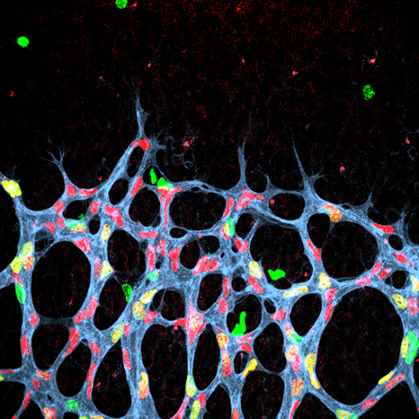
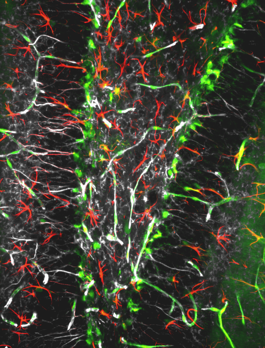
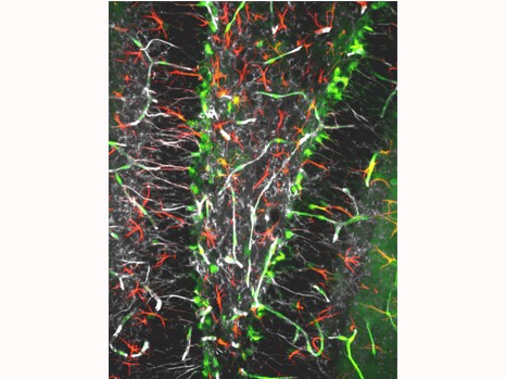
©Inserm, Jinah Han. Among the hippocampal neural stem cells (characterised by expression of Nestin [in white] and GFAP [in red]), those that possess receptor VEGFR3 (in green) are very numerous.

In mice that produce VEGFR3-deficient NSC, this neurogenesis phenomenon is abolished. The scientists thus show that VEGF-C/VEGFR3 signalling is not just involved, but is absolutely required for the neural stem cells to “awaken,” and is hence required for the creation of new neurons.

This mutant model also enabled the team to observe a correlation between mood disorders and deterioration of the neurogenesis function of the hippocampus. As suspected, these mice, in which NSC activation is compromised, develop exaggerated anxiety on ageing, similar to that seen in patients with Alzheimer’s disease. This result suggests that VEGF-C/VEGFR3 neural signalling participates in the maintenance of cognitive functions in the murine model.

Observations validated in humans

As a logical continuation of this work in rodents, the researchers wondered about the presence of similar mechanisms in humans. They then discovered that this signalling pathway is conserved in human nerve cells, where it also promotes cell proliferation and survival in vitro.

Although these results are still preliminary, they provide arguments to support the idea that adult NSC activity could have a central role in physiological and behavioural control in the body. Similarly, the decline in this activity during ageing could be associated with the onset of mood disorders such as anxiety and depression.

From the therapeutic point of view, these studies are encouraging: VEGF-C might be a good candidate for improving the production of new neurons and offsetting cognitive decline in people with Alzheimer’s disease.

Brain and spatial attention: Can the left hemisphere compensate for a lesion in the right hemisphere?

Dr Paolo Bartolomeo, Inserm Research Director and head of the PICNIC LAB[1] team at the Brain and Spinal Cord Institute (ICM, an Institute supervised by Inserm, CNRS and UPMC) and his collaborators have published the results of their research on “unilateral spatial neglect,” also known by the term “hemineglect,” in the journal Brain. People with this disorder act as if they did not know about the left side of their world. This disorder occurs primarily after an injury to the right cerebral hemisphere, for example following a stroke (cerebrovascular accident or CVA), and exacerbates the resulting disability by impeding rehabilitation and recovery. The scientists therefore searched for factors predicting the persistence of this disorder, in order to offer patients appropriate rehabilitation.

The published study shows that the two hemispheres can partially compensate for one another in the event of an injury, through incompletely understood mechanisms known as “brain plasticity.” However, results suggest that this compensation requires both hemispheres to “speak to one another” via intact connections—bundles of white matter formed by extensions of the neurons.

In the acute phase of a stroke affecting the right hemisphere, the vast majority of patients show signs of left-sided neglect (since the left side of our body is controlled by the right hemisphere and vice versa). These patients behave as if the left side of their world no longer existed. They do not eat food from the left side of their plate, they collide with furniture located on their left, and do not shave or apply make-up to the left side of the face. They also recover less well from their motor deficits than patients affected in the left hemisphere. Some of them recover with time, but spontaneous improvement of neglect is far from being the rule: at least one-third of patients showing this disorder in the acute phase continue to show signs of it over a year after acquiring their lesion. This highlights the clinical imperative to identify factors that predict the persistence of negligence, in order to offer appropriate rehabilitation to patients for whom this disorder risks becoming chronic.

Dr Paolo Bartolomeo and his collaborators monitored changes in neglect over time in 45 patients with vascular lesions in the right hemisphere. Advanced magnetic resonance imaging methods made it possible to study the condition of the fibres in the white matter that enable the different regions of the brain to communicate with one another, as well as the two hemispheres. All neglect patients had damage to the lines of communication between the anterior and posterior parts of the right hemisphere; patients with persistent neglect lasting one year after acquiring the lesion also showed damage to the posterior part of the corpus callosum, the connection that allows the two cerebral hemispheres to communicate with one another. The (healthy) left hemisphere must therefore be able to communicate with the injured (right) hemisphere, in order to learn to compensate for the visuospatial defects provoked by the brain lesion. Patients with damage to the corpus callosum are at risk of developing chronic neglect, and should therefore have priority access to rehabilitation treatments.

PhotoCP web

A patient with spatial neglect fails to copy the elements located on the left side of a picture. © PICNIC LAB – ICM

[1] Physiological Investigations of Clinically Normal and Impaired Cognition

fermer