Menu

Stroke (CVA) in young adults: discovery of a susceptibility gene associated with bleeding of the cervical arteries

Researchers from the joint research unit “Public health and molecular epidemiology of aging related diseases” (Inserm/Institut Pasteur Lille/Université Lille 2), in collaboration with Lille Regional University Hospital (CHRU), have discovered a susceptibility gene involved in this major cause of stroke (cerebrovascular accident) in young subjects. This gene, PHACTR1, is also known to be associated with the occurrence of migraine and myocardial infarction. This international study, carried out by the CADISP[1] international consortium and published in the journal Nature Genetics, shows that a form of the gene is associated with a reduced risk of developing a dissection of the cervical arteries responsible for the bleeding that leads to stroke. This work opens up new possibilities for identifying people at risk, and trying to prevent the occurrence of stroke in young adults.
PhotoCP web

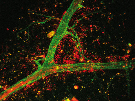
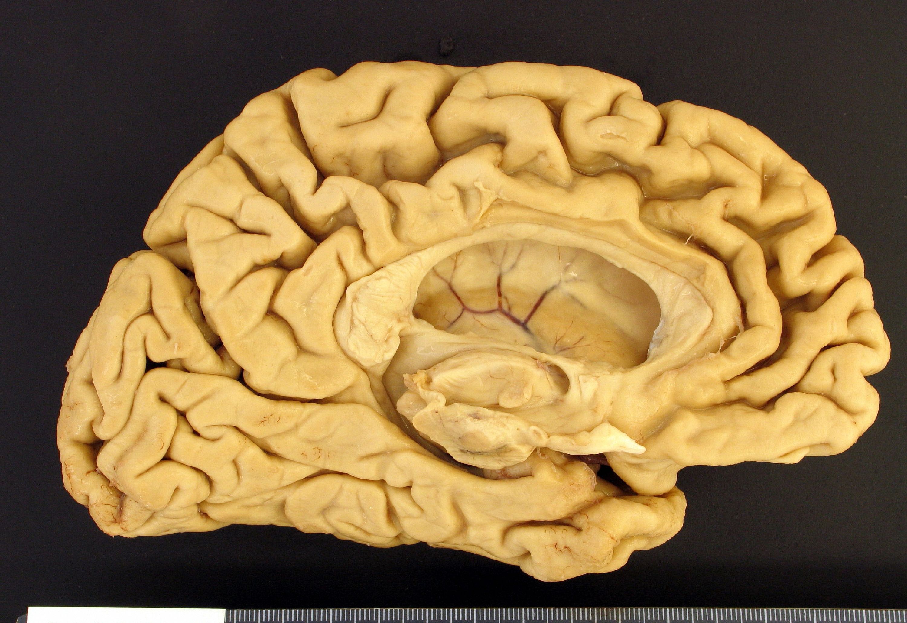
Stroke (CVA) ©Inserm/U919

Cervical artery dissection is a major cause of stroke in young subjects. It involves bleeding within the lining of the carotid or vertebral artery walls, which causes longitudinal “splitting” (hence the term dissection) without rupturing the vessel. This bleeding gives rise to a haematoma, which reduces the diameter of the artery and may lead to its closure. Often, the formation of a blood clot inside the artery thus completely stops the flow of blood to the brain, resulting in a cerebrovascular accident, or stroke.

The causes of these dissections are still unknown. The current prevailing hypothesis is of a multifactorial disease, possibly related to a pre-existing abnormality in the elasticity of the blood vessel walls. We observe associated factors in these patients, such as migraine, high blood pressure, infections or recent, sometimes minor, trauma (carrying heavy loads, a fall from a bicycle, neck extension due to acceleration in some roller-coaster rides, blows to the nape of the neck, etc.). In the vast majority of cases, cervical artery dissection occurs in the absence of any family history or underlying hereditary disease. However, several hypotheses favour individual susceptibility to arterial dissection, encoded by the genome. This was the context for launching the CADISP consortium, in order to initiate the largest study ever conducted in the area, and thereby systematically screen our genome and discover the basis for this individual genetic susceptibility.

A total of twelve countries, comprising ten European countries, the United States and Russia, were able to bring together 2,052 patients with dissection, and compare their genomes with those of 17,064 unaffected individuals. The researchers and physicians were able to demonstrate that a particular form of the PHACTR1 gene was associated with a reduced risk of developing a cervical artery dissection. This same form of the PHACTR1 gene has been associated with a reduced risk of migraine and an increased risk of myocardial infarction in other studies. The researchers also identified two other genes that are potentially associated with the risk of dissection: the LRP1 gene, already associated with migraine and abdominal aorta aneurysm, and the LNX1 gene, both of which need further confirmation.

“Given the difficult diagnosis and seriousness of this disease, the characterisation of the genetic susceptibility gene PHACTR1 will help to provide a better understanding of the mechanisms leading to the occurrence of these dissections,” explains Stéphanie Debette, neurologist, first author of the article and coordinator of the CADISP international consortium.

“By pooling research efforts on a worldwide scale, we hope to succeed in more rapidly identifying people at risk, and find solutions in order to prevent the main functional consequences associated with the occurrence of stroke in the young adult,” concludes Philippe Amouyel, epidemiologist, director of the joint research unit involving Inserm, Institut Pasteur Lille and Université Lille 2.

These results have been obtained with the help of all the clinicians and their patients, and the genotyping and analysis facilities of the French National Genotyping Centre of the French Atomic Energy and Alternative Energies Commission (CEA), the Human Polymorphism Study Center (CEPH) and the Institut Pasteur Lille.

[1] Cervical Artery Dissections and Ischemic Stroke Patients

To find out more :

Cervical artery dissection: what are the signs?
This pathological phenomenon affects young adults, both men and women, around the age of 40 years, with few or no vascular risk factors. It presents as pain at the location of dissection, generally in the neck region, or unusual headaches. In most cases, concurrently or in the following hours, characteristic symptoms of stroke (paralysis, loss of sensation, loss of speech, reduced field of vision, etc.) appear. The frequency of these dissections is approximately 2.5 to 3 per 100,000 inhabitants per year, which makes it a rare disease, and now thought to be underestimated.

A means for autistic people to restore correct interpretation of sensory information

In autistic children, information coming from the 5 senses – touch, hearing, sight or other stimuli – are not correctly interpreted in the brain, leading to inappropriate behaviour and sometimes uncontrollable reactions. Inserm researchers led by Andréas Frick in Inserm Unit 862 ‘Magendie Neurocentre’ have recently understood why by studying a mouse model mimicking the disorder. They have even found a molecule that could reverse these effects and restore ‘normal’ behaviour in these mice.

These findings are published in the journal Nature Neuroscience

cerveau© Fotolia
Autistic Spectrum Disorders (ASD) affect more than 3 million people in the European Union, including about 650,000 in France. Recent estimates from the Centre for Disease Control (in the USA) suggest that one child in 68 is affected by this disorder. ASDs are neuro-developmental disorders that affect children from all ethnic and socio-economic origins, and are characterised by a spectrum of symptoms including both difficulties in social interactions and communication as well as stereotypical repetitive behaviours.

Another common aspect of neuro-developmental disorders is the problem of processing sensory information. Nearly 90% of children with ASDs are affected by different types of sensory problems. Problems of sensory interpretation derive from the fact that peripheral information, whether from touch, hearing, sight or other stimuli, are not interpreted or organised correctly in the brain, leading to inappropriate behaviour. Such problems can be extremely disabling in daily life for people affected by autism and they create a challenge for parents and teachers. For example, during a visit to the supermarket, simple fluorescent lights can be an unpleasant sensory experience. Unfortunately, alterations of sensory interpretation in these disorders and their pharmacological treatments are little studied, even if these alterations are also frequently observed in a related neuro-developmental disorder, Fragile X Syndrome.

In a study published in Nature Neuroscience, Inserm researchers (working with researchers from the French CNRS – national centre for scientific research) have shown that Fragile X mice display disorders in the manner that sensory information is processed by the neocortex, which is one of the parts of the brain responsible for sensory perception. The researchers have shown that the neocortex of these mice is hyperexcited in response to tactile sensory stimulation. They then performed a variety of detailed tests showing that this neocortical hyperexcitability is linked to the way the neurons in this region of the brain interpret sensory information. With this study, the researchers found that the function of certain ionic channels (molecules that determine the manner in which neurons process electrical signals) is altered in the dendritic compartment (the structure that interprets information and really behaves as the ‘brain’ of neurons).

By using a pharmacological molecule mimicking the function of one of these channels, they were able to correct this neocortical hyperexcitability as well as the neuron interpretation anomalies.

Furthermore, they were also able to correct a behavioural consequence, particularly of hypersensitivity to sensory stimuli (healthy mice were not affected by this treatment). These findings offer new hope for personalised treatment for the sensory aspects of Fragile X Syndrome and autistic spectrum disorders, especially because these treatments could be applied to adult or adolescent patients.

Repeated episodes of depression: what are the effects on the brain?

Depression is not an ordinary case of the blues—it causes lasting changes to the intellectual functions unless it is managed. According to the results of a study carried out by Philip Gorwood (Inserm Unit 894, “Psychiatry and Neurosciences Center”, Mental and Brain illness Clinic – CMME, Saint Anne’s Hospital, Paris), people who have already experienced two or more depressive episodes perform routine cognitive tasks that require attention, concentration and speed abnormally slowly. These results, published in the journal European Neuropsychopharmacology, appear to confirm that depression may be a “neurotoxic” illness. Preventing recurrences is therefore clearly essential.

 Fotolia_12376795_S

fotolia


Depression is a common illness that has affected, affects or will affect at least one person out of ten. it is characterised by a permanent sadness, a loss of motivation and pleasure, and altered appetite, sleep and libido. Diagnosis corresponds to specific criteria established by international psychiatric standards. Although different types of management, based on drugs and psychotherapy, have been shown to be effective, the risk of recurrence is high, even after several years of remission.

The consequences of these repeated recurrences are a source of concern for physicians and researchers. Although it has already been proven that there is psychomotor retardation in depressed people (this is indeed one of the diagnostic criteria for the illness), there was until now no indication that this change could persist following a depressive episode.

To find out more, researchers at Inserm carried out a study in more than 2,000 patients who had experienced between 1 and over 5 depressive episodes during their lives. In order to assess their cognitive abilities, they measured the speed in performing a simple test (TMT: trail making test), which consists of linking numbered circles distributed in a disordered manner on a sheet of paper. The test was performed twice for each patient: during the depressive episode, and then 6 weeks later, by which time a good proportion of these patients were in complete remission (with no residual depressive symptoms).

Just after a first episode of depression, the time to perform this test was 35 seconds. These performances were almost identical among people undergoing the second depressive episodes in their lives. However, for people who had already had a history of 2, 3 or more depressive episodes, this time was considerably longer, even in subjects who had recovered (1 min 20 sec instead of 35 sec).

“Several other variables offer a potential explanation (age, education level, occupation, etc.), but if these parameters are adjusted, our results remain extremely robust,” explains Philip Gorwood.

Trails_Screenshot2 test TMA

Fig 1: Sample TMT    
Fig 2: Speed of performing TMT during and after depression, according to the number of depressive episodes previously experienced.

This result is the first to show in such a simple manner the “neurotoxic” effects of depression. It also supports the daily observations made by physicians, and the conclusions of earlier epidemiological studies indicating that depression is an illness that becomes worse with time. The researchers therefore judge that after treatment, prevention of recurrence must be one of the priorities for management.

Moreover, this study might also provide an explanation for this vicious circle: the more depressive episodes I have experienced, the more likely I am to have a recurrence. If speed and efficacy are increasingly altered with the number of recurrences, we imagine that it will become more difficult to adapt to new situations. For example, a worker at a computer, showing limited attention span, oversights in the tasks required, and general slowness in carrying out his/her work, will have lower self-esteem and less recognition from his/her work colleagues, which could make him/her more vulnerable to recurrences of his/her depression in the event of any stress.

Finally, the fact that these cognitive impairments may occur as a sequela of depression might also be considered as an argument in favour of the use of “cognitive remediation.” This therapy is based on the controlled stimulation of the impaired cognitive functions in order to reduce the risk of recurrence. It is widely used in schizophrenia and addiction, but is rarely used to treat depressive disorders.”

The INSIGHT study: to better understand the factors that trigger Alzheimer’s disease

Over 35 million people worldwide suffer from dementia, including Alzheimer’s disease [1]. In France, in 2014, approximately 900,000 people are affected, and the prevalence of the disease will increase considerably: an estimated 1.3 million people will be affected in 2020 [2], and over 2 million in 2040 [3], with more than 225,000 new cases reported each year.

World Alzheimer’s Day, which will take place on Sunday, 21 September, is an opportunity to review the progress of research and to present INSIGHT, a new study launched by teams from Inserm and the city of Paris public hospital system (AP-HP) at the Institute of Memory and Alzheimer’s Disease (IM2A) and the Brain and Spinal Cord Institute (ICM), working together in the Research Institute for Translational Neuroscience (IHU-A-ICM), and in collaboration with Pfizer, in order to observe and understand the natural course of Alzheimer’s disease. INSIGHT is an ancillary study of the MEMENTO national cohort.

Etude de la maladie d'Alzheimer
A study of Alzheimer’s disease – Atrophy of the cerebral hemisphere due to Alzheimer’s disease

INSIGHT is an innovative study of Alzheimer’s disease, and one of the first in the world to monitor healthy subjects at-risk. This study holds great hope for understanding the disease, and its results may hold promise of future treatments for patients.

“Research on Alzheimer’s disease, and more generally on pathologies associated with memory disorders, is evolving rapidly. Today we can recognise the disease very early, through a biological signature that can be identified in all patients. What we now want to explore are the endogenous and exogenous conditions for progression of the disease: why and how it occurs in some subjects but not in others,” explains Professor Bruno Dubois, Director of IM2A (Institute of Memory and Alzheimer’s Disease, located at La Pitié Salpêtrière Hospital in Paris), Director of the Inserm Team “Cognition, Neuroimaging and Brain Diseases” at the ICM, and principal investigator of the study.

Research method
The project involves monitoring 400 healthy volunteer subjects aged 70-85 years, with normal memory function. Its objective is not to detect or treat disease, but to observe changes in these healthy subjects. Do they have brain lesions? Will they develop the disease? How long does it take for the initial symptoms to appear? For the duration of the study, these subjects benefit from the monitoring and support of one of the best teams in the world working on memory-related diseases. To date, nearly 220 subjects have already been recruited.
This single-centre study, which will be conducted entirely at the IM2A (Institute of Memory and Alzheimer’s Disease at La Pitié Salpêtrière Hospital, AP-HP) is itself an unprecedented challenge.

INSIGHT is an ambitious study made possible through an innovative new scheme in medical research, a multi-partner foundation that focuses the Investissements d’Avenir (Investment for the Future) programme via IHU-A-ICM and Pfizer, together with the teams at IM2A and ICM, on a shared objective, that of better understanding Alzheimer’s disease

[1] For information on this subject, see the 2013 report from Alzheimer’s Disease International
[2] Figures from the Inserm information pack on Alzheimer’s disease
[3] More information on the France Alzheimer and Related Diseases website

Alim Louis Benabid receives the 2014 Lasker Award

The 2014 Lasker Award has just been won by Alim Louis Benabid, Director of Inserm Unit 318 “Preclinical Neurosciences” from 1988 to 2006, and winner of the 2008 Inserm Prix d’Honneur for his work on deep brain stimulation for the treatment of Parkinson’s disease.

Since 1945, the Lasker Award has distinguished the most brilliant researchers who have contributed to major advances in the diagnosis, treatment and prevention of human disease. In the international community, it is widely considered a precursor to the Nobel Prize. Prof. Benabid is the 8th French person to receive this prize.

Prix d'Honneur Inserm 2008 : Alim-Louis Benabid

© Inserm/Latron, Patrice

Born on 2 May 1942 in la Tronche, near Grenoble, Alim Louis Benabid spent his childhood in Sétif in Algeria. He returned to France to attend secondary school in Grenoble, and received his higher education in medical and science faculties in Paris.
Following his hospital residency, Doctor of Medicine (1970), Doctor of Science in Physics (1978) and a period as Professor of Biophysics at Université Joseph-Fourier in Grenoble, he directed the Inserm Unit 318 “Preclinical Neurosciences” from 1988 to 2006.

Alim-Louis Benabid focused his research on several brain pathologies, especially tumours and abnormal movements, and developed the surgical technique known as stereotactic (or stereotaxic) surgery, which makes it possible to target certain areas of the brain with a high degree of accuracy. He extended its applications to the treatment of patients with drug-resistant Parkinson’s disease and the treatment of other brain disorders.

From 1987 to 1991, Alim-Louis Benabid and his team developed a technique that involves implanting electrodes directly into the brains of patients with Parkinson’s disease in order to apply high-frequency electrical stimulation. They were thus able to eliminate the motor symptoms (tremor, akinesia, rigidity) of the disease. This intervention shows remarkable efficacy, with very low morbidity, and allows a reduction in drug-based treatments. It presently constitutes the most effective surgical treatment for Parkinson’s disease, and also provides basic data of considerable theoretical value.

Alim-Louis Benabid has also extended the indications for deep electrical stimulation to other pathologies, namely dystonia, refractory epilepsy, and obsessive compulsive disorder.

The introduction of electrodes into the brain, a technology developed under his direction, was greeted as a major discovery by the international scientific community.

Alim-Louis Benabid has subsequently focused his efforts on understanding the mechanisms of action of high-frequency deep brain stimulation and demonstrating its potential long-term effects, including its neuroprotective effects, on the natural course of Parkinson’s disease. He is also eager to identify in detail the effective targets and neuronal networks involved in this intervention.

Yves Lévy, Chairman and CEO of Inserm, wishes to convey his warmest congratulations to Alim Louis Benabid: “This prestigious award is a fitting tribute to his brilliant work on Parkinson’s disease, which has significantly improved the everyday lives of patients. The awarding of this internationally recognised prize confirms the excellence of French biomedical research. It also emphasises the importance of carrying out basic research and high-level clinical research in parallel.”

A scientific advisor for the French Atomic Energy Commission (CEA) since 2007, today he combines his research in this area with research carried out in the field of nanotechnology for the CLINATEC project. Developed by the leaders of technological research at CEA, in partnership with Grenoble University Hospital, Inserm and the Université Joseph Fourier in Grenoble, this biomedical research laboratory, devoted to the health-related applications of micro-nanotechnologies, is responding to a major public health challenge: developing new therapeutic approaches for brain diseases.

Read and view the profile of Alim Louis Benabid, winner of the 2008 Inserm Prix d’Honneur :

Ultrasound tracks odor representation in the brain

A new ultrasound imaging technique has provided the first ever in vivo visualization of activity in the piriform cortex of rats during odor perception. This deep-seated brain structure plays an important role in olfaction, and was inaccessible to functional imaging until now. This work also sheds new light on the still poorly known functioning of the olfactory system, and notably how information is processed in the brain. This study is the result of a collaboration between the team led by Mickael Tanter at the Institut Langevin (CNRS/INSERM/ESPCI ParisTech/UPMC/Université Paris Diderot) and that led by Hirac Gurden in the Laboratoire Imagerie et Modélisation en Neurobiologie et Cancérologie (CNRS/Université Paris-Sud/Université Paris Diderot). Their findings are published in NeuroImage dated July 15, 2014.

How can the perception of the senses help represent the external environment? How, for example, does the brain process food or perfume related olfactory data? Although the organization of the olfactory system is well known it is similar in organisms ranging from insects to mammals its functioning remains unclear. To answer these questions, the scientists focused on the two brain structures that act as major olfactory relays: the olfactory bulb and the piriform cortex. In the rat, the olfactory bulb is located between the eyes, just behind the nasal bone. The piriform cortex, meanwhile, is deep seated in the brain of rodents, which made it impossible to obtain any functional images in a living animal until now.

Yet the neurofunctional ultrasound imaging technique developed by Mickael Tanter’s team, called fUS (functional Ultrasound), allows the monitoring of neuronal activity in the piriform cortex. It is based on the transmission of ultrasonic plane waves into the brain tissue. After data processing, the echoes returned by the structures crossed by these waves can provide images with unequalled spatial and temporal resolution: 80 micrometers and a few tens of milliseconds. The contrast on these images is due to variations in the brain’s blood flow. Indeed, the activity of nerve cells requires an input of energy: it is therefore coupled to an influx of blood into the zone concerned. By recording volume variations in the blood vessels irrigating the different brain structures, it is therefore possible to determine the location of activated neurons.

Several imaging techniques, such as MRI, are already based on the link between blood volume and neuronal activity. But fUS offers advantages in terms of cost, ease of use and resolution. Furthermore, it provides easier access to the deepest structures that are often located several centimeters beneath the cranium. The recordings performed by Hirac Gurden’s team using this technique made it possible to observe the spatial distribution of activity within the olfactory bulb. When an odor was perceived, blood volume increased in clearly defined areas: each odor thus corresponded to a specific pattern of activated neurons.

In addition to these findings, and for the first time, the images revealed an absence of spatial distribution in the piriform cortex. At this level, two different odors triggered the same activation throughout the region. The cellular mechanisms responsible for the disappearance of a spatial signature are not yet clearlydefined, but these findings lead to the formulation of several hypotheses. The piriform cortex could be a structure that serves not only to process olfactory stimuli but rather to integrate and memorize different types of data. By making abstraction of the strict odor induced patterns, it would be possible to make associations and achieve a global concept. For example, based on the perception of the hundreds of odorant molecules found in coffee, the piriform cortex would be able to recognize a single odor, that of coffee.

This work opens new perspectives for both imaging and neurobiology. The researchers will now be focusing on the effects of learning on cortical activity in order to elucidate its role and the specificities of the olfactory system.

pomme banane

Alzheimer’s disease: simplified diagnosis, with more reliable criteria

How many patients receive an incorrect diagnosis of Alzheimer’s disease? The answer is a surprisingly high number: over a third! To reduce the number of errors, the diagnostic criteria must be the most reliable possible, especially at the very early stages of the disease. For the last decade, an international team of neurologists, coordinated by Bruno Dubois (Inserm/Pierre and Marie Curie University/AP-HP Joint Research Unit 975) has been working towards this. In the June issue of The Lancet Neurology journal, we see how the researchers have developed a simplified diagnosis based on the most specific criteria of the disease. A challenge primarily for research, but also for clinical practice.

Alzheimer’s disease is a neurodegenerative disease. It is the most common (70%) form of dementia. In France, the number of people with Alzheimer’s disease and other forms of dementia is estimated at between 750,000 and one million, and is expected to reach 1.29-1.40 million patients by 2030. Alzheimer’s disease results from a loss of neurons. The lesions are caused by an accumulation of some brain proteins. The pathology begins with memory problems. This is followed by problems of orientation in space and in time, behavioural problems and loss of autonomy. However, these symptoms are not specific to Alzheimer’s disease. The real challenge is to know how to distinguish this disease from other types of dementia, and establish the diagnosis as reliably and as early as possible.

In 2005, an international group of neurologists, coordinated by Bruno Dubois at Inserm, came together to redefine the diagnostic criteria established in 1984. Until then, it had been necessary to await the death of a patient in order to establish a diagnosis of Alzheimer’s disease with certainty by examining the lesions in his/her brain. And in the living, only a probability of disease could be inferred, and only at a late stage, based on a certain threshold of severity of dementia.

In 2007, the international team shattered these concepts. The researchers introduced new diagnostic criteria, particularly biomarkers. These are genuine signatures of the disease, and are present from the initial symptoms (prodromal stage).

The publication of these results constituted a revolution. Researchers then observed that with these new criteria, “36% of their patients included in a therapeutic trial based on previous clinical criteria did not have Alzheimer’s disease,” reports Bruno Dubois. And although this analysis involved only a subgroup of patients, the implications are serious. Patients did not receive the correct treatment and/or care. And flawed patient selection might have had an impact on the lack of efficacy observed for the new treatment.

Since 2007, many studies have been published. And the international group decided to analyse this literature to make the diagnostic algorithm for Alzheimer’s disease simpler and more reliable. 

“We have reached the end of the road; we have arrived at the essence, something refined, resulting from an international consensus”, indicates Prof. Dubois. The diagnosis of Alzheimer’s disease will henceforth rely on “just a couple of clinical-biological criteria for all stages of the disease” (see box).

Most of the time, the diagnosis of Alzheimer’s disease is based primarily on a suggestive clinical picture. It is subsequently confirmed or rejected using a biomarker.

As regards the clinical picture, there are three scenarios:

–      typical cases (80-85% of all cases): impairment of episodic long-term memory (known as amnestic syndrome of the hippocampal type and corresponding to difficulty remembering a list a words, even with clues, for example)

–      atypical cases (15-20% of cases): atrophy of the posterior part of the cerebral cortex or logopenic aphasia (impairment of verbal memory where the patient inverts the syllables of a word when repeating it, for example), or frontal brain damage (which results in behavioural problems)

–      preclinical states: asymptomatic at-risk (patients without symptoms, but who are fortuitously discovered to have positive biomarkers during scientific studies), and presymptomatic (with a genetic mutation)

One of the following two biomarkers is required:

–      in the cerebrospinal fluid (obtained by lumbar puncture): abnormal levels of brain proteins (reduced beta amyloid protein and increased tau protein)

–      in the brain by PET (positron emission tomography) neuroimaging: elevated retention of amyloid tracer

This simpler and more reliable algorithm is important, primarily for research (therapeutic trials, characterisation of the disease, monitoring of patient cohorts, etc.). Outside of research, the use of biomarkers, which is expensive and/or invasive, currently remains limited to young patients or difficult or complex cases in expert centres.

Neurons are oversensitive to cellular stress from the outset in Huntington’s disease

Neurons cannot properly defend themselves against Huntington’s disease, right from the onset of the pathology. This has been discovered by a team of Inserm researchers from the Paris-Seine Biology Institute (IBPS) (Inserm/CNRS/Pierre and Marie Curie University) and their American and Australian colleagues. The cause is the failure of an important mechanism involved in cellular longevity. In addition to this result, the present study shows the importance of restoring the ability of the neurons to resist stress in order to delay the manifestations of the disease. This work is leading to a new approach for treating neurodegenerative diseases. The results of this work are published in PLoS Biology.

Rôle de la huntingtine au cours de la mitose 
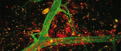
©Inserm/Elias, Salah

Huntington’s disease is a hereditary neurodegenerative disease, the symptoms of which include involuntary movements (chorea), and cognitive and psychiatric problems. The disease can become apparent at any age, but generally does so at around 40 years. It develops over many years, with progressive loss of autonomy, and death after an average of 15-20 years. There is currently no curative or preventive treatment. However, appropriate care can improve a patient’s condition.

Approximately 6,000 people would be affected by this disease in France, and there are probably more who carry the genetic mutation that causes the disease. Genetic, epigenetic and environmental factors may help to explain the differences between patients, both in terms of diversity and severity of the symptoms encountered and in the age at which the disease appears.

Huntington’s disease is caused by mutations in the huntingtin gene and the production of abnormally formed proteins of the same name. The mutant huntingtin generates continuous cellular stress that leads to the death of some neurons, especially in the striatum (central part of the brain). From this postulate, the researchers proposed a hypothesis that studying this disease might help in understanding how neurons resist stress in neurodegenerative diseases.

“Up to now, we thought that neurons were able to adapt to this stressful situation, and to resist it by fully mobilising their abilities to compensate. What we discovered was that resistance to stress is defective in these neurons from the very outset, and that it is lower than that of a normal cell,” points out Christian Néri, an Inserm Research Director at the Paris-Seine Biology Institute (IBPS) (Inserm/CNRS/Pierre and Marie Curie University). “This is an unexpected situation, caused by early abnormalities that block the mechanisms responsible for the longevity of adult neurons. This raises the question of the “biological age” of neurons in neurodegenerative diseases,” he adds.

The scientist and his French, American and Australian colleagues worked on a small worm (Caenorhabditis elegans) which, because it is transparent, means that its neurons are easy to manipulate and observe. Using versions of these animals in which the mutant huntingtin gene is introduced into the neurons, they studied the effects of huntingtin at genome level with the help of mathematical modelling. They demonstrated an inhibition of the FOXO proteins, which are known to play an important role in longevity and resistance to stress. In centenarians, the FOXO genes may be effective in fighting cellular stress. The secret of their longevity might be explained by better “equipment” for resisting stress.

The researchers confirmed these results in other models of the disease, bringing to light the close relationships between cellular longevity and resistance to neurodegenerative diseases.

Above all, however, this research, which is central to Inserm’s International Associated Laboratory (LIA) for “Neuronal Longevity,” shows the importance of restoring neuronal capacity for resistance to stress in delaying the manifestations of the disease.


This therapeutic avenue might be of particular relevance for combating the very initial stages of Huntington’s disease and other neurodegenerative diseases.

neri 1

neri 2

Picture 1 The researchers used systems biology to decipher the complexity of the toxic effects of mutant huntingtin on the neuronal genome. They analysed the expression of this genome using mathematical models based on gene interaction networks. The result is illustrated here in stylised 3D form.

Picture 2 The researchers worked on small transparent worms. These are transgenic C elegans nematodes that model the neuronal pathology found in Huntington’s disease. Morphological abnormalities in the neuronal axons of the live animal can be seen with the help of fluorescent markers.


Mild hearing impairment may indicate greater underlying problems

Scientists from the Institut Pasteur, INSERM, Collège de France, and Pierre and Marie Curie University, in collaboration with a team from the University of Auvergne, identified mice models that mimic high-frequency hearing impairment in humans, with a strong low-frequency sound interference. Their work sheds light on the anomalies causing the hearing impairment and reveals cochlear defects that profoundly affect the way sound frequencies are processed. This work could explain the pronounced masking effect experienced by some hearing-impaired individuals when trying to discriminate high-frequency sounds in noisy environments. The scientists suggest that more substantial auditory assessments would enable clinicians to improve diagnosis of these auditory impairments and provide better care for individuals who, despite showing only a mild hearing impairment using standard audiometric evaluations, should be fitted with hearing aids that appropriately target the defective sound frequencies and correct the hearing impairment.

surdite

Despite an audiometric evaluation revealing mild hearing impairment only, some patients experience severe difficulties in understanding speech because of profound sound interference in noisy environments. This represents a major handicap in their daily lives. By studying mice carrying a mutation that affects a subpopulation of auditory hair cells, scientists were able to observe a clinical profile similar to that of these patients. The project was led by Dr. Aziz El-Amraoui and Professor Christine Petit, head of the Genetics and Physiology of Hearing Unit (Institut Pasteur/INSERM UMRS 1120/Pierre and Marie Curie University/Collège de France), in tight collaboration with the neurosensory biophysics team from the INSERM joint research unit UMR 1107 at the University of Auvergne led by Professor Paul Avan.

The mice being studied presented a misleadingly mild hearing loss, observed only for high-frequency sounds, sound frequencies that are normally processed at the cochlear base. However, scientists found significant morphological defects in this very area. The outer hair cells at the cochlear base were unable to respond to high-frequency sounds and their corresponding hair bundles, which act as antennas for sound reception, were markedly misshaped (see photo).

Subjecting the mice to interfering sounds led the scientists to discover an unexpected phenomenon: the low frequency sounds (two octaves lower, and at much lower intensity) significantly masked the high-frequency sounds. The scientists were then able to identify that the fully functioning cochlear apex, which normally processes low-frequency sounds, was behind the masking effect. However, in the mutant mice where the cochlear base had lost all sensitivity, the cochlear apex encoded both low- and high-frequency sounds. In a healthy cochlea, high-frequency sounds cannot even reach the apical region because of the base-to-apex varying physical characteristics of the basilar membrane. However, hair bundle anomalies in mutant mice seem to allow high-frequency sound vibrations to travel along another pathway with very different physical characteristics.

Transposed to humans, this study suggests that some individuals with seemingly optimistic audiograms should indeed be urged to use hearing aids. To avoid the misleading interpretations of standard audiometric tests, individuals with high–frequency hearing impairment vulnerable to interference by low-frequency sounds should undergo complementary clinical evaluations. This would notably allow for a more detailed study of the frequency responses of their auditory sensory cells. Accordingly, the prescribed auditory hearing aids should appropriately focus on the selective restoration of high-frequency sound detection, while taking into account the prevention of low-frequency sound interference.

Illustration: Hair bundles of the auditory outer hair cells from a normal mouse (upper) and a mutant mouse (lower) observed by scanning electron microscopy. – Copyright Institut Pasteur – V. Michel & K. Kamiya
This study was funded by the ERC-hair bundle grant (2011-ADG_20110310), LABEX Lifesenses, the Japan Society for the Promotion of Science, the Uehara Memorial Foundation, Réunica-Prévoyance, Humanis, Irène Errera-Hoechstetter, the Fondation BNP-Paribas and the Fondation Voir et Entendre.

Epileptic seizure: a primitive brain activity with mechanisms that are conserved across species

Everything seems to distinguish a fly from a man. However, as remarkable as it may seem, Inserm researchers led by Christophe Bernard and Viktor Jirsa at the Institute of Systems Neuroscience (INS) – Inserm U1106 in Marseille have just shown that epileptic seizures follow simple mathematical rules that are conserved across species. The epileptic seizure is a form of neuronal activity that is encoded in every healthy brain, but expressed only in pathological situations. By identifying these basic principles, the researchers were able to precisely classify seizures into 16 distinct types, a classification which will be very useful to clinicians for planning increasingly personalised treatments and seeking new drugs. This research is published in the journal Brain.

Photo CP Brain epilepsieWhat is an epileptic seizure? For centuries this question has represented an enigma for patients and those close to them, as well as for researchers and physicians. Seizures often occur without any warning sign, with clinical manifestations that can be spectacular (examples?) For researchers and physicians, the seizure is considered a very difficult problem to solve, involving highly complex mechanisms.

A major breakthrough has just been made by two teams from the Institute of Systems Neuroscience (INS) – Inserm U1106 in Marseille. Combining theoretical neuroscience with basic and clinical research, the researchers have provided evidence that the principles governing the onset, middle and end of focal epileptic seizures (a very common form of epilepsy) are very simple and do not vary from one region of the brain to another or from one species to another, from fly to man.

The starting point is simple: any healthy brain can have a seizure, for instance after an electric shock, head trauma, etc., without being or without necessarily becoming epileptic. In other words, this brain activity which constitutes a seizure exists in a latent state in every one of us. The seizure is naturally encoded in our neurons. It is always possible, but in a “healthy” brain, the probability of occurrence is very small.

We will therefore use a metaphor to describe the main outcome. Let us represent the brain’s activity as a character travelling in a country made up of mountains, valleys, plains, beaches, etc. The different areas of the country represent as many activities in which the brain is involved (e.g. reading a book, riding a bicycle, etc.). In this country, there is one very particular place—a forbidden zone surrounded by a very high barrier. This forbidden zone is always there; it forms part of the landscape, but our character cannot enter it. This forbidden zone is the epileptic seizure. Extreme conditions are required in order to enter it—after an electric shock for example.

The researchers at the Institute of Systems Neuroscience have constructed a mathematical model that describes what happens from the time the barrier is crossed (onset of seizure) until the time the character finishes by leaving the forbidden zone (end of seizure) and returns to normal activity outside of it. They showed that the trajectories for entering and exiting the seizure follow simple and precise mathematical rules. They also showed that the seizure is the simplest—or most primitive—form of activity that the brain can generate.

“The mathematical model predicts the existence of 16 types of seizures, which enables a precise classification of seizures, a classification which will be highly useful to clinicians treating these seizures and seeking new treatments,” explains Christophe Bernard, a Research Director at Inserm.

The researchers then verified the predictions of the mathematical model experimentally, by analysing the seizures recorded for different species, including man (using an international database).

They were thus able to show that the rules for entry into and exit from the seizure were invariable, from fly to man. The same forbidden zone is therefore present in most regions of the brain across species.

Why is epilepsy so difficult to treat?

By using an experimental mouse model of epilepsy, the researchers have demonstrated that the forbidden zone can be entered at many locations. Furthermore, there are many ways of damaging the barrier. This multiplicity of opportunities explains why treatments must be tailored to every patient, because crossing the barrier does not necessarily take place at the same location from one individual to another.

This research is of major importance, not only because it contributes to demystifying epilepsy, but also because it supplies a conceptual framework for better understanding the mechanisms of seizures and proposing new solutions for treatment.

What is happening in patients who have seizures?
In patients, the barrier is damaged, and it is much easier to enter the forbidden zone. This destruction of the barrier is a very common phenomenon. It occurs naturally in the course of ageing, and this is why the frequency of epilepsy increases with age. Children are also very prone to epileptic seizures, because their barrier is not high enough. Many pathological conditions, such as autism, Alzheimer’s disease and Huntington’s disease, are associated with epileptic seizures, because the destructive effects generated in the brain by these pathologies ultimately erode the barrier.
For this reason, epilepsy may exist alone or in association with other pathologies. Pathological conditions and diseases only unmask an activity that potentially exists in every one of us.

How does the brain adapt to different situations?

© CC BY-SA 2.0 by ZeroOne

When we are confronted with an uncertain, changeable or new situation, our brain, after a moment’s reflection, will opt for one course of action over another. A team of scientists led by Etienne Koechlin, Director of the Cognitive Neuroscience Laboratory (Inserm/ENS), has just decoded the reasoning process behind the human ability to adapt. The scientists have discovered the algorithm used by the prefrontal cortex to enable human beings to think rationally and hence to adapt to different situations by means of two distinct processes.

The results have been published in the 29 May 2014 issue of Science Express.

Decision making occurs in the frontal lobe of the brain, in an area known as the prefrontal cortex. We already knew that this area was involved in decision making and behavioural control. However, we did not understand how it endows human beings with their highly-developed reasoning and analysis skills, which are strongly solicited in new situations.

In the study published in Science Express, researchers at the Cognitive Neuroscience Laboratory (Inserm/ENs) analysed the brain activity of 40 healthy young people (aged between 18 and 26), according to a protocol inspired by the board game Mastermind. They were placed in an uncertain and changeable situation much like in the game, where players have to use their powers of deduction to find a hidden combination of coloured pegs using fragmented information. They also had to adapt because, in the protocol used, the combination could change without the participants’ knowledge.

Using neuroimaging techniques, the researchers have discovered how the problem-solving algorithm in the prefrontal cortex works and explained how human beings reason and adapt to uncertain, changeable and new situations.

The study reveals the key role played by two areas of the prefrontal cortex. The first area, which is located between the ventro and dorsomedial regions of the prefrontal cortex, is able to analyse the situation and arbitrate between adjusting the individual’s current behaviour or exploring new strategies coming from the individual’s long-term memory.

The second area, known as the ‛frontopolar’ cortex is found in the most anterior, lateral part of the frontal lobes and is believed to be absent in non-human primates. It is capable of analysing two or three alternative strategies at the same time. “The frontopolar cortex enables individuals to assess several concurrent hypotheses simultaneously, to judge their reliability and to develop new hypotheses based on long-term memories”, explains Etienne Koechlin, Inserm research director and the principal author of the study.

These two areas operate jointly and are responsible for the reasoning process that consists in comparing and testing hypotheses and deciding whether to accept them or to reject them in favour of other, newly created strategies.

Our findings are a major step forward, since it is the first time that the problem-solving algorithm in this part of the brain has been mathematically modelled and updated”, he concludes.

Neuropsychiatric disorders massively impair the function of the prefrontal cortex. Its development is delayed late into adolescence and it becomes impaired with age. These findings open up many possibilities, as they will help us to better understand how the development, ageing and impairment of the prefrontal cortex affect the judgement of individuals and how to remedy these effects.

fermer