Menu

ChroMS – the brain as never seen before

©3D brainbow astrocytes

Developed by researchers from École Polytechnique, Sorbonne Université, Inserm and the CNRS, ChroMS is a new microscopy technique bringing together color, 3D and high-resolution imaging, and is nothing short of a revolution in vertebrate brain imaging. The ChroMS technique is described in detail in a recently published article in Nature Communications.

Until now, researchers have had to choose between resolution and volume when performing vertebrate brain imaging. They could either obtain very high resolution over small volumes using three-dimensional electron microscopy or an image of the whole brain at resolutions that are far too low to understand the details.

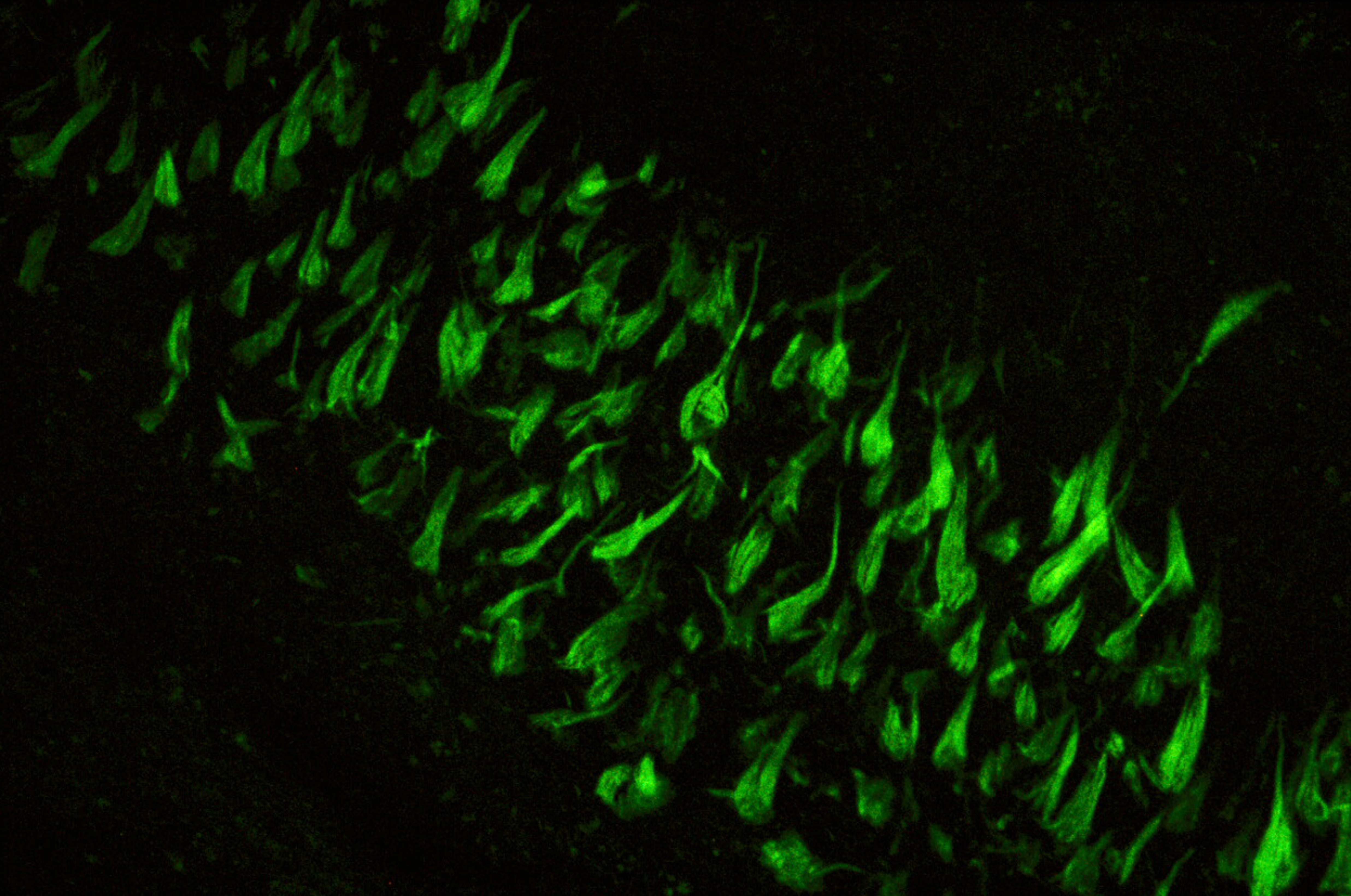
The main advantage of the ChroMS (Chromatic Multiphoton Serial imaging) technique is that it provides a truly high-resolution virtual view (at the cellular level) of certain parts of the brain that are essential for understanding the development of neuronal circuits. Although the visit is virtual, the data are real. They are obtained from the brains of transgenic mice whose neurons produce fluorescent markers originating from jellyfish or coral. When stimulated by an infrared laser, these markers show up as colors.

This instrument is ideal for making extremely precise, 3D reconstructions of regions of the brain with a volume of a few cubic millimeters, which is a breakthrough with this image quality, and this is the appropriate scale for what we want to observe”, explains Emmanuel Beaurepaire, researcher from the Laboratory for Optics and Biosciences (LOB – a joint research lab between École Polytechnique, the CNRS and Inserm). “Using the current version of our instrument, we can also reconstitute a complete mouse brain, albeit at a lower level of precision”.

We are particularly interested in cell lineage,” states Jean Livet, researcher from the Institut de la Vision (Sorbonne Université, Inserm, CNRS). “In other words, the manner in which the brain develops from neural stem cells: what are the daughter cells from a given stem cell, how can a stem cell mutation influence their development, and how are groups of cells generated by different stem cells organized in relation to one another? The high-volume, color-coded images produced by ChroMS reveal the developmental history of an individual region of the brain”.

ChroMS should enable us to answer questions that neuroscientists have been asking for a long time, such as whether neurons arising from the same stem cell connect to each other preferentially to fulfil a given function, and whether pathologies such as epilepsy could be linked to localized problems affecting certain neural stem cells.

Although the ChroMS technique is ideally suited to the study of highly complex organs such as the brain, it can be used on all organs and should also prove a very effective tool for embryogenesis studies.

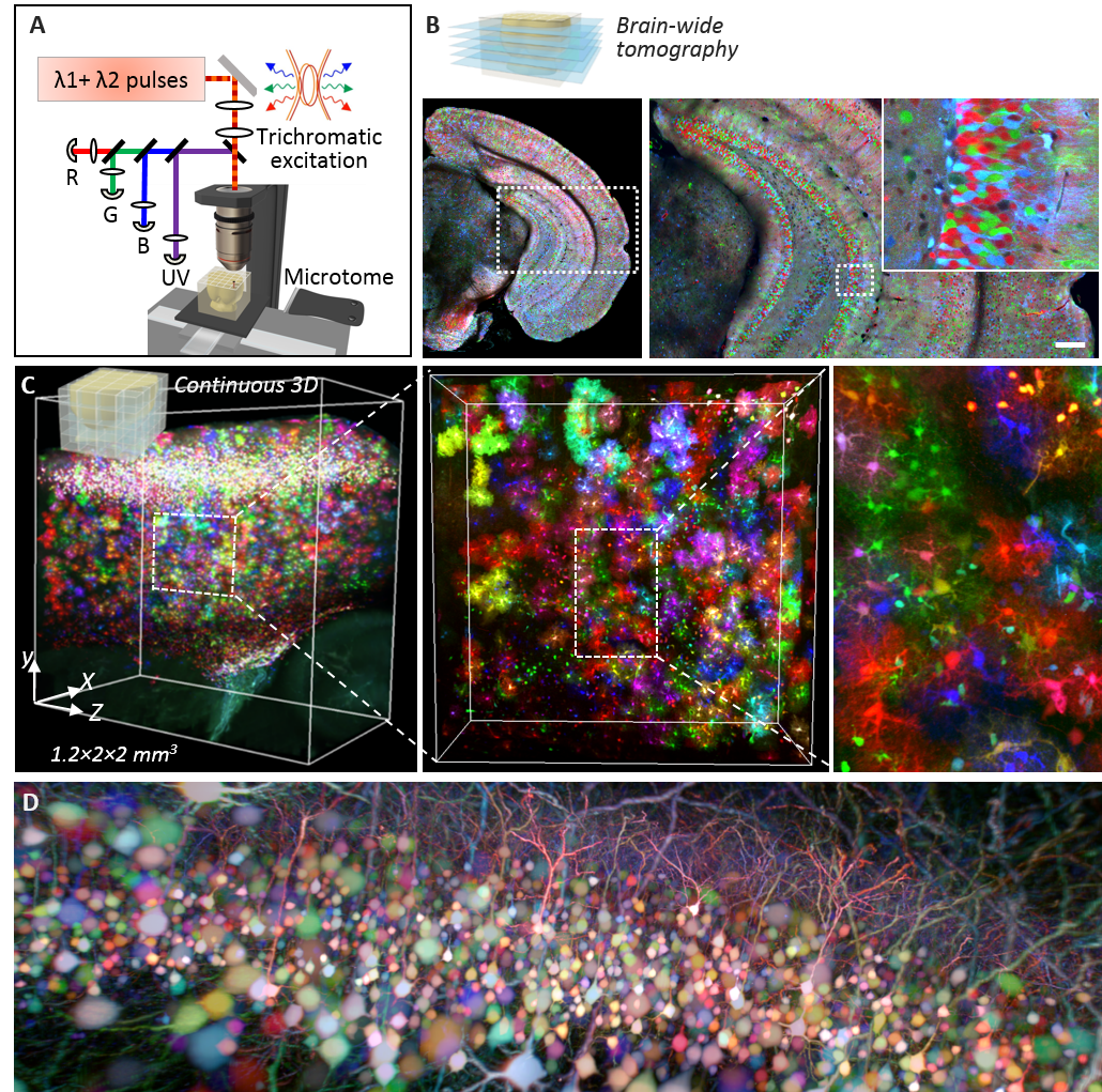
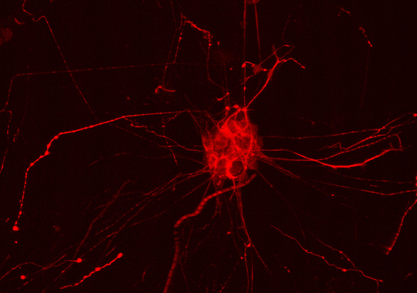
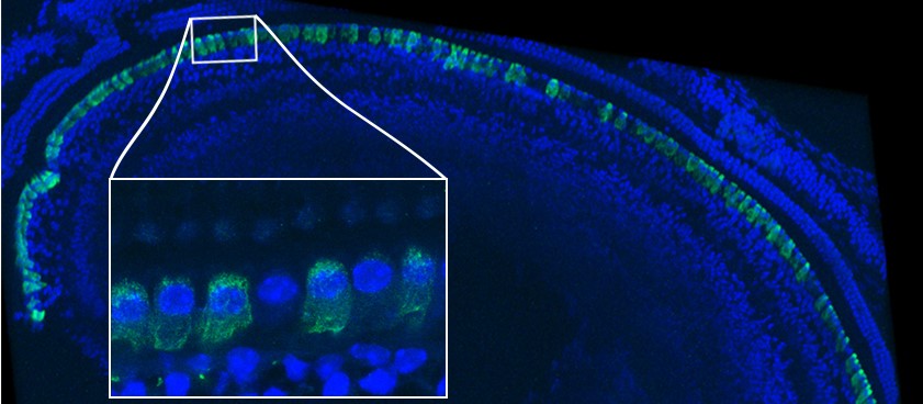
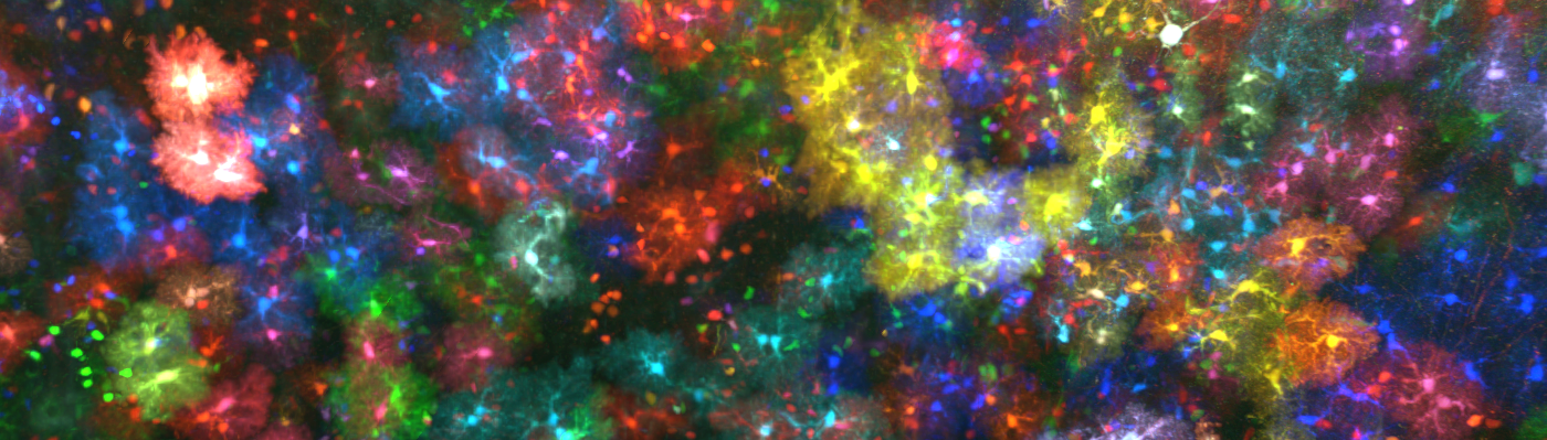
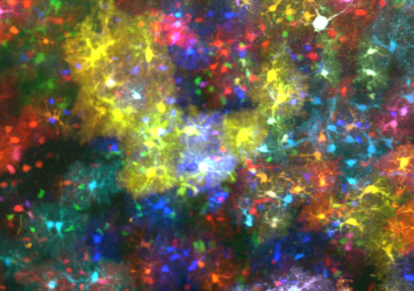
(A) Principle of ChroMS microscopy, combining color two-photon excitation by frequency mixing and automated serial slicing of brain tissue. (B) Image acquired with the “whole brain tomography” mode showing the cortex and hippocampus of a Brainbow mouse. (C) 3D reconstruction and view at different scales of a 4.8 mm3 volume of mouse cortex in which astrocytes are marked with fluorescent proteins of different colors. (D) 3D view of color-marked neurons in the mouse cortex. Adapted from: Abdeladim et al, Nat Commun 2019.

Narcolepsy: A New Drug to Fight Sleepiness

©Photo by Cris Saur on Unsplash

How can the quality of life of patients with narcolepsy, the severest sleep disorder in humans, be improved? An international scientific team led by Yves Dauvilliers, a researcher at Inserm and Université de Montpellier, is working on Solriamfetol – a promising new drug that stimulates alertness and improves resistance to sleepiness. The results of the Phase 3 clinical trial published in Annals of Neurology show that when compared with existing treatments, Solriamfetol is not just more effective and long-lasting, but also has fewer side effects.

Narcolepsy is a chronic rare neurological disorder caused by a loss of neurons that synthesize the protein hypocretin. It is characterized by excessive daytime sleepiness and difficulty staying awake. Being the severest sleep disorder in humans, it is an excellent model for studying other pathologies of the same type.

Existing treatments to improve the symptoms of narcolepsy are thin on the ground, inconsistent in their efficacy and sometimes linked to side effects. And they only treat the symptoms, not the root cause. Since research is as yet unable to produce synthetic hypocretin, these treatments make do with compensating the lack of hypocretin: they stimulate alertness by acting primarily on dopamine transporters.

It is on the development of a more effective drug in improving narcolepsy symptoms that is working Yves Dauvilliers, researcher at Inserm and Université de Montpellier in the “Neuropsychiatry: Epidemiological and Clinical Research” laboratory (Inserm/Université de Montpellier), in collaboration with international teams. The research he is leading is centered around Solriamfetol[1], a drug that not only inhibits the transporters of dopamine but also those of norepinephrine – another neurotransmitter involved in the regulation of waking.

In this Phase 3 clinical trial, 240 narcoleptic patients were followed for 12 weeks in order to evaluate the efficacy and safety of Solriamfetol in humans. The tests were performed under double-blind conditions on groups of 60 patients receiving different doses of Solriamfetol or placebo. In addition to patient feedback on the day-to-day changes in their sleepiness, the trial also involved tests in which the patients were required to try to stay awake in an atmosphere conducive to sleep.

The research team observed that those patients receiving a daily dose of 150 mg or 300 mg Solriamfetol managed to fight sleepiness for around 20 minutes versus 10 without treatment, i.e. for twice as long. The currently-prescribed treatments extend this alertness by only 2 to 3 minutes. This efficacy was maintained throughout the 12 weeks of treatment, with few side effects and no need to increase dosage.

“By enabling them to better resist sleepiness, Solriamfetol therefore proves to be highly promising when it comes to improving the quality of life of people with narcolepsy and also in the other disorders linked to sleepiness, such as sleep apnea syndrome – in which it offers the same efficacy”, states Dauvilliers. In order to evaluate its efficacy and safety over time, the researchers have launched a new clinical trial lasting one year.

[1]The study protocol was developed, in collaboration with the authors, by Jazz Pharmaceuticals, which is funding the trial and holds a license to develop and commercialize Solriamfetol

Go for a Run or Eat Chocolate: A Choice Dictated by the Cannabinoid Receptors


©AdobeStock

Physical inactivity is a common factor in lifestyle diseases – and one that is often linked to the excessive consumption of fatty and/or sugary foods. The opposite scenario of excessive physical activity at the expense of caloric intake can also be harmful, as cases of anorexia nervosa illustrate. These data therefore point to the crucial need to research the neurobiological processes that control the respective motivations for exercise and food intake. A study by Inserm and CNRS researchers published on March 7, 2019 in JCI Insight reveals that the cannabinoid type 1 (CB1) receptors play an essential role in the choice between running and eating chocolatey food.

The authors of this paper had previously reported that the cannabinoid type-1 (CB1) receptors, present on several types of neurons, play a key role in performance during physical activity in mice. A conclusion based on the performances achieved by animals with free access to an exercise wheel – a model in which it was not possible to distinguish the mechanism involved (motivation, pleasure…). Given that the motivation for a reward can only be estimated by measuring the efforts that the individual – whether human or animal – is prepared to make to get that reward, the researchers devised a model in which each access to the wheel was conditional on a prior effort. This involved the animal repeatedly introducing its snout into a recipient, an essential prerequisite for unlocking the wheel. After a training period during which the level of effort required to unlock the wheel remained the same, the mice were confronted with a test in which the effort required was gradually increased. When exposed to this test, the mice lacking CB1 receptors showed an 80 % deficit in the maximum effort they were prepared to make to unlock the wheel, and without a decrease in performance during their access to it. This finding indicates that the CB1 receptors play a major role in controlling motivation for exercise. The use of other genetically-modified mice also enabled the researchers to demonstrate that these CB1 receptors controlling motivation for exercise are located on GABAergic neurons.

The researchers then examined whether the CB1 receptors in the GABAergic neurons control the motivation for another reward: chocolatey food (like humans, mice love it even when they are otherwise well-fed). While the CB1 receptors also play a role in motivation for food – albeit to a lesser extent than in motivation for exercise – the CB1 receptors located on the GABAergic neurons are not implicated in the motivation for eating chocolatey food.

In our daily life, we are faced with an ongoing choice between various rewards. A fact which has encouraged the researchers to develop a model in which following a learning period the mice had the choice – in return for the efforts described above – between exercise and chocolatey food. The motivation for exercise was greater than that for chocolatey food, with the exception of the mice lacking CB1 – whether generally or just on GABAergic neurons – whose preference was for the food.

In addition to these findings indicating that the cannabinoid receptor is essential for the motivation for exercise, this study opens up avenues for researching the neurobiological mechanisms behind pathological increases in this motivation. One illustration is provided by anorexia nervosa which often combines the decreased motivation to eat with an increased motivation to exercise.

Alzheimer’s disease: five new genetic markers identified

Fluorescent marking of the Tau protein in a human cell hNT; the Tau protein, has a role in Alzheimer’s disease, particularly in its familial forms ©Inserm/U837

Knowledge of the genetic component of Alzheimer’s disease continues to deepen. The aim is not to predict the disease but reveal its pathophysiological mechanisms in order to develop new drugs. At Lille’s Institut Pasteur, an Inserm team led by Jean Charles Lambert within Inserm Unit 1167 “Risk factors and molecular determinants of diseases linked to aging” directed by Philippe Amouyel recently hit a new milestone thanks to the Genomics of Alzheimer’s Project (IGAP), in which the genomes of 94,000 people were analyzed. This research revealed five new genetic variants linked to the disease and has been published in Nature Genetics.

Out of the forty genetic variants now linked to Alzheimer’s disease, five have recently been discovered as part of the Genomics of Alzheimer’s Project (IGAP) co-led by the “Identification of molecular determinants of neurodegenerative diseases” team at Inserm unit 1167 at Institut Pasteur in Lille. This major project conducted in partnership with four international consortia*[1] studied 94,000 genomes – 35,000 of which from people with Alzheimer’s disease and 59,000 from healthy controls. The vast majority of cases concerned late-onset Alzheimer’s disease, with the researchers having excluded the early-onset familial forms.

The patients’ genomes were compared with those of the healthy individuals presenting an equivalent average age. The objective was to look for variants, namely distinct DNA sequences between these two groups statistically linked to the development of the disease. The scientists reviewed 11 million of them, revealing five new variants linked to Alzheimer’s disease either in or in proximity to the genes IQCK, ACE, ADAM10, ADAMTS1 and WWOX. One of the five was a rare variant found in less than 1% of the patients. “The discovery of this rare variant validates the strategy of building increasingly large cohorts to obtain a very high statistical power. By increasing their size yet further, we will be able to look for more rare variants which probably represent a large part of the as yet undiscovered genetic component of this disease”, considers Jean-Charles Lambert, Inserm researcher and co-leader of this research.

Some of these new variants are concentrated in regions implicated in the metabolism of the Tau and amyloid precursor proteins, whose role in Alzheimer’s disease is already known – particularly in its familial forms. “This suggests shared mechanisms in the early or later forms”, explains Lambert. Treatments targeting these mechanisms could therefore be effective against these two forms. But variants are also found in regions implicating immunity, which is very interesting because it could validate a current research avenue. “One hypothesis is that the microglia, made up of immune cells related to the macrophages and playing a role in defending the brain, is implicated in the disease”, clarifies Lambert. For the researchers, it is probable that these variants control shared biological pathways which would lead to multiple impairments and the onset of the disease.

Given the repeated failures of the therapeutic trials in Alzheimer’s disease, better knowledge of the variants linked to the disease and the pathophysiological mechanisms they control is fundamental if new therapeutic strategies are to be developed. A new European project coordinated by Lambert – in which a yet larger number of patients will be enrolled to continue this mission – is already on the drawing board: the European DNA bank for deciphering the missing heritability of Alzheimer’s disease (EADB).

[1] *CHARGE (Heart and Aging Research in Genomic Epidemiology Consortium, USA), EADI (The European Alzheimer’s Disease Initiative), ADGC (Alzheimer Disease Genetics Consortium, USA) and GERAD/PERADES (Cohorts for and Genetic and Environmental Risk in AD/Defining Genetic, Polygenic and Environmental Risk for Alzheimer’s Disease Consortium, France)

Brain Prize 2019 a French team receives international award for his research on CADASIL, a hereditary cerebrovascular disease

Left to right : Pr Elisabeth Tournier-Lasserve, Pr Hugues Chabriat, Pr Marie-Germaine Bousser, Dr Anne Joutel

Awarded by the Danish Lundbeck Foundation, the “Brain Prize” is a major international award for scientific importance of their research in neuroscience. It has a total of one million euros.

He puts this year in honor of the work begun there nearly forty years by four French scientists on CADASIL, a hereditary cerebrovascular disease, which causes migraine attacks, stroke and cognitive decline. Today is the genetic disease of small cerebral vessels most frequently diagnosed.

The four winners of the French “Brain Prize 2019” are:

> Professor Elisabeth Tournier-Lasserve , head of the hospital neurovascular genetics department Lariboisière AP-HP, medical genetics professor at Paris Diderot University and head of the research team “cerebrovascular diseases, genomics , imaging and personalized medicine “within the unit Paris Diderot – Inserm 1141” NeuroDiderot “.

> Professor Hugh Chabriat , head of the neurology department of the hospital Lariboisière AP-HP, coordinator of the reference center for rare vascular diseases of the brain and the eye (CERVCO), professor of neurology at the University Paris Diderot and researcher at the research team “cerebrovascular diseases, genomics, imaging and personalized medicine” in the unit of Paris-Diderot – Inserm 1141 “NeuroDiderot”. Prof. Chabriat also co-coordinates the University Hospital Department (DHU) neurovasc Sorbonne Paris Cité.

> Professor Marie-Germaine Bousser , former head of the neurology department at the hospital Lariboisière AP-HP and emeritus professor of neurology at the University of Paris Diderot.

> Dr. Anne Joutel, Research Director Inserm, director of the team “pathophysiological mechanisms of diseases of the small cerebral vessels” at the Institute of Psychiatry and Neurosciences Paris Descartes – UMR1266 Inserm and professor in the pharmacology department University of Vermont (USA).

The four scientists showed that CADASIL (for “Cerebral Autosomal Dominant arteriopathy with sub-cortical Infarcts and Leukoencephalopathy”) is an inherited cerebrovascular disease caused by mutations in the NOTCH3 gene on chromosome 19. This condition is responsible for migraine attacks and stroke, can lead to severe motor and cognitive disorders. Patients may also suffer from depression, difficulty concentrating, a slowdown and balance disorders.

The discovery of CADASIL has allowed the development of diagnostic tests and the development of mouse models of the disease, essential to understand the mechanisms of brain damage and the development of therapeutic. The identification and clinical and preclinical study of this disease are also a major step to identify and better understand other diseases of cerebral vessels.

Expertise caregivers helps improve patient diagnosis in altered state of consciousness

©AdobeStock

Hospital teams Pitié-Salpêtrière Hospital AP-HP, Inserm and the Institute of the brain and spinal cord – AP-HP / CNRS / Inserm / University-Sorbonne showed appreciation caregivers (nurses and nursing auxiliaries) for the state of consciousness of patients represented a real added value to medical and electrophysiological exams and conventional brain imaging diagnostics. This work, published in the journal The British Medical Journal Open, apply the principle of “collective intelligence” (or “wisdom of the crowds”).

The waning of severe brain injury, initially a coma patient can progress to altered state of consciousness such as vegetative or minimally conscious state. The determination of the level of consciousness is important both to better assess the patient’s condition, to explain to his family, and also because of the prognostic value of that information.

However it is sometimes difficult to establish and then requires an approach called “multimodal” combining clinical expertise and neuroimaging. The recent international recommendations stress in particular the need to repeat clinical evaluations using a specific scale (the “Coma Recovery Scale – Revised”), and the utility of complement by specialized brain imaging examinations (EEG, evoked potentials cognitive, PET scans and functional MRI).

It is in this context that researchers from Inserm, a medical team of the neurological intensive care hospital Pitié-Salpêtrière AP-HP, led by Dr. Sophie Demeret and a team of the neurophysiology department clinic Pitié-Salpêtrière hospital AP-HP and laboratory “PICNIB lab” at the Institute of the brain and spinal cord led by Prof. Lionel Naccache, professor of physiology at Sorbonne University, wanted to add to this multimodal approach an additional source of information on the principle of “collective intelligence” (or “wisdom of the crowds”): the expertise of the medical staff is constantly in contact with patients throughout their hospital stay.

The tool, called “DoC-feeling” (DoC for Disorders of Consciousness) using a visual analog scale (as used for pain assessment) to collect the subjective perception vis-à-vis the status of carers awareness of the patient quickly and easily. The synthesis of all the measurements taken over a week and provides a “collective” score between 0 and 100.

Forty-nine patients hospitalized for expert evaluation of the level of consciousness in the neurological intensive care unit of the hospital of the Pitié-Salpêtrière AP-HP were included in a year and a half. Nearly 700 evaluations by more than 80 carers were collected. The study, supervised by two referent nurses and supported by health managers, has shown that the median value of individual assessments by caregivers was closely correlated with depth specialized clinical assessments. Another advantage is that this approach allowed to significantly increase the number of patient observations, the state of consciousness may fluctuate over time.

The teams conclude that this approach, in addition to the medical clinic assessment and examinations electrophysiological and brain imaging, should improve the diagnostic accuracy of patients’ state of consciousness. This work puts more and before the interest of the collective intelligence and a collaborative approach to a complex known clinical question.

“This work initiated by two nurses in neurological intensive care hospital Pitié-Salpêtrière AP-HP: Gwen Goudard and Karine Courcoux, which involved more than 80 carers over a period of more than a year, demonstrates extraordinary motivation and the enormous potential of research in allied unity, “ says Dr. Benjamin Rohaut who oversaw the study. He added : “The support of service managers, Louise Richard Gili and Julie Bourmaleau and the help of Dr. Bertrand Hermann, Inserm researcher at the PICNIC-lab for data analysis and writing of the article, were key assets for conducting the study to completion. “

Gene therapy durably reverses congenital deafness in mice

Immunofluorescence imaging of the cochlear sensory epithelium in a mouse treated with gene therapy © Institut Pasteur

In collaboration with the universities of Miami, Columbia and San Francisco, scientists from the Institut Pasteur, Inserm, CNRS, Collège de France, Sorbonne University and the University of Clermont Auvergne have managed to restore hearing in an adult mouse model of DFNB9 deafness – a hearing disorder that represents one of the most frequent cases of congenital genetic deafness. Individuals with DFNB9 deafness are profoundly deaf as they are deficient in the gene coding for otoferlin, a protein which is essential for transmitting sound information at the auditory sensory cell synapses. By carrying out an intracochlear injection of this gene in an adult DFNB9 mouse model, the scientists successfully restored auditory synapse function and hearing thresholds to a near-normal level. These findings, published in the journal PNAS, open up new avenues for future gene therapy trials in patients with DFNB9.

Over half of nonsyndromic profound congenital deafness cases have a genetic cause, and most (~80%) of these cases are due to autosomal recessive forms of deafness (DFNB). Cochlear implants are currently the only option for recovering hearing in these patients.

Adeno-associated viruses (AAVs) are among the most promising vectors for therapeutic gene transfer to treat human diseases. AAV-based gene therapy is a promising therapeutic option for treating deafness but its application is limited by a potentially narrow therapeutic window. In humans, inner ear development is completed in utero and hearing becomes possible at approximately 20 weeks of gestation. In addition, genetic forms of congenital deafness are generally diagnosed during the neonatal period. Gene therapy approaches in animal models must therefore take this into account, and gene therapy efficacy must be demonstrated following a gene injection when the auditory system is already in place. In other words, therapy must reverse existing deafness. The team led by Saaïd Safieddine, a CNRS researcher in the Genetics and Physiology of Hearing Unit (Institut Pasteur/ Inserm) and coordinator of the project, used a mouse model of DFNB9, a form of human deafness that represents 2 to 8% of all cases of congenital genetic deafness.

DFNB9 deafness is caused by mutations in the gene coding for otoferlin, a protein that plays a key role in transmitting sound information at the inner hair cell synapses. Mutant mice deficient in otoferlin are profoundly deaf as these synapses fail to release neurotransmitters in response to sound stimulation, despite the absence of detectable sensory epithelial defects. DFNB9 mice therefore constitute an appropriate model for testing the efficacy of viral gene therapy when it is administered at a late stage. However, as AAVs have limited DNA packaging capacity (approximately 4.7 kilobase (kb)), it is difficult to use this technique for genes whose coding region (cDNA) exceeds 5 kb, such as the gene coding for otoferlin, which has a 6 kb coding region. The scientists have overcome this limitation by adapting an AAV approach known as dual AAV strategy because it uses two different recombinant vectors, one containing the 5’-end and the other the 3’-end of the otoferlin cDNA

A single intracochlear injection of the vector pair in adult mutant mice was used to reconstruct the otoferlin coding region by recombining 5′ and 3′-end DNA segments, leading to long-term restoration of otoferlin expression in the inner hair cells, and then restored hearing.

The scientists have therefore obtained initial proof of the concept of viral transfer of fragmented cDNA in the cochlea using two vectors, showing that this approach can be used to produce otoferlin and durably correct the profound deafness phenotype in mice.

The outcomes achieved by the scientists suggest that the therapeutic window for local gene transfer in patients with DFNB9 congenital deafness could be wider than thought, and offers hope of extending these findings to other forms of deafness. These results are the subject of a patent application filed

In addition to the institutions mentioned in the first paragraph, this research was funded by the French Foundation for Medical Research, the European Union (TREAT RUSH) and the French National Research Agency (EargenCure and Lifesenses LabEx).


Oreille humaine anglais

The left panel is a schematic representation of the human ear. Sound waves are collected by the outer ear made up of the pinna and ear canal. The middle ear, composed of the eardrum and ossicles, transmits sound waves to the inner ear, which features the cochlea – the hearing organ responsible for transmitting auditory messages to the central nervous system. The right panel shows an immunofluorescence image of the auditory sensory epithelium within an injected cochlea. The inner hair cells have been stained for otoferlin in green. Otoferlin is detected in almost all of these cells. The inset is a high magnification area showing an inner hair cell that has not been transduced. © Institut Pasteur

[1] Research published by the Institut Pasteur and Inserm Genetics and Physiology of Hearing Unit (UMRS1120): “Otoferlin, defective in DFNB9 deafness, is essential for synaptic vesicle exocytosis at the auditory ribbon synapse” Cell, October 20, 2006

A nanomedicine which relieves pain without the risk of addiction

©Adobe Stock


A painkiller nanomedicine has just been developed by Patrick Couvreur’s team at Institut Galien Paris-Sud (Université Paris-Sud/CNRS)[i]. This new drug specifically targets the area of painful inflammation without causing the addiction phenomena of current medication.

In pain management, morphine and synthetic opioids are currently effective drugs. Unfortunately, their side effects are serious, in particular respiratory depression, but especially addiction. Opioid addiction is indeed a global healthcare scourge. The United States is particularly affected with 11 million dependent patients and around 175 daily deaths as a result of overdoses.

The use of certain neuropeptides, natural peptides used by neurons to communicate with each other, is undoubtedly an interesting alternative to these drugs. In fact, acting mainly on the neurotransmitter receptors that specifically modulate the pain response function, these natural molecules do not cause these side effects. Unfortunately, after administration these molecules degrade in a few minutes without having time to act.

An effective mode of action without side effects

The “Innovative Nanomedicines for the Treatment of Serious Illnesses” team, led by Patrick Couvreur at the Faculty of Pharmacy at Université Paris-Sud, had the idea of synthesising samples of nanoparticles made up of Leu-enkephalin and coupling them to squalene, a naturally occurring lipid in the body, whose specificity is to be able to provide a protective membrane for the whole compound.

Together with scientists from the Paris Institute of Psychiatry and Neuroscience (Inserm/Université Paris Descartes) and the Neuropharmacology Laboratory (Université Paris-Sud/Inserm), the team demonstrated that these nanomedicines induced a significant and prolonged pain-relief effect in rats, with a higher efficacy than morphine.

The researchers particularly noted that, unlike morphine, the Leu-enkephalin-squalene nanoparticles spared brain tissue and acted solely on peripheral receptors. Furthermore, imaging showed that the nanoparticles were capable of delivering the neuropeptide specifically to the area of painful inflammation, thus avoiding the central effects responsible for addiction phenomena.

Biochemical and histological investigations conducted on the treated animals, have moreover demonstrated that this new painkiller medicine did not cause any toxicity or side effects.

This study, published in the journal Science Advances, on 13 February 2019, represents a breakthrough in pain treatment due to the nanomedicine. Further research is still necessary before being able to move to clinical trials. A start-up could be established in the coming months to raise the funds needed for their financing.

[i]  Together with scientists from the Paris Institute of Psychiatry and Neurosciences (Inserm/Université Paris Descartes) and the Neuropharmacology Laboratory (Université Paris-Sud/Inserm).


Images en cryo-TEM des nanoparticules de Leu-Enképhaline-squalène d’une taille comprise entre 70 et 100 nm.© Institut Galien Paris-Sud, Faculté de Pharmacie  


Effet antidouleur des nanoparticules (courbe bleue) aboli après traitement par l’antagoniste naloxone méthiodide (courbe verte) et l’antagoniste naloxone (courbe rouge). Le traitement par la leu-enképhaline libre ou le vecteur seul est sans effet (courbe noire et grise) © Institut Galien Paris-Sud, Faculté de Pharmacie  

Nanoparticules de Leu-enképhaline-squalène libérant le peptide spécifiquement au niveau du site de la douleur inflammatoire (zone rouge). © Institut Galien Paris-Sud, Faculté de Pharmacie   

Miniaturized Chemical Sensors to Monitor Brain Function

©Stéphane Marinesco / Inserm, Photograph of an implantable chemical sensor (bottom right) made with platinized carbon fiber and coated with a recognition enzyme, placed next to a human hair (top).

A team of Inserm and CNRS researchers has succeeded in developing new-generation chemical sensors to monitor the brain’s metabolism, particularly during stroke, trauma or epileptic seizure. Measuring less than 15 µm in diameter, these minimally-invasive tools monitor what is happening in the brain in order to obtain data that are much more reliable and representative of the neurochemical exchanges. This research has been published in ACS Central Science.

Analyzing the interstitial fluid of the brain can reveal important chemical information about the state of the latter. In the clinic or in laboratory animals, the ability to detect, over time, the levels of metabolites characteristic of brain energy (such as glucose) can help detect the onset of brain lesions, enabling doctors to act before it is too late. In addition, the activation of neuronal networks leading to a release of neurotransmitters can be detected in interstitial fluid. However, up until now the size of the probes and the local injury caused by their implantation were parameters which disrupted the quality of the measurements obtained. In particular, the rupture of small cerebral blood vessels during implantation represents a major trigger for inflammation. Within the first hour after implantation, local chemical brain tissue composition can be affected.

The first innovation presented by the scientists in this research consisted of developing miniature sensors.

Invisible to the naked eye, they measure less than 15 microns in diameter (compared with 50 to 250 microns, currently), making them narrower than a strand of hair. The major advantage of being able to miniaturize the sensors to this extent is that implanting them no longer causes lesions in the nervous tissues. “Their size is smaller than the average distance between two brain capillaries, meaning that they are not damaged by the device” explains Stéphane Marinesco, Inserm researcher in charge of the study.

The second innovation was to coat the carbon fibers with platinum followed by a very thin layer of enzyme.

Up until then, electrochemical analysis using carbon fiber microelectrodes was limited to a highly-restrictive number of so-called “oxidable” molecules. Coating them with platinum makes it possible to attach enzymes and detect a potentially unlimited number of molecules. For Marinesco, “while platinum deposition is a commonly used technique in the field of microelectronics, it is usually performed with flat silicon substrates. Our results show that, despite their unusual cylindrical geometry, carbon fibers could be successfully covered with a platinum layer. The sensitivity achieved is similar or better than that of the thicker solid platinum wires which are commercially available.”

When these sensors were implanted in the brains of rats during laboratory testing, no injuries to the brain tissue or blood vessels were detected.

In addition, these microelectrodes supplied more precise and reliable evaluations of glucose, lactate and oxygen concentrations compared with conventional sensors (in which one sensor per parameter is necessary by implanting a multi-microelectrode “comb”). Numerous tests were performed on these new microelectrodes, in particular on their stability over time because they were also tested after 6 months of storage (room temperature in darkness).

Marinesco clarifies that: “This minimally invasive device represents a major advance in our ability to analyze the brain interstitial fluid, paving the way for the measurement of new physiological parameters and multiple applications. This novel tool could be used to test the effect of certain medicinal products on the brain. Finally, in the longer term, monitoring the human brain could provide invaluable information to doctors in order to better understand how a patient with brain lesions recovers after a head injury or stroke. This device could also help them to take the best therapeutic decisions depending on the patient’s condition”.

Alzheimer’s: identification of potential target protein aggregates for treating the disease

Aggregates of Tau protein in Alzheimer’s disease. Inserm/U837, 2008

The propagation of tau protein aggregates in the brain contributes to the progression of Alzheimer’s disease. Researchers at the Neurodegenerative Diseases Laboratory: mechanisms, therapies, imaging (CNRS/CEA/Université Paris-Sud, MIRCen), working in collaboration with the Ecole Normale Supérieure, Sorbonne University and Inserm, have just identified the targets of these aggregates. Published in the EMBO Journal on 10 January 2019, this work will enable the development of tools capable of blocking these key elements of aggregate propagation and thus combating their pathological effect.

The aggregation of alpha-synuclein proteins in Parkinson’s disease and tau proteins in Alzheimer’s disease is intimately linked to the progression of these neurodegenerative diseases. These aggregates propagate from one neuronal cell to another, attaching themselves to the cells.

They multiply[1] during this propagation. It has already been shown that the propagation and amplification of these protein aggregates are harmful and contribute to the progression of these diseases.

Understanding the formation of these aggregates, their propagation and their multiplication in the cells of the central nervous system offers potential for treatments: it would make it possible to target these processes and to act on their consequences.

Protein propagation

The key step in the propagation of the pathogenic aggregates is the attachment of aggregates released from affected neuronal cells to the membranes of unaffected cells. Having already identified the targets of pathogenic aggregates of the alpha-synuclein protein (Shrivastava et al., 2015 EMBO-J), the team at the Neurodegenerative Diseases Laboratory (CNRS/CEA/Université Paris-Sud , MIRCen, Fontenay-aux-Roses), in collaboration with the Ecole normale supérieure, Sorbonne University and Inserm, has just identified the targets of tau protein aggregates. The targets are the sodium / potassium pump and glutamate receptors, two essential proteins for the survival of neurons The experiment was carried out on mouse neurons in culture.

Neuron membrane modification

The researchers also showed that the pathogenic aggregates modify the neuron membranes by redistributing the membrane proteins. The integrity of the membrane—and particularly of the synapses, the essential nodes for communication between neurons—is affected. These changes have a deleterious effect on the neurons because they cause abnormal communication between the neurons, as well as their degeneration.

This work therefore explains the early malfunctioning of the synapses and the degradation of normal communication observed in the neuronal networks as the disease progresses.

Towards new treatments

It also paves the way for the development of new treatment strategies based on protecting the integrity of the synapses, restoring the activity of the tau protein membrane receptors through the use of decoys to prevent harmful interaction between the pathogenic tau protein aggregates and their neuron membrane targets. These therapeutic approaches could be developed using human neurons, since researchers at the laboratory have just developed cultures of this type in collaboration with the I-Stem (Institute for Stem Cell Therapy & Exploration of Monogenic Diseases, AFM-Téléthon/Insem/Génopole/University of Evry-Val-d’Essonne) laboratory and Sorbonne University. This latter study is also published on 10 January 2019, in Stem Cell Reports[2].


[2] Propagation of α-Synuclein strains within human reconstructed neuronal network. Simona Gribaudo, Philippe Tixador, Luc Bousset, Alexis Fenyi, Patricia Lino, Ronald Melki, Jean-Michel Peyrin, Anselme Louis Perrier, Stem Cell Reports, 10 January 2019.

[1] They multiply by recruiting the endogenous alpha-synuclein and tau proteins from the affected cells during this propagation


About the Neurodegenerative Diseases Laboratory: mechanisms, therapies, imaging (LMN), a joint research unit of the CEA, CNRS and Université Paris-Sud.

The laboratory brings together nearly 60 scientists with research interests in neurosciences covering the mechanisms of degeneration, animal models, brain imaging and the study of gene-, cell- and drug-based strategies for treating neurodegenerative diseases such as Alzheimer’s disease, Parkinson’s disease and Huntington’s disease.

The LMN is located at MIRCen (Molecular Imaging Research Centre), a preclinical research facility developed by the CEA and Inserm. MIRCen is one of the departments of the CEA’s François Jacob Institute of Biology, on the Fontenay-aux-Roses site at CEA Paris-Saclay. 


Filming the brain to shed light on sleep

©Adobe Stock

What happens inside our brains when we sleep? To answer this question, French researchers have, for the first time, filmed the entire brain in sleeping rats, thanks to innovative ultrasound imaging technology. They were thus able to closely observe brain function in rodents, particularly during the REM sleep phase. These results were obtained in shared Inserm, ESPCI Paris, CNRS, and Sorbonne University laboratories. Published in Nature Communications, these findings allow this period to be redefined as a brain hypersynchronization phase, characterized by massive peaks in blood flow, particularly in the hippocampus. These new data, which question the role currently ascribed to REM sleep, have yet to be confirmed in humans.

REM sleep is a specific sleeping state in which brain activity is similar to the waking state, while being associated with inhibition of muscle activity. It is notably characterized by rapid eye movements, and has long been considered to coincide only with dreaming and emotional processes. However, recent studies showed that it also played a major role in neuronal plasticity in the hippocampus, i.e., the ability of neurons to reconfigure their connections.

To have a better understanding of brain function during REM sleep, researchers from Inserm unit 979 “Wave Physics for Medicine” managed by Mickaël Tanter at Institut Langevin (ESPCI Paris/CNRS), and recently accredited as a “Biomedical ultrasound” technology research accelerator in partnership with the Paris-Seine Neuroscience Laboratory (Sorbonne University/CNRS/Inserm), studied brain activity in sleeping rats. To do so, they combined electroencephalography (EEG), which records neuronal electrical activity, with an ultrarapid ultrasound imaging technique known as fUS (functional ultrasound). This innovative technique, developed by the team led by Mickaël Tanter, allows highly precise visualization of variations in blood flow related to neuronal activity throughout the brain in alert, moving rats.

The research team observed that REM sleep in rats is associated with substantially increased blood flow in the brain, in the form of waves which first reach the subcortical regions and then move along the hippocampus, followed by the cortex. In comparison, non-REM sleep and waking phases in inactive rats show relatively low cerebral blood volumes.

This vascular hyperactivity during REM sleep is characterized by two phases: one, similar to the findings observed when recording active rats, and the other, unknown until now, consisting of sudden elevations in blood flow which the researchers describe as “vascular surges”. Although lasting 5 to 30 seconds on average, these can persist for 1 minute in the cortical regions, and are particularly strong in the hippocampus.

The researchers successfully identified an electrical signal in the hippocampus (a crucial zone for memory), characteristic of these elevated blood flow peaks. This signal – high-frequency gamma oscillations – is ordinarily observed in alert rats. Their intensity during REM sleep is directly correlated with vascular surge intensity, which suggests that these local oscillations could control vascular flow throughout the brain. “This information is crucial,” points out Antoine Bergel, co-author of the study, “as it allows us to target very precise regions of the brain potentially involved in the genesis of these intense vascular events.”

The scientists also noted that, during REM sleep, a vascular synchronization phenomenon exists between distant areas of the brain (cortex, hippocampus, and thalamus), which is much more extensive compared to any other sleeping or waking states.

These studies reveal the very first films of the entire brain during REM sleep and confirm the benefit of neurofunctional ultrasound to fundamental neuroscience research. At present, the fUS technique is still difficult to apply in adult humans. However, these results can now be confirmed in neonates, although caution should be exercised in terms of extrapolation to human physiology. These results nonetheless represent major progress in understanding the connection between electrical and vascular activity (a phenomenon involved in numerous human disorders, such as stroke or epilepsy), and challenge our understanding of REM sleep, the function of which has yet to be elucidated.

Images obtained using the fUS technique and EEG signals generated in a rat brain during the waking, deep sleep and REM sleep phases. In contrast to the highly similar electrical signals between waking and REM sleep, vascular brain activity is much more intense and more “synchronized” than during the waking state. The brain structures are identified by superimposing a brain atlas onto the vascular network images.

fermer