Menu

Yes, you can make your children eat vegetables!

As part of the European project HabEat (2010-2014), coordinated by INRA and involving 10 scientific partners, researchers have made a step forward in the understanding of how eating behaviours and preferences form in early life. On March 31st and April 1st in Dijon (France), a symposium presents the results and recommendations for early childhood professionals and parents.

When looking at eating behaviours and preferences, even if it’s not all over at the end of early childhood, the first two years of life are of utmost importance regarding the development of eating behaviours in children, with a key period when solid foods are introduced. The collaborative project HabEat had the aim of better understanding the determinants of the formation of eating habits through two distinct approaches, epidemiological on one side and experimental on the other.
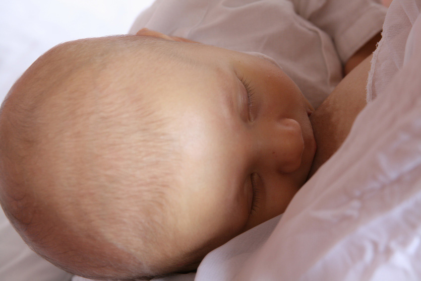
Food pyramid isolated on white

© Fotolia

Data from 18 000 mother-child pairs followed from birth to at least 4 years of age

Four large European cohorts have given insight into usual early feeding practices. The factors associated to the frequency of consumption of fruits and vegetables at various times during the establishment of the food repertoire were particularly investigated.

18 studies involving, in total, nearly 2000 children between 6 months and 6 years of age

The experimental part of the project comprised two sections. The first one primarily focused on key learning mechanisms, and concerned children from the beginning of complementary feeding (around 6 months) to 3-year-olds. The second studied different strategies aimed at increasing vegetable consumption beyond 3 and until 6 years of age.

Main results

Longer breastfeeding was associated with higher frequency of fruit and vegetable consumption in later childhood.

Introducing a variety of pureed vegetables at the beginning of the complementary feeding period facilitates the acceptance of novel vegetables in the short and medium term.

Repeatedly giving a novel vegetable is sufficient for increasing its consumption even in children who are ‘picky’ about food. However, giving already known vegetables repeatedly to kids between 3 and 6 is not efficient for increasing their consumption.

It is difficult to increase intake of an already familiar vegetable in older children (3 to 6). One strategy that seems to help is to let the child choose between two vegetables.

As early as 3 years of age, if a snack is offered before a meal, or if palatable foods are available after a meal, some children increase their total energy intake. This could lead over time to the child becoming overweight.

Recommendations for early childhood professionals and parents

The scientific results of this project have been translated into recommendations that will be addressed to early childhood professionals, pediatricians, politicians in charge of nutritional policies, and to agri-food industrials. These recommendations will be discussed during the symposium.

A guide for parents has been designed on the basis of the project’s results and of prior data.

HabEat received funding from the European Community’s Seventh Framework Programme (FP7/2007-2013) under the grant agreement n°245012-HabEat.

The cohorts:

ALSPAC (UK), 14 000 mother-child pairs, pregnant women were recruited from 1991 to 1992 and their children followed-up until 13 years of age

EDEN (FR), 2 000 mother-child pairs, pregnant women recruitedfrom 2003 to 2006, and their children followed-up until 5 years of age

Generation XXI (Portugal), about 8 500 mother-child pairs, pregnant women recruited from 2005 to 2006, and their children followed-up until 4 years of age

Europrevall (Greece), about 1 000 mother-child couples recruited from  2005 to 2007, and their children followed-up until 4 years of age.

Breast milk and diet up to 2 years old: a means of preventing the risk of child obesity

Many studies have focused on the influence of breast-feeding on child health. From analysis of data from the ELANCE cohort, Marie Françoise Rolland-Cachera, former researcher at Inserm and her co-workers in the Nutritional Epidemiology Research Team (EREN)[1] have shown that breast-feeding has a protective effect on the risk of obesity at 20 years of age. Researchers also emphasise that nutritional intake at the age of 2 years are critical in providing this beneficial effect. The results of the study are published in The Journal of Pediatrics

Recent studies have focused on the influence of breast-feeding on the risk of the child developing obesity: results showed beneficial but still inconclusive trends. They adjusted their results by considering various factors such as social categories, the weight of parents, age of diversification, etc. but until now no study had made adjustment for nutritional intakes subsequent to breast-feeding. It has now been shown that nutrition during the first two years of life had long-term consequences on health that can persist into adulthood.

fotolia_3869723_allaitement

© Fotolia

Researchers therefore studied relationships between breast-feeding and the risk of excess weight in adulthood by considering diet at 10 months and 2 years for children included in the ELANCE cohort.

The ELANCE Cohort started with children in good health, born in 1984 and 1985, recruited in Child Health Assessment Centres. Information on breast-feeding was gathered and nutritional intakes was assessed at ages 10 months and 2 years, then every two years up to the age of 20. At 20 years, several measurements were taken, including height, weight and body composition (measurements of lean mass and fat mass determined by impedancemetry).

The results show that the beneficial effect of breast-feeding is clearly seen when nutritional intake up to the age of 2 is considered and is significantly linked to a reduction in body fat at 20 years old. Furthermore, in the statistical model, higher fat intake at 2 years are linked to a reduction in fat mass at 20 years.

“Our study has therefore shown, for the 1st time, that if we take account of diet after the period of breast-feeding, the protective role of breast milk over the risk of obesity is clearly apparent,” explains Marie Françoise Rolland-Cachera, former Inserm researcher.

The diet of young children is often characterised by high protein intake and low fat intake; breast milk is rich in fat and contains a small proportion of protein. According to official recommendations, fats should not be restricted in young children in order to meet their high energy requirements for growth and rapid development of their nervous system. In particular, low-calorie dairy products with low fat content and a high proportion of protein are not indicated before the age of 2-3 years. Restricted fats may programme the child’s metabolism to deal with this deficit, but this adaptation will make it more likely become overweight when the fat intake increases later on.

“The beneficial effect of breast milk may be masked by a low-fat diet following breast-feeding, while a diet following official recommendations (no restriction in fats before the age of 2-3 years) allows its beneficial effect to appear” emphasises Sandrine Péneau, co-author of this work.

Researchers agree about the benefit of breast-feeding reducing the risk of future obesity and highlight the importance of a diet following official recommendations in relation to young children. A poorly-balanced diet after breast-feeding can compromise the benefit provided by breast milk and explain the controversies over its protective role against the risk of obesity.


[1] Joint research unit 1073 ‘Epidemiology and Biostatistics’

Ribavirin – an effective and safe treatment for hepatitis E

Hepatitis E causes acute or chronic inflammation of the liver. It is an emerging disease which can be fatal and has no known treatment. Vincent Mallet, Stanislas Pol and their team at the Institut Cochin (Paris Descartes University, Assistance Publique – Hôpitaux de Paris, CNRS, Inserm) and French hospital-based teams* have proved the efficacy of a treatment for patients suffering from chronic hepatitis E virus infection. Most patients can be cured when treated with ribavirin in monotherapy for 3 months. This work was published in The New England Journal of Medicine of 20 March 2014.

The hepatitis E virus is the biggest cause of viral hepatitis in the world and it is estimated that a third of the global population is at risk of being infected with this virus. Although most cases occur in developing countries, there has been an increase in the number of cases reported in France and other industrialised countries where the virus is spread to humans by contaminated, undercooked food and blood. Until now there has been no treatment for patients suffering from hepatitis E.

Like the other hepatitis viruses, hepatitis E causes inflammation of the liver. In its acute form, the infection can be fatal for elderly people, pregnant women and people with liver disease. In immunosuppressed people (transplant patients, patients on chemotherapy or people living with HIV), the hepatitis E virus infection can progress to chronic hepatitis and cause cirrhosis.

Developing a treatment for hepatitis E

Ribavirin is a drug prescribed for some types of viral respiratory infections in children and some forms of haemorrhagic fever. It is also used to treat hepatitis C. Vincent Mallet, professor at Paris Descartes University and hospital practitioner at the Cochin teaching hospital (AP-HP) and Nassim Kamar, professor at Paul Sabatier University and hospital practitioner at the Rangueil teaching hospital previously proved its efficacy for immunosuppressed patients suffering from a chronic hepatitis E virus infection. **

In this new study, data from 59 ribavirin-treated transplant patients suffering from hepatitis E was collected at 13 French transplant centres by Nassim Kamar (from the Rangueil teaching hospital in Toulouse) and Vincent Mallet. The researchers confirmed that “ribavirin prescribed as monotherapy for 3 months is an effective treatment for chronic hepatitis E virus infection”. For most of the patients (46 out of 59 patients), the virus remained undetectable 6 months after treatment was discontinued. A longer treatment period appears desirable for highly immunosuppressed patients or those with detectable traces of the virus in the blood after a month of treatment. The only identified and foreseeable side effect of ribavirin is anaemia which was managed in most patients without any difficulty.

“This work is the result of a genuine partnership between a large number of centres in France. It represents a major advance in this area” states Vincent Mallet. We hope our results will pave the way for further prospective studies designed to assess the efficacy of ribavirin for severe forms of hepatitis E virus infection, especially in countries in the southern hemisphere where the disease is a serious problem”.

* CHU Rangueil et CHU Purpan  Toulouse –  Hôpital Cochin, AP-HP), Pitié Salpêtrière (AP-HP) – Hôpital Saint Eloi de Montpellier – Hôpital Foch de Suresnes – CHU Lyon et CHU de la Croix Rousse de Lyon – Hôpital Paul Brousse (Villejuif, AP-HP) – Hôpital Lapeyronie de Montpellier – Hôpital Bretonneau et CHU Trousseau de Tours – CHU Bordeaux – Hôpital Claude Huriez et CHU de Lille – CHU Le Bocage de Dijon – CHU de Nantes

** Mallet V, Nicand E, Sultanik P, Chakvetadze C, Tesse S, Thervet E, Mouthon L, Sogni P, Pol S. Brief communication: case reports of ribavirin treatment for chronic hepatitis E. Ann Intern Med. 2010 Jul 20;153(2):85-9 et Kamar N, Rostaing L, Abravanel F, Garrouste C, Lhomme S, Esposito L, Basse G, Cointault O, Ribes D, Nogier MB, Alric L, Peron JM, Izopet J. Ribavirin Therapy Inhibits Viral Replication on Patients With Chronic Hepatitis E Virus Infection. Gastroenterology. 2010 Nov;139(5):1612-8).

Stopping the vicious circle of tumour progression in children with bone cancer

Primary bone cancer develops when the cells which are continually forming our bones get out of control. If cancer takes hold, these cells can degenerate and form bone haphazardly without any specific organisation. Researchers in Inserm Unit 957 “Bone Resorption Physiopathology and Primary Bone Tumour Therapy” in Nantes have recently developed an innovative treatment which stops the vicious circle that allows bone cancer to progress.

Published in today’s issue of Nature Communications, their study shows an inhibition of tumour progression and a reduction in bone degradation combined with extended lifespan in animals.

Survival rates for primary bone cancer, which mainly affects children and teenagers and whose incidence peaks around the age of 15, are 50 to 70% in the best cases for localised forms and 20 to 30% in the event of metastasis, relapse or treatment resistance. This prognosis has not changed over the last 30 years. Although these forms of cancer, which include osteosarcoma, Ewing’s sarcoma and chrondrosarcoma, are caused by a diverse range of factors of which little is yet known, they seem to involve similar types of cell dysfunction. However, there have been no major advances in the treatment of these types of cancer for about fifteen years.

Bone is living tissue

Bone tissue in physiological condition is constantly reforming, a process which entails phases of bone destruction and bone formation. Bone consists mainly of two types of cells: osteoclasts and osteoblasts which are constantly interacting to maintain a balance between bone destruction and formation. Osteoblasts are the cells responsible for bone formation. Osteoclasts are responsible for bone resorption. Any disruption of the balance in bone formation/destruction can cause cancer.

Thanks to work carried out by the Inserm research team, it has been clearly established that an imbalance between the action of osteoblasts and osteoclasts is involved in the development of primary bone tumours. Indeed, when a tumour cell grows on a bone site, major resorption occurs causing bones to weaken (with lesions, fractures etc.).

Stopping the vicious circle: a challenge overcome

Primary bone tumours ‘use’ the bones’ microenvironment in order to spread. Tumour cells disrupt the system’s natural balance by releasing proteins known as ‘growth factors’. These molecules are capable of activating osteoclasts/osteoblasts, not only causing major bone degradation but also releasing other growth factors normally trapped inside the bone. Once released these stimulate tumour growth. The larger the quantities of growth factors present in the tumour’s microenvironment, the more the tumour spreads. This is what we refer to as the ‘vicious circle’.

No treatment is yet capable of inhibiting these three components of the vicious circle, namely the tumour, osteoblasts and osteoclasts.

The researchers decided to focus on the fact that cells can turn tumorous if the expression of certain genes known as ‘tumour facilitators’ gets out of control. A number of proteins are involved in controlling the expression of these genes, in particular the BRD protein family. The researchers have proven for the first time that an innovative treatment targeting these transcription-controlling BRD proteins inhibits the three components of the vicious circle, namely the tumour cells and the differentiation of osteoclasts and osteoblasts. By chemically inhibiting the BRD4 protein (which belongs to the BRD family), researchers limited the spread of primary bone tumours while maintaining bone architecture.

Further tests were performed on patient biopsies to illustrate and confirm the results achieved with animals.

Tibias3D

3D reconstruction of a mouse tibia with a primary bone tumour (left) and post-treatment (right)©Inserm/F Lamoureux 

“Our study clearly demonstrates inhibition of tumour progression and bone degradation combined with extended lifespan in animals”, comments Inserm researcher François Lamoureux. At 32 days, all the mice in the control group had died while the treated mice were still alive after 40 days. “On the basis of work, we can now seriously consider the possibility of developing a new treatment for primary bone tumour patients which tackles both the tumour and the associated bone degradation.” Moreover, since bone architecture is preserved, we could envisage a wider range of indications including bone metastases in patients with prostate or breast cancer and also non-tumorous bone diseases (e.g. osteoporosis)”.

Schéma

Diagram: treatment tackles the 3 components of the vicious circle

Neonates are able to make connections between space, time and number

From birth, babies already have a representation of space, time and number. This has been proven by Dr Maria Dolores de Hevia, Dr Véronique Izard, Aurélie Coubart, Professor Elizabeth Spelke and Professor Arlette Streri from the Psychology and Perception Laboratory (Paris Descartes University/CNRS/Inserm) in a study published in PNAS.

The origin of our understanding of space, time and number is a subject examined by various disciplines such as philosophy, experimental psychology, developmental psychology and cognitive science. Space, time and number are connected both in the real world and the human mind, but how do we come to understand these connections? Do we learn to connect these concepts based on sensory experience by observing their correlations in the world around us or are our minds capable of understanding them innately from birth?

4 month old baby


© Fotolia

To answer this question, a test protocol was set up in the maternity unit of Bichat Hospital which recorded the visual attention of 96 neonates with an average age of 2 days (aged between 7 and 96 hours). The experiment put them in a situation where they had to use two of their senses: sight and hearing. In the first phase, a sequence of sounds evoking a numerical quantity (6 or 18 syllables) and/or a time period (1.4 or 4.2 seconds) was played to the babies over a period of one minute while they watched a moving line on a screen. In the second phase, the researchers presented new visual and auditory events, which were different to the first phase. These events were changed either congruently (1) in the same direction (e.g. longer line, larger number of sounds) or incongruently in opposite directions (e.g. longer line, smaller number of sounds).

The results show that neonates respond when these values change congruently. They are therefore able to make the connection between a numerical quantity and/or a time period and a length in space.

This test protocol proved that just hours after birth, humans are already sensitive to the common structure of time, space and number, which corroborates philosophical theories such as those espoused by Kant.

We still need to establish whether this applies to other quantitative dimensions such as light and sound and to determine the cerebral bases of these predispositions.

(1) With different elements matching and corresponding

Inserm is celebrating its 50th anniversary


In preface to this book, I would like to take a moment to acknowledge those who paved our way, certain as I am that there is no better way of honouring the past than to focus resolutely on a future which Inserm has and will continue to shape in the service of science and health.”


(Excerpt from the book Au Coeur du vivant (At the heart of life))


portrait officiel André Syrota

Prof André Syrota, Inserm Chairman and CEO


INSERM SINCE 1964

Resources
Researchers, engineers, technicians and administrative staff have all helped make Inserm a universally recognised biomedical research organisation.

Values
Inserm has overcome some major scientific challenges to be at the cutting edge of health research and, since it was founded, has never stopped reaffirming its priorities and goals.

50 years of scientific excellence – The institute has been involved in some important medical advances (cancer treatments, gene therapy, new imaging technologies, discovery of HIV). In the past thirty years, two of its researchers have received the Nobel Prize in Physiology or Medicine. The fact that Inserm has become a producer and disseminator of important scientific work and an expert on health issues is beyond dispute.

50 years of dialogue with society – Keen to make science accessible to a large section of the population, the institute has continuously sought to diversify its awareness-raising activities. Its aim is to familiarise non-specialists with the research sector and the issues it currently faces. In this spirit of openness, Inserm has spent the past ten years working to bring patient associations and researchers together.

50 years of international fame – Over the years, the institute has become firmly established in France and developed its international reputation. It is the top European biomedical research organisation. Inserm has a long tradition of national, European and international cooperation, which has resulted in an increasing number of joint publications.


Highlights marking 50 years of Inserm

To mark its 50th anniversary, Inserm has decided to deliver its key messages at around a hundred events which will take place all over France and involve all its staff, partners and supervisory bodies throughout the year.

To mark its 50th anniversary

      • The book Au coeur du vivant
      • Science Tour
      • Virus Attack
      • Destination Labo
      • Meetings “Researchers meet patients”
      • Famelab competition
      • 50th anniversary symposium (Detailed symposium programme)

      • Please contact the Inserm press office for any further information, interviews, contact details, illustrations etc. at: rf.mresni@esserp

Non-coding genomic regions ameliorate the severity of beta-thalassemia and sickle cell anemia

Beta-thalassaemia and sickle cell anaemia are genetic disorders caused by mutations in a single gene but non-coding genomic regions seem to have a strong influence on disease severity. The teams of Eric Soler, researcher at Inserm unit 967, Fontenay-aux-Roses, France (Inserm / CEA), Swee Lay Thein, Clinical Director of the Red Cell Centre in King’s college London and King’s College Hospital, London, UK, and Frank Grosveld, professor of Cell Biology at the Erasmus Medical center, Rotterdam, unraveled the molecular mechanisms explaining how non-coding genomic variants, located far away from genes, were able to modify the clinical severity of beta thalassaemia and sickle cell anaemia. To reach this goal, the researchers have combined different expertise, including the study of spatial chromosome architecture. This work will be published in The Journal of Clinical Investigation and will be accessible online starting from March 10th 2014.

It is now clear that the vast majority of mutations and genetic variations uncovered by Genome-Wide Association Studies (GWAS), associated with common and rare diseases (diabetes, cardiovascular diseases, cancers) lie within non-coding sequences in the genome, i.e. do not directly hit the structural part of genes. Surprisingly, these genomic variants, found in the human population may be located at considerable distances from genes. This fact strongly complicates their functional analysis, and illustrates the complexity of the human genome organization and its relationship with diseases.

Etude de la drépanocytose. © C Feo/Inserm

Beta-thalassaemias and sickle cell anaemia are among the most common inherited disorders affecting red blood cells. In particular, sickle cell anaemia which affects 300,000 newborns annually, is fast becoming the most common serious genetic disease in UK, France, and the rest of Europe. These disorders are caused by mutations affecting a single gene – the beta globin gene- leading to alterations of the adult haemoglobin. Despite being single gene mutations, both disorders display an extremely variable range of disease severity, many factors modify the disease severity including the ability to produce fetal haemoglobin. Fetal haemoglobin is normally ‘silenced’ in adults, but some adults are able to escape this silencing and continue to produce fetal haemoglobin, and although harmless in healthy adults, fetal haemoglobin  can compensate for the altered adult haemoglobin in patients with beta thalassaemia and sickle cell anaemia, reducing the severity of the anaemia. In 2007, Swee Lay Thein’s lab identified a number of variants in a “gene desert” on chromosome 6q23, at tens of thousands of base-pairs from the closest genes, as affecting the ability to produce fetal haemoglobin in adults. How these genetic variants and the mechanism involved in ‘reactivating’ fetal haemoglobin were the focus of the three labs.

The researchers used a combination of chromosome conformation technologies and high throughput DNA analyses on thalassemia patient samples to elucidate the molecular mechanisms of how non-coding variants exert their action and improve the symptoms of thalassaemia and sickle cell anaemia.

The researchers showed that these variants, in a normal context, physically interact with the MYB gene, located at a distance of more than 80 000 base-pairs, via chromosome folding. In individuals carrying the variants, chromosome folding is diminished leading to a decrease in expression of the MYB gene. 

The decrease of MYB expression in patients carrying the variants leads to a reactivation of fœtal haemoglobin which compensates for the defect of adult haemoglobin and significantly ameliorates sickle cell and beta thalassaemia severity “, the authors say.

These data identify directly and for the first time the MYB gene as the target of chromosome 6q23 non-coding variants. Thus, the MYB gene represents a major therapeutic target for the induction of fœtal haemoglobin, a potential therapeutic approach for beta thalassaemia and sickle cell anaemia, Eric Soler and Swee Lay Thein suggest.

How does light affect our brain’s performance?

Who would have thought that our brains are better equipped to process cognitive tasks if we are exposed to light several hours beforehand? Researchers in Inserm Unit 846 “Stem Cells and Brain” and the Cyclotron Research Centre at the University of Liege (Belgium) have recently proven that this ‘delayed effect’ is due to a sort of light memory (or photic memory). The results of this work are published in the PNAS journal.


It has long been known that light exerts powerful effects on the brain and on our well-being. Light is not only required for vision but is also essential for a wide range of “non-visual” functions including  synchronization of our biological clock to the 24h day-night cycle. Light also conveys a powerful stimulating signal for human alertness and cognition and has been routinely employed to improve performance, counter the negative impact of sleepiness or “winter blues”.

The mechanisms underlying these positive effects of light still remain largely unknown.

However, within the last decade or so, researchers have discovered a new type of light sensitive cell (photoreceptor) in the eye called melanopsin. This novel photoreceptor has been shown to be an essential component for relaying light information to a set of so-called non-visual centers in the brain. In the absence of this photoreceptor, animal research showed that non-visual functions are disrupted, the biological clock becomes deregulated and ‘free-runs” independent from the 24 day-night cycle and the stimulatory influence of light is impaired.

The melanopsin photopigment is unusual in many aspects and differs from our rods and cones since it shows invertebrate-like properties and is maximally sensitive to blue light. In humans, it has not been possible to apply genetic tools to selectively isolate the precise role of this new photoreception system and consequently the role of melanopsin in human cognition and alertness has not been established.

However, researchers from the Cyclotron Research Centre of the University of Liège (Belgium) and of the Department of Chronobiology of the INSERM Stem Cell and Brain Research Institute (Bron, France) have just provided evidence demonstrating the involvement of melanopsin in the impact of light on cognitive brain function.

By exploiting the specific invertebrate-like responses of melanopsin combined with state-of-the-art functional Magnetic Resonance Imaging (fMRI) recording strategies, they were able to show that impact of light on brain areas recruited to perform an ongoing cognitive task depended on the specific color of previous light exposures.

Prior exposure 1-hr earlier to an orange light enhanced the subsequent impact of the test light, while prior exposure to blue light had the reverse outcome.

image IRM

FIGURE: 16 young and health participants performed an auditory cognitive task while exposed to a test light. Brain areas in orange responded more to the test light if participants had been exposed to orange light 70 minutes earlier. 1. Thalamus; 2. Dorsolateral prefrontal cortex; 3. Ventrolateral prefrontal cortex.© Inserm/ Howard Cooper

These areas are important in the regulation of alertness and complex cognitive processes.

The phenomenon of prior light effects on a subsequent response to light is typical of melanopsin as well as certain photopigments of invertebrate and plant, and has been referred to as a “photic memory”.

“Humans may therefore have an invertebrate or plant-like machinery within the eyes that participates to regulate cognition. It may also explain what human chronobiologists have described as “previous light history effects”, a form of long term adaptation to previous lighting conditions,” explains Howard Cooper.

These findings emphasize the importance of light for human cognitive brain functions and constitute compelling evidence in favor of a cognitive role for melanopsin. More generally, the continuous change  of light throughout the day also changes us. Ultimately, these findings argue for the use and design of lighting systems to optimize cognitive performance.

Europe is joining forces against neglected parasitic diseases

The international consortium A-PARADDISE (Anti-Parasitic Drug Discovery in Epigenetics), coordinated by Inserm, has just obtained funds of €6 million from the European Commission to conduct large-scale testing of innovative therapies against four neglected parasitic diseases: schistosomiasis, leishmaniasis, Chagas disease and malaria. The researchers have a common objective: to develop new drugs against the parasites that cause these diseases. The project involves 10 European partners, 5 Brazilian partners (who operate in the region where the diseases are endemic) and 2 Australian partners. They will all be meeting on 17 and 18 March at the Institute of Genetics and Molecular and Cellular Biology (Inserm / CNRS / University of Strasbourg Joint Research Unit), to get the project started.

Schistosomiasis, leishmaniasis, Chagas disease and malaria are regarded as neglected diseases because the effort and funds put into developing new treatment and control methods have not been commensurate with their catastrophic human impact. They affect people in developing countries, essentially in Africa, the Middle East, South America and eastern Asia, in tropical and sub-tropical regions. Around one billion people are regularly exposed to these diseases, which cause almost one million deaths every year.

At present, there is no vaccine against these parasites. Furthermore, the efficacy of existing treatments is limited, either by the side effects or by the current or potential development of resistance. Consequently, the A-PARADDISE consortium, which is coordinated by Inserm and headed by Raymond Pierce – Director of Research at the Centre for Infection and Immunity in Lille – is focusing on developing new drugs against these parasitoses.

The A-PARADDISE project will use a methodology that has already been tried and tested during a previous project of a similar scale (SEtTReND), which aimed to develop drugs against schistosomiasis. The researchers investigated histone-modifying enzymes (HME), which determine the structure of the parasite’s chromosomes. It was demonstrated that some HME inhibitors induce cell death, which makes them toxic to this parasite. This research provided the proof of concept that HMEs act on the schistosomiasis parasite, and has led to the development of a bank of candidate compounds which can rapidly be tested against other human parasites.

Thanks to the new project A-PARADDISE, researchers will be able to put the basic principle into practice and build on it by creating a unique platform for testing anti-parasitic drugs targeting HMEs, with a view to incorporating them into a clinical development programme.

The experimental method consists in physically and virtually testing the efficacy and the toxicity of the compounds, in vitro and in vivo.

The ultimate objective of the A-PARADDISE project is to provide several candidate treatments against the four parasites and to pave the way for clinical trials in the near future.

To ensure the success of the project, the participants were all selected for their high level of expertise in their respective fields: high-throughput screening, computer-aided screening, the production of recombinant proteins, next generation sequencing, phenotypic tests, toxicology and pharmacology.

A-PARADDISE: Anti-Parasitic Drug Discovery in Epigenetics

The A-PARADDISE project began on 1 February 2014 and will be backed by the European Union for three years (FP7, grant agreement no. 602080). It is coordinated by Inserm and includes 17 partners based in 5 European countries, Brazil and Australia:

Institut National de la Santé et de la Recherche Médicale, Group Avenir, Paris, France
Centre Européen de Recherche en Biologie et Médecine (CERBM*), France
Martin Luther Universität Halle- Wittenberg (MLU), Germany
Universidade Federal do Rio de Janeiro (UFRJ), Brazil
Universidade de Sao Paulo (USP), Brazil
Albert Ludwigs Universität Freiburg  (ALU-FR), Germany
Fundação Oswaldo Cruz, Centro de Pesquisas René Rachou (Fiocruz), Brazil
Fundação Oswaldo Cruz, Instituto Carlos Chagas (Fiocruz), Brazil
Inserm Transfert SA (IT), France
KANCERA AB (KAN), Sweden
Adlego Biomedical AB (Adlego), Sweden
GriffithUniversity (GU), Australia
University of Queensland (UQ), Australia
Università degli Studi di Roma La Sapienza  (UNIROMA1), Italy
University of East Anglia (UEA), Great Britain
Fundação Arthur Bernardes – Universidade Federal de Viçosa (UFV), Brazil
Institut Pasteur Paris (IPP), France

* The CERBM is the European branch of the Institute of Genetics and Molecular and Cellular Biology (IGBMC, Inserm/CNRS/Université de Strasbourg)

fermer