Menu

Space and Health In the presence of the President of France, CNES and Inserm sign a framework agreement at the Elysée Palace

JMH-4-4

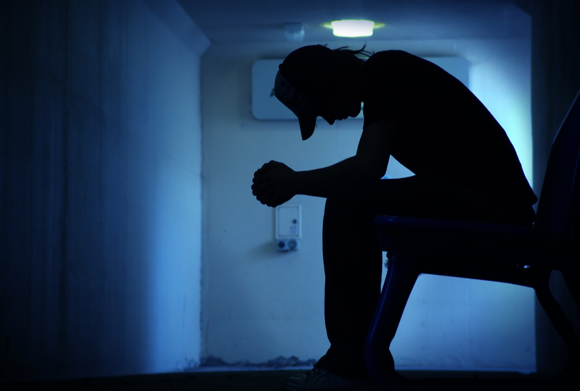
 ©Jean Marie Heidinger/Inserm

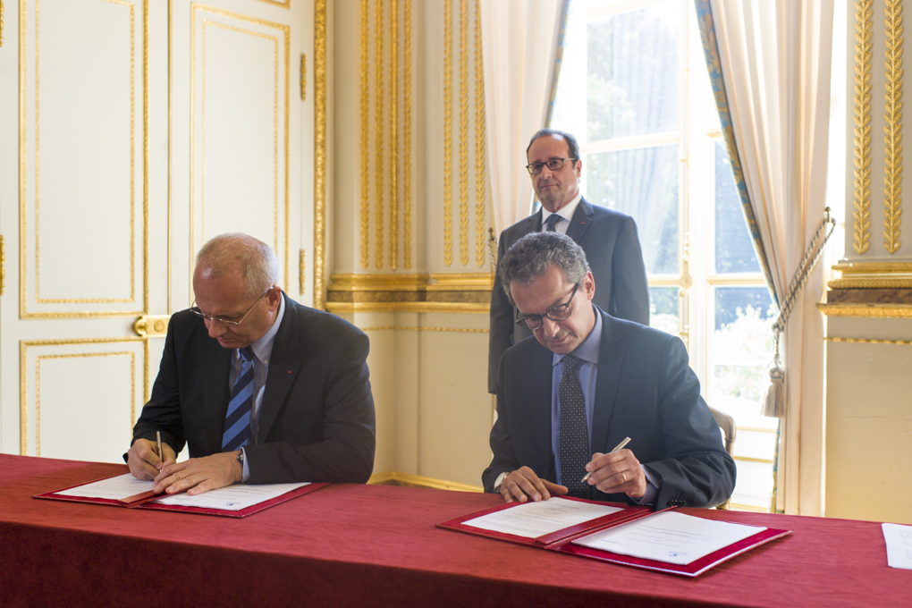
CNES and Inserm, respectively the institution in charge of proposing and implementing French space policy and the leading biomedical research organisation in Europe, have decided to expand their cooperation in the area of space and health. For the first time, they have signed a framework agreement that will cover the advances in basic research made possible by studying the human being in space, as well as the applications of findings from space research in matters of health.


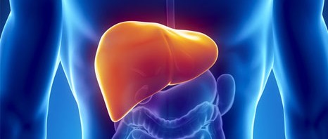
Space is an extraordinary laboratory for medicine and medical research on Earth. The loss of muscle mass and bone density, and the accelerated ageing of the arteries or disruption of the internal body clock observed in astronauts affected by zero gravity have led to improved knowledge of the human body. There has been a human presence in space almost continuously for over 40 years, and the main concern of the space agencies has always been to ensure astronauts’ health by taking appropriate measures. But in recent years, there have been instruments that allow comprehensive medical monitoring of astronauts, opening the opportunity for basic studies in physiology and medicine, conducted by Inserm and CNES. Moreover, these instruments may also find applications in medical research and public health.


This agreement, signed in the presence of the President of France by Yves Lévy, Chairman and Chief Executive Officer of Inserm and Jean-Yves Le Gall, President of CNES, provides for collaboration in the area of health in order to gain a better understanding of:

  • the impact of space conditions on physiology and its consequences for health (particularly the sensory, cognitive, biomechanical and immunological effects)

Its aim is to develop methods, tools and services based on, among other things, space technologies in the area of health, particularly:

  • the development of connected devices, particularly those designed for astronaut flight,
  • the development of medical instruments

It will also give rise to a large number of experiments during the period spent by Thomas Pesquet on board the international space station. Access to simulation experiments conducted on earth will also be possible, as well as the opportunity to conduct research projects during parabolic flights or in recoverable capsules.


“Thomas Pesquet’s space mission will make it possible to write a new chapter in French excellence in the area of manned flight. With this framework agreement signed with Inserm, medical research will benefit from all the advances enabled by studying the human being in space, and major advantages both for applications and for routine health matters,” stated Jean-Yves Le Gall, President of CNES.

“This agreement is a first between our two institutions, each of which embodies French excellence in Europe and worldwide in its own domain. This is an important step, which allows us to combine exploration of extreme environments and the use of the most advanced technologies for research on human health,” emphasised Yves Lévy, Chairman and Chief Executive Officer of Inserm.

Saturday 10 September 2016: World Suicide Prevention Day

desperate

(c) Fotolia

Suicide causes more than 800,000 deaths every year according to estimates by the World Health Organisation, i.e. one death every 40 seconds. It affects all age categories, and spares no region of the world.

To find out more about suicide mortality data for France, you may contact Grégoire Rey, director of CépiDc-Inserm (Epidemiological Center on Medical Causes of Death).


For those aged 15-29 years, suicide is the second leading cause of mortality. Moreover, a large survey, coordinated by Inserm Unit 1178, “Mental Health and Public Health,” and the University Division of Fondation Vallée, conducted in 2013 among 15,235 young students aged 13-18 years, shows that suicide attempts appear to be more common than before: 7.8% of young people have already attempted suicide once, and 3.7%, more than once. Alarming figures, which highlight the need for action to prevent suicidal behaviours.

See the press release “The new face of our adolescents.


World Suicide Prevention Day, which will be held on Saturday 10 September 2016, at the initiative of the International Association for Suicide Prevention and WHO, is aimed at emphasising the scale of this public health problem, and at making the public aware of the existing preventive measures.

Caffeine and its analogues revert memory deficits by normalizing stress responses in the brain

Roasted coffee beans background concept

(c) Fotolia

A study published in the journal Scientific Reports from Nature publishing group, describes the mechanism by which caffeine counteracts age-related cognitive deficits in animals.

The study coordinated by Portuguese researchers from Instituto de Medicina Molecular (iMM Lisboa) and collaborators from Inserm in Lille, France, along with teams from Germany and United States, showed that the abnormalexpression of a particular receptor – the adenosine A2A, target for caffeine – in the brain of rats induces an aging-like profile namely memory impairments linked to the loss of stress controlling mechanisms.

“This is part of a larger study initiated 4 years ago in which we identified the role of this receptor in stress, but we did not know whether its activation would be sufficient to trigger all the changes. We now found that by altering the amount of this receptor alone in neurons from hippocampus and cortex – memory related areas – is sufficient to induce a profile that we designate as ‘early-aging’ combining the memory loss and an increase in stress hormones in plasma (cortisol)” – explains Luisa Lopes, Group Leader at iMM Lisboa and the coordinator of the study.

When the same animals were treated with a caffeine analogue, which blocks the action of adenosine A2A receptors, both memory and stress related deficits were normalized.

David Blum, from Inserm research director, adds:

“In elderly people, we know there is an increase of stress hormones that have an impact on memory. Our work supports the view that the procognitive effects of A2AR antagonists, namely caffeine, observed in Alzheimer’s and age-related cognitive impairments may rely on this ability to counteract the loss of stress controlling mechanisms that occurs upon aging”

This is important not only to understand the fundamental changes that occur upon aging, but it also identifies the dysfunctions of the adenosine A2A receptor as a key player in triggering these changes. And a very appealing therapeutic target” – concludes Luisa Lopes.


About Luísa Lopes

Luísa V. Lopes is a Group leader@iMM Lisboa, Portugal since 2013.  Luisa graduated in Lisbon in 1998 in Biochemistry and then pursued a PhD in Neurosciences being trained in the University of Cambridge, UK and at the Karolinska Institute, Sweden. Her current work focuses on understanding the mechanisms inducing the “early-aging” of the cognitive function. The team has provided evidence for an important contribution of adenosine receptors in pathophysiological context, and their impact in noxious brain conditions such as stress, aging and neurodegeneration. Her team provided crucial evidence of a possible glucocorticoid-adenosine link in Alzheimer’s disease (with D. Blum) following previous groundwork suggesting circadian disorders as a trigger for accelerated cognitive loss.


About David Blum

David Blum is Inserm Research Director at UMR-S 1172 Inserm/Université Lille2/CHRU Lille in the laboratory “Alzheimer & Tauopathies” headed by Dr. Luc Buée. David graduated in Strasbourg University and got his PhD at Inserm in Grenoble. Current work of David Blum focus on the understanding of the role of caffeine and adenosine receptors in cognitive disorders, particularly Alzheimer’s Disease. In this field, the laboratory has recently provided evidence, with the group of Luisa Lopes, that A2A receptors are important for the development of AD-like Tau pathology. Our current work is interested on the instrumental role of sub-population of A2A receptors in the brain on the occurence of cognitive disorders in neurological disorders.


About iMM Lisboa

Instituto de Medicina Molecular (iMM Lisboa) is a reference biomedical research centre in Portugal, having acquired the special status of Associate Laboratory of the Portuguese Ministry of Science and Technology. iMM’s mission is to promote basic, translational and clinical biomedical research, with the aim of understanding the mechanisms of disease and developing new therapeutic approaches.

For more information please visit the iMM Lisboa’s website: www.imm.medicina.ulisboa.pt


 “Alzheimer & Tauopahies” laboratory at UMR-S 1172 Inserm/Université Lille 2/CHRU-Lille  is a world class research laboratory interested in the molecular, cellular, and physiological aspects of Alzheimer’s Disease and dementia. The lab is part of the LabEx DISTALZ (national consortium on AD) and of the LICEND excellence centre for neurodegenerative diseases.

Artificial retinas: promising leads towards clearer vision

Visuel chavanne

Activation (colored circles at the level of the visual cortex) of the visual system by prosthetic stimulation (in the middle, in red, the insert shows an image of an implanted prosthesis) is greater and more elongated than the activation achieved under natural stimulation (on the left, in yellow). Using a protocol to adapt stimulation (on the right, in green), the size and shape of the activation can be controlled and are more similar to natural visual activation (yellow).

© F. Chavane & S. Roux.


A major therapeutic challenge, the retinal prostheses that have been under development during the past ten years can enable some blind subjects to perceive light signals, but the image thus restored is still far from being clear. By comparing in rodents the activity of the visual cortex generated artificially by implants against that produced by “natural sight”, scientists from CNRS, CEA, Inserm, AP-HM and Aix-Marseille Université identified two factors that limit the resolution of prostheses. Based on these findings, they were able to improve the precision of prosthetic activation. These multidisciplinary efforts, published on 23 August 2016 in eLife, thus open the way towards further advances in retinal prostheses that will enhance the quality of life of implanted patients.


A retinal prosthesis comprises three elements: a camera (inserted in the patient’s spectacles), an electronic microcircuit (which transforms data from the camera into an electrical signal) and a matrix of microscopic electrodes (implanted in the eye in contact with the retina). This prosthesis replaces the photoreceptor cells of the retina: like them, it converts visual information into electrical signals which are then transmitted to the brain via the optic nerve. It can treat blindness caused by a degeneration of retinal photoreceptors, on condition that the optical nerve has remained functional1 . Equipped with these implants, patients who were totally blind can recover visual perceptions in the form of light spots, or phosphenes. Unfortunately, at present, the light signals perceived are not clear enough to recognize faces, read or move about independently.

To understand the resolution limits of the image generated by the prosthesis, and to find ways of optimizing the system, the scientists carried out a large-scale experiment on rodents. By combining their skills in ophthalmology and the physiology of vision, they compared the response of the visual system of rodents to both natural visual stimuli and those generated by the prosthesis.

Their work showed that the prosthesis activated the visual cortex of the rodent in the correct position and at ranges comparable to those obtained under natural conditions. However, the extent of the activation was much too great, and its shape was much too elongated. This deformation was due to two separate phenomena observed at the level of the electrode matrix. Firstly, the scientists observed excessive electrical diffusion: the thin layer of liquid situated between the electrode and the retina passively diffused the electrical stimulus to neighboring nerve cells. And secondly, they detected the unwanted activation of retinal fibers situated close to the cells targeted for stimulation.

Armed with these findings, the scientists were able to improve the properties of the interface between the prosthesis and retina, with the help of specialists in interface physics. Together, they were able to 1 This is the case of patients with Retinitis Pigmentosa or Age-related Macular Degeneration (AMD). Artificial retinas: promising leads towards clearer vision generate less diffuse currents and significantly improve artificial activation, and hence the performance of the prosthesis.

This lengthy study, because of the range of parameters covered (to study the different positions, types and intensities of signals) and the surgical problems encountered (in inserting the implant and recording the images generated in the animal’s brain) has nevertheless opened the way towards making promising improvements to retinal prostheses for humans.

This work was carried out by scientists from the Institut de Neurosciences de la Timone (CNRS/AMU) and AP-HM, in collaboration with CEA-Leti and the Institut de la Vision (CNRS/Inserm/UPMC).

The 2016 Summer Olympic Games in Rio

The world’s finest athletes will meet at the 2016 Summer Olympic Games in Rio de Janeiro to try to win Olympic medals, thus testing their bodies to the limit.

What are the risks for the top athletes? What methods would enable them to optimise their physical abilities? What lines of prevention can be developed? How does our environment influence performances? What are the effects of athletic activity on ageing?

Such are the questions that the researchers at Inserm, who are involved all year round in studying the interactions between sport, physical activity and health, are able to answer.


At Inserm Unit 1093, “Cognition, Motor Activity and Sensorimotor Plasticity,” Romuald Lepers focuses on the performances of centenarian athletes so as to better understand how the human body can fight the deleterious effects of ageing. In a recent study, he compared the records of these athletes in certain disciplines with world records for all age categories combined. Although the record of centenarian cyclist Robert Marchand, with nearly 27 kilometres covered in an hour, appears the most impressive, the researcher also emphasises that age-related decline in performance is less marked for cycling than for athletics or swimming.

Read the news item “Centenarian athletes: flawless performances?


Meanwhile, the research undertaken by Mathias Pessiglione, Inserm Research Director at Unit 1127, “Brain and Spine Institute”? deals with the brain mechanisms involved in motivation. He has modelled the motivational aspects of athletic performance according to a cost/benefit calculation, i.e. how much the athlete is prepared to invest as a function of how much the objective is worth.

For your interviews and reporting needs, see the “Physical Activity press-kit,” available as a download opposite, for a listing of the contact details of specialists in this discipline, together with the latest news from Inserm on the subject.

Athletisme *** Local Caption ***

(c) Fotolia

Unconscious processing operates under conscious influence

Opened book

(c) Fotolia

In a study published in the new international scientific journal Neuroscience of Consciousness, Benjamin Rohaut, Inserm researcher, and Lionel Naccache, leader of the team “Picnic lab: Physiological Investigation of Clinically Normal and Impaired Cognition,” both of whom are also clinicians attached to the Neurology Department at Pitié-Salpêtrière Hospital, AP-HP, provide proof that unconscious semantic processing of a word exists, but that it is subject to very strong conscious influences. The work was conducted in collaboration with their colleagues at the Brain and Spine Institute – AP-HP/CNRS/Inserm/UPMC and the Laboratory of Cognitive Psychology (CNRS/AMU).


Experimental psychology is full of situations that make it possible to probe the depth and variety of mental operations performed unbeknown to us, i.e. unconsciously. For example, using subliminal visual presentation techniques, it is possible to “inject” a word into the brain of someone, and then track its fate at psychological and brain levels with the help of behavioural measurements and functional brain imaging. Since the late 1990s, several high-profile studies have also demonstrated that the meaning of a subliminal image, number or word may be unconsciously represented in our mind/brain.

In the study conducted by Lionel Naccache, the researchers provide proof that unconscious semantic processing of a word genuinely exists, but that it is subject to very strong conscious influences.

To do this, they used words such as: “palm, bank, jam, bail, date, ball, tire, bark, bowl, spit  …”. All these words share a common semantic property, have you noticed?

In reality each of these words is “polysemous,” and therefore has two (or more) different meanings. Each time such as word is presented to you, you can therefore understand it in two different ways. Consciously, we perceive only one meaning at a time, as stated by Descartes back in 1649 in Les Passions de l’âme : “We have only one thought of one thing at a time.” The meaning of the word we access consciously at a given moment is likely to be influenced.


Thus, if you read: TREE then PALM you are very likely to access the botanic meaning of the word PALM: “an unbranched evergreen tree with a crown of long feathered or fan-shaped leaves, and typically having old leaf scars forming a regular pattern on the trunk. Palms grow in warm regions, especially the tropics” whereas the HAND – PALM pair will strongly orient your semantic analysis toward the “the inner surface of the hand between the wrist and fingers”.

In this experiment authors presented the volunteers with words triplets while recording their brain activity using an electrode headset. Each test began by presenting the first word, which was always visible and which allowed a particular semantic context to be defined (e.g. HAND). Then the second word was flashed on the screen and was either subliminal or consciously visible. The third word then appeared, and was always consciously visible. It served as a target stimulus to which the subjects had to respond by pressing a button in order to indicate whether it was a real word (e.g. WRIST) or a pronounceable chain of letters which did not correspond to a lexicon word (known as a pseudo-word, such as DRAIE). When the middle word was semantically related to the target word, the subjects responded faster. This is known as a priming effect. This priming effect also appeared in the analysis of brain activities.

When the polysemous word (the middle word of the triplet) was consciously visible, a priming effect was present only when the meaning was consistent with the contextual word presented at the start of each test (Word 1). For example, when the following triplet was presented: HAND-PALM-WRIST, a priming effect of the word WRIST by the word PALM was found, but this effect was absent from triplets such as: HAND-PALM-TREE. Analysis of the electrical activity in the brain confirmed and clarified this result. The absence of priming for the non-contextualised meaning of the polysemous word indicates that this meaning was simply not analysed by the subjects. Conscious semantic processing is therefore strongly influenced by the conscious context.

The core result of this work lies in the discovery that it is the same for the unconscious perception of polysemous words. When the polysemous word (Word 2) was presented in a subliminal manner, the authors found semantic priming effects comparable to those observed under conscious reading conditions: only those meanings of the polysemous word that were consistent with the contextual word were unconsciously analysed.

This series of experiments thus demonstrates that unconscious cognition is not only highly complex, since it can reach the level of semantics (the meaning of words), but also shows that it seems to be extremely sensitive to conscious influences. At every moment, our conscious position influences the nature of the mental operations unconsciously unfolding within us.

“This work, which combines neurosciences with the psycholinguistics of the French language, also illustrates the potential of multidisciplinary scientific approaches,” conclude Lionel Naccache and his collaborators

One hour of physical activity per day may attenuate the increased risk of death associated with sitting time

A meta-analysis of data from over one million people, published on July 27th in a new Lancet Series ahead of the 2016 Summer Olympic Games, emphasize the importance of physical activity to attenuate the harmful effect of sitting time.

The researchers classified individuals into four groups according to how active they were, less than 5 mins a day, up to 60-75 mins a day for the most active. The study suggest that doing at least one hour of moderate intensity physical activity per day such as brisk walking at 5.6 km/h or cycling at 16 km/h, may attenuate the increased risk of death associated with sitting for 8h a day. Furthermore authors’ recommendations are superior to World Health Organization guidelines, which recommend at least 150 minutes of physical activity per week.

If we are active enough, it is not necessary to go to the gym for one hour per day, according to Ulf Ekelund, lead author (Norwegian School of Sport, Norway and the University of Cambridge, UK).

To comment on this work, please contact Romuald Lepers, Inserm Unit 1093 “Cognition, Motor Activity and Sensorimotor Plasticity”.

Velo

(c) Fotolia

A virtual brain helps decrypt epilepsy

cerveau virtuel

The Virtual Brain: reconstruction of brain regions and where they are connected. The green cubes indicate the center of brain regions that are connected

©INS UMR1106 INSERM/AMU.

Researchers at CNRS, INSERM, Aix-Marseille University and AP-HM have just created a virtual brain that can reconstitute the brain of a person affected by epilepsy for the first time. From this work we understand better how the disease works and can also better prepare for surgery. These results are published in Neuroimage, on July 28, 2016.


Worldwide, one percent of the population suffers from epilepsy. The disease affects individuals differently, so personalized diagnosis and treatment are important. Currently we have few ways to understand the pathology’s mechanisms of action, and mainly use visual interpretation of an MRI and electroencephalogram. This is especially difficult because 50% of patients do not present anomalies visible in MRI, so the cause of their epilepsy is unknown.

Researchers have succeeded for the first time in developing a personalized virtual brain, by designing a base “template” and adding individual patient information, such as the specific way the brain’s regions are organized and connected in each individual. Mathematical models that cause cerebral activity can be tested on the virtual brain. In this way, scientists have been able to reproduce the place where epilepsy seizures initiate and how they propagate. This brain therefore has real value in predicting how seizures occur in each patient, which could lead to much more precise diagnosis.


Moreover, 30% of epileptic patients do not respond to drugs, so their only hope remains surgery. This is effective if the surgeon has good indications of where to operate.

The virtual brain gives surgeons a virtual “platform.” In this way they can determine where to operate while avoiding invasive procedures, and especially prepare for the operation by testing different surgical possibilities, seeing which would be most effective and what the consequences would be, something that is obviously impossible to do on the patient.

In the long run, the team’s goal is to provide personalized medicine for the brain, by offering virtual, tailored, therapeutic solutions that are specific for each patient. The researchers are currently working on clinical trials to demonstrate the predictive value of their discovery. This technology is also being tested on other pathologies that affect the brain, such as strokes, Alzheimer’s, degenerative neurological diseases, and multiple sclerosis.


This work involves researchers at the Institut de Neurosciences des Systèmes (INSERM/AMU), the Centre de Résonance Magnétique Biologique et Médicale (CNRS/AMU/AP-HM), the Département Epileptologie et du Département Neurophysiologie Clinique at AP-HM, and the Epilepsy Center of Cleveland. It was done in the Fédération Hospitalo-Universitaire Epinext (www.epinext.org).

cerveau

The Virtual Epileptic Patient: brain regions and their connections are rebuilt by computer. Digital simulations generate an electric signal similar to that generated by the brain during seizures. These simulations allow digital testing of new therapeutic strategies

©INS UMR1106 INSERM/AMU.

Inserm, France’s rising research star according to Nature

1024x512

(c) Macmillan Publishers Ltd

The French National Institute of Health and Medical Research (Inserm) is the only research institution in France listed in the World Top 100 institutions with the most progress in the last 3 years. This is indicated by the Nature Index 2016 Rising Stars, which highlights the big contribution of Inserm researchers to publications in the best 68 journals selected by Nature. This ranking identifies the most brilliant institutions to watch in 2016 and in the coming years.


Of 8,000 institutions reviewed, Inserm appears in 75th place among the institutions with the strongest progress. In three years, of a selection of 68 scientific journals rated best by Nature, the number of publications increased by 17.43%. Contributions from Inserm researchers published in the journal Science and the Nature Group Journals almost doubled in three years. Inserm is the only research institution in France listed in the Nature Index 2016 Rising Stars. Its presence in this index is all the more exceptional given that the index is naturally dominated by countries, such as China, that have very recently entered into international competition, and have exceptional growth in their number of publications.


For Professor Yves Lévy, Chairman and Chief Executive Officer of Inserm: “This indicator reflects our research policy, which encourages Inserm researchers to publish in the best journals because they are talented. This index didn’t take into account medical journals specialising in clinical research, such as NEJM, JAMA or The Lancet, in which growth is 37%. Theses results confirm the place of Inserm in basic science and in clinical research.”


About Inserm

Founded in 1964, the French National Institute of Health and Medical research (Inserm) is a public science and technology institute, jointly supervised by the French Ministry of National Education, Higher Education and Research and the Ministry of Social Affairs, Health and women’s Rights. Inserm is the only French public research institute to focus entirely on human health with nearly 15000 researchers, engineers, technicians, post-doctoral students and more than 300 laboratories. The mission of its scientists is to study all diseases, from the most common to the rarest. Inserm is a member of Aviesan, the French National Alliance for Life Sciences and Health founded in 2009. Other founding members of Aviesan : CEA, CNRS, CHRU, CPU, INRA, INRIA, Institut Pasteur, IRD.

Omega-3 fatty acids and stress management: growing links

nutrition poisson légumes aliments

Fotolia

The relationship between omega-3 fatty acid intake and adaptation to stress or anxiety is becoming clearer. Back in 2011, a team of researchers from INRA and Inserm showed that reducing the intake of omega-3 fatty acids in mice increased their stress. This phenomenon may be linked to impairment of the brain’s ability to produce endogenous cannabinoids, the “endocannabinoids,” brain lipids that control synaptic memory. To better understand the endocannabinoid-dependent links between anxiety and synaptic plasticity, the research team continued its experiments by testing different models of behavioural stress on the rodents. These studies are the subject of an article published in the journal Cell Reports on 21 July.

Mice do not respond equally to stress. An observation made by an INRA/Inserm research team who, after submitting the rodents to a battery of tests related to behaviour stress – isolation, maze or anxiogenic environment – found that some subjects were naturally resilient, or in other words, more resistant to stress. This ability may be related to better plasticity in the neurons of the accumbens nucleus, an area of the brain associated with regulation of the emotions and of stress, where endocannabinoids play a key role in memory at synapse level.

To confirm this relationship, mice showing several anxiety-related symptoms received a treatment to stimulate endocannabinoid production in the accumbens nucleus. Result: the scientists observed an attenuation of anxiety in these mice. For the first time, it has been proven that there is a relationship between anxiety and the levels of endocannabinoids produced by the brain. These results further emphasise the therapeutic potential of molecules that modulate the natural production of endocannabinoids, including dietary omega-3 fatty acids.

A milestone has thus been reached in demonstrating the protective effect of omega-3 fatty acids on the impairment of the brain’s ability to produce endocannabinoids. Ultimately, the idea is to understand how omega-3 fatty acids exert a protective action in response to the impairment of endocannabinoid-dependent plasticity in the accumbens nucleus, a part of the brain that constitutes the neurobiological substrate for anxiety.

This opens new perspectives in understanding the role of omega-3 fatty acids in the management of stress by the brain. The next studies will be aimed at better understanding the role of dietary omega-3 fatty acids in protecting the plasticity of the accumbens nucleus, and hence their ability to stimulate endocannabinoid production in a situation of stress.

Long term correction of hyperbilirubinemia in animal models of Crigler-Najjar syndrome after AAV vector-mediated liver gene transfer

Dr. Federico MINGOZZI, Team Leader of the Immunology and Liver Gene Transfer unit at Généthon, (Inserm U951/UPMC), France, has demonstrated the long-term efficacy of an optimized AAV-UGT1A1 vector for the correction of Crigler-Najjar syndrome (CN) in two different animal models of the disease. This work, published in July 20, 2016 in Molecular Therapy Methods and Clinical Development was supported by the AFM-Telethon.


Crigler-Najjar syndrome (CN) is a rare inherited liver disorder characterized by a deficiency of the enzyme UDP-glucuronosyltransferase 1A1 (UGT1A1). This enzyme converts bilirubin, a yellow pigment, into conjugated bilirubin. After conjugation, bilirubin becomes water soluble and is excreted in the bile and further eliminated from the organism. UGT1A1 enzyme deficiency leads to the accumulation of bilirubin in all body tissues, especially in the brain. If not treated, the brain accumulation of bilirubin leads to neurological damage and death.

Currently, treatment of CN is based on the ability of blue light to degrade bilirubin. Based on that, patients are exposed to phototherapy for 10 to 12 hours per day. Although the treatment is relatively efficacious and keeps bilirubin under the toxicity level it presents several disadvantages. In fact the long phototherapy sessions prevent patients from conducting a normal life, resulting in lack of compliance and other issues. Additionally, the time of exposure to phototherapy increases with the age due to the decreased body surface/mass ratio, and the therapy does not prevent potentially lethal bilirubin spikes associated for example with trauma or infections. The only curative treatment for CN is liver transplantation, which also presents significant potential risks and long-term complications.
Hence, gene therapy represents an alternative curative option to correct the genetic defect, restore the enzyme expression and the consequent bilirubin conjugation. Researchers at Genethon have designed an optimized AAV vector expressing the UGT1A1 transgene and tested it in two animal models of Crigler-Najjar syndrome, the Gunn rat and in UGT1A1 deficient mouse.

Following a single administration of the therapeutic vector, the researchers observed safe and efficient correction of the disease, with disappearance of bilirubin from serum consequent to restoration of the UGT1A1 activity in liver. Animals were followed for more than one year after vector injection, demonstrating long-lasting efficacy of the approach.
This work is the result of the coordinated efforts of a European Network of collaborators that includes the laboratory of Dr. Mingozzi at Genethon, the laboratories of Dr. Bosma at the Academic Medical Center in the Netherlands and Dr. Muro at the International Centre for Genetic Engineering and Biotechnology in Italy, the Crigler-Najjar patients associations of France, Italy and the Netherlands, and several clinical centers in Europe.
Based on these results, Genethon researchers are now preparing a phase I/II clinical trial that will open for enrollment in the near future.

fermer