Menu

Effects of the gut microbiota revisited

cp-serino-synopsis-1

© Matteo Serino

Matteo Serino, Inserm Research Fellow at the Digestive Health Research Institute (IRSD, an Inserm/University of Toulouse III Paul Sabatier/National Veterinary College of Toulouse/Inra joint research unit), and his collaborators show that alteration of the intestinal microbiota, whether nutritional or genetic in origin, may have beneficial effects on liver metabolism. These results contradict results previously obtained in the area, which showed that transferring an altered microbiota inevitably had negative consequences for health. They have just been published in Molecular Systems Biology.

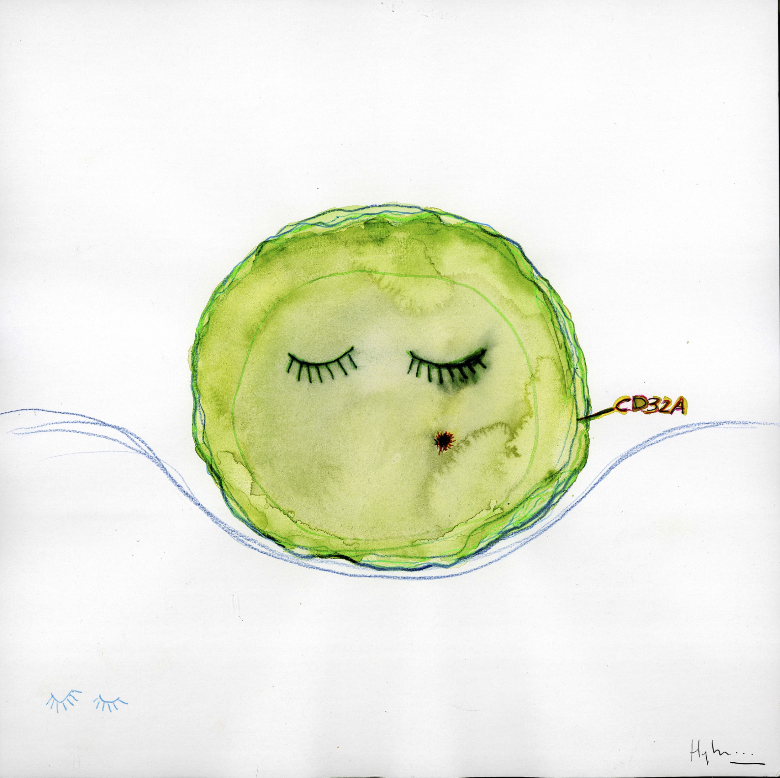
The gut microbiota is involved in the occurrence of metabolic diseases such as obesity and liver diseases. However, the molecular mechanisms are still incompletely known, and its role as a cause or consequence of these diseases remains a matter of debate.

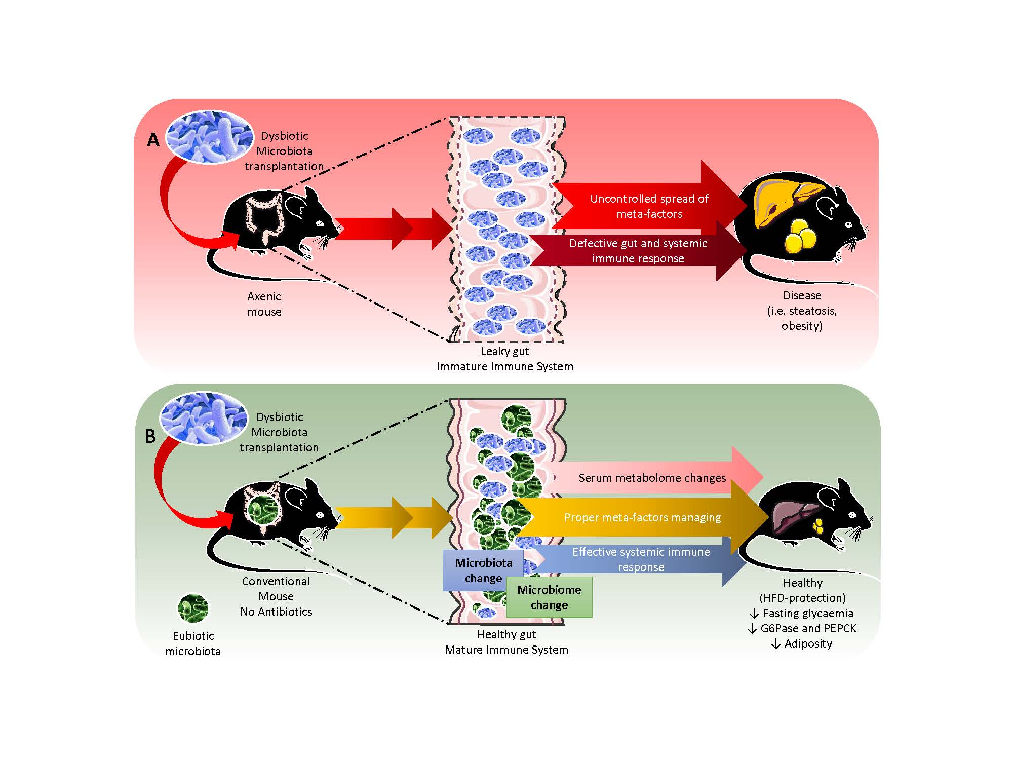
To understand this, Matteo Serino and his collaborators transferred the gut microbiota from obese mice to wildtype mice fed a normal diet and unexposed to any antibiotic treatment. Indeed, it has been shown that antibiotic treatment prior to transplantation of the gut microbiota could limit the occurrence of metabolic disease, and affect the efficacy of gut microbe transfer.

The researchers observed that on a normal diet, mice that received a microbiota from obese mice underwent a sharp reduction in liver glucose production and fasting blood glucose. Transfer of the microbiota from obese mice not only modified the gut microbiota, but also the function of the bacteria (or microbiome) of the recipient mice.

Unexpectedly, when mice were transplanted with a microbiota from obese mice, and then fed a high-fat diet, their liver metabolism and overall carbohydrate metabolism seemed to be protected. Moreover, their fat mass was less developed and their fat cells were smaller in size.

Conversely, transferring a microbiota from lean mice did not affect liver glucose production.

These research results show that alteration of the microbiota, subsequent to a high-fat diet, is not always harmful. Insofar as the intestinal barrier is intact and the immune system functional, an altered gut microbiota may even protect against the harmful effects of a high-fat diet. “Our observations have opened a new debate on the role of alteration of the gut microbiota in the occurrence of metabolic diseases,says Matteo Serino.

Discovery of an HIV reservoir marker: A new avenue toward eliminating the virus

L1bis

© Fabrice Hyber, pour Organoïde/Institut Pasteur

French researchers have identified a marker that makes it possible to differentiate “dormant” HIV-infected cells from healthy cells. This discovery will make it possible to isolate and analyze reservoir cells which, by silently hosting the virus, are responsible for its persistence even among patients receiving antiviral treatment, whose viral load is undetectable. It offers new therapeutic strategies for targeting infected cells. This research is part of the ANRS strategic program “Réservoirs du VIH”. It is the result of a collaboration between the CNRS, Montpellier University, Inserm, the Institut Pasteur, the Henri-Mondor AP-HP hospital in Créteil, the Gui de Chauliac hospital (CHU de Montpellier) and the VRI (Vaccine Research Institute), and is published in the journal Nature on March 15, 2017. A patent owned by the CNRS has been filed for the diagnostic and therapeutic use of the identified marker.

Since 1996, there has been consensus among the scientific community that a cure for HIV will involve targeting “reservoir cells” that host the virus in the organisms of patients undergoing triple therapy. HIV can remain hidden in these reservoirs, in latent form, for several decades, eluding the immune system’s response and antiviral treatments, without any viral protein being expressed. But if treatment ceases, the virus massively proliferates and the disease progresses again. Patients must therefore receive treatment for life. To envisage eliminating this dormant virus, a first stage consists in distinguishing the HIV-infected reservoir cells from their healthy counterpart cells, which resemble them to a very large degree. This is what has been achieved by a team of researchers, who have identified a marker of reservoir cells: a protein present only on the surface of infected cells.

Hypothesizing that HIV might leave a mark on the surface of its host cell, researchers from the Institut de génétique humaine (CNRS/Montpellier University) first worked in vitro on an infection model developed in their laboratory. After comparing infected cells and healthy cells,[1] they noticed one particular protein, coded by a gene among the hundred of those expressed in a specific way by infected cells. Present only on the surface of the infected cells, the CD32a protein thus met, in vitro, the criteria of a reservoir cell marker. This was then confirmed by experiments on clinical samples. By studying blood samples from 12 patients living with HIV and receiving treatment,[2] the researchers isolated the cells expressing the marker and observed that almost all were HIV carriers. In vitro, the activation of these cells induced a production of viruses capable of reinfecting healthy cells whereas their elimination entailed a significant delay in viral production.

In the fight against HIV, this discovery paves the way to a better fundamental understanding of viral reservoirs, which it will now be possible to isolate more easily and analyze directly. In the longer term, it should lead to therapeutic strategies aiming to eliminate the latent virus from the organism and make remission—at least temporary—possible in the absence of antiviral treatments.

This research received support from the ANRS, MSD Avenir, the European Commission, the Fondation Bettencourt Schuelle, the Fondation pour la recherche médicale and the Vaccine Research Institute (VRI).

[1] The cells studied are T CD4 lymphocytes, whose numbers are progressively reduced upon infection by HIV. The number of these cells is thus used by doctors to monitor the progression of the disease and the efficacy of treatments.

[2] Patients monitored by the Clinical Immunology and Infectious Disease Department at the Henri-Mondor AP-HP hospital in Créteil and by the Infectious and Tropical Disease Department at Gui de Chauliac hospital (CHU de Montpellier).

Toward the synthesis of antibiotics by a new bacterial enzyme

cp-enzymebact_synthantib

©Inra

Researchers at the French National Institute for Agricultural Research (Inra) and Inserm have discovered a new family of bacterial enzymes that can produce hitherto unidentified peptides with antibiotic activity. Published in Nature Chemistry, this work holds promise for the synthesis of molecules of pharmaceutical interest and the design of new antibiotics.

As part of research involving study of the enzymes of the gut microbiota, researchers from Inra and Inserm studied the model bacterium Bacillus subtilis. Genetic analysis of the organism showed the presence of genes conserved by common bacteria from the gut microbiota, such as enterococci. *

The scientists were especially interested in two Bacillus subtilis genes potentially coding for a peptide and an enzyme from the superfamily known as “radical SAM enzymes.” Their work has made it possible to describe a new enzyme mechanism that can transform a peptide into a bioactive molecule. Known as epimerisation, this enzymic transformation brings about a change in the configuration of certain amino acid residues from the L- configuration (normally found in peptides) to the D-configuration. The researchers discovered how this enzyme works: it removes a hydrogen atom from the alpha-carbon atom of the amino acid residues and donates another, bringing about their epimerisation. It is a mechanism not previously described in nature.

It is the first time that researchers have demonstrated the ability of “radical SAM” enzymes to catalyse epimerisation within a peptide in vitro. Surprisingly, the peptide thus modified and called an “epipeptide” is able to inhibit growth of Bacillus subtilis very effectively. These epipeptides thus represent a new class of natural products that might be used to develop new antibiotics against Gram positive bacteria (such as staphylococci, enterococci or streptococci), which are becoming increasingly antibiotic resistant, constituting a major public health problem.

A warning on taking ibuprofen during pregnancy

fotolia_4518648 Suivi médical grossesse

©fotolia

A new study conducted by Inserm researchers at Irset (Institute of Research in Environmental and Occupational Health)[1] shows that ibuprofen is liable to cause disruptions in the hormone system in the human foetal testis, with possible implications for the development of the male urogenital tract. This drug suppresses the production of various testicular hormones, including testosterone, which controls the primary and secondary sex characteristics and the descent of the testes. These effects are obtained at doses similar to the standard dosage. These results are published in Scientific Reports.

Ibuprofen, which can be obtained without prescription, is one of the drugs most commonly consumed by pregnant women. Although nearly one woman in ten reports having taken ibuprofen during her pregnancy, studies indicate that in reality up to 3 in 10 have self-medicated with it.

Epidemiological studies conducted in recent years have shown a link between taking analgesics during pregnancy and the occurrence of adverse effects in the child (low birthweight, asthma, premature birth etc.). Other research combining epidemiology, in utero experimentation in rats and ex vivo on rat and human organs, undertaken at Irset in collaboration with Danish researchers from the University of Copenhagen, showed that paracetamol and aspirin could disrupt the endocrine system of the foetal testis, resulting in an increased risk of failure of the testes to descend (cryptorchidism). Only the effects of ibuprofen had not yet been tested.

To do that, the Irset researchers – with the support of colleagues at Rennes University Hospital and the University of Copenhagen, researchers from Laberca (Laboratory for the Study of Residues and Contaminants in Food) in Nantes, and Scots colleagues from MRC Edinburgh – conducted two series of tests to study the effects of ibuprofen on the human foetal testis. In the first series of studies, the testes were cultured; in the second, they were grafted onto mice[2]. The effects of ibuprofen were studied over periods corresponding to the 1st and 2nd trimesters of pregnancy.

When testes corresponding to the 1st trimester of pregnancy were exposed to ibuprofen, there was a sharp drop in testosterone production by the Leydig cells. During the same period (up to 12 weeks of development), the researchers observed for the first time that ibuprofen also affected the production of antimüllerian hormone by the Sertoli cells. This hormone plays a key role in genital tract masculinisation.

Moreover, expression of the genes needed for the germ cells, the progenitors of spermatozoa, to function is considerably reduced in the presence of ibuprofen.

Finally, production of prostaglandin E2 (known to be produced by the testes, and known to be involved in many biological processes) and the corresponding genes are also inhibited by the presence of ibuprofen at the same developmental age.

All these effects are observed very early in the first trimester, and none of them is found in tests conducted during the second trimester.

For Bernard Jégou, Inserm Research Director and coordinator of this study, and Séverine Mazaud-Guittot, Inserm Research Fellow, the conclusions of this work, which was supported by the French National Agency of Medicine and Health Products Safety (ANSM), are to be taken seriously: “There is a well-defined window of sensitivity during the 1st trimester of foetal development during which ibuprofen seems to present a risk for the future genital and reproductive system of the child. All the indications point to the need for great prudence regarding the use of this drug during the 1st trimester of pregnancy. Furthermore, if we now take the body of available data into account, it seems that taking several analgesics during pregnancy represents an even greater danger for the hormonal balance of the male foetus.

[2] Xenografting is the transplantation of cells or organ fragments from one living organism (e.g. human cells) into the body of another species (here the mouse) in order to understand their development.

[1] Institute of Research in Environmental and Occupational Health; Inserm; French School of Public Health (EHESP), University of Rennes 1.

13–19 March: Brain Awareness Week

capture_decran_2017-01-27_a_15-12-06

© Institut de la vision

The 19th edition of Brain Awareness Week, coordinated by the French Society for Neuroscience, will take place from 13 to 19 March 2017, in France and 62 other countries. During the many events, the general public is invited to come and meet the researchers in order to gain a better understanding of the workings of the brain and find out about advances in research.

Researchers from research organisations, including Inserm, neuroscience institutes and the teaching hospitals, are coming together to participate in 400 free events in 32 towns and cities in France.

“It is an opportunity for many researchers, physicians and students – all volunteers – to meet the public and let it share the advances in research and neuroscience, and to present the challenges and implications of these advances for our society,” says Jean-Antoine Girault, President of the French Society for Neuroscience.

Brain Awareness Week will open with an inaugural lecture given by Jean-Antoine Girault, President of the French Society for Neuroscience, titled À la recherche des traces de la memoire dans le cerveau” (In search of traces of memory in the brain), Monday 13 March at 6:30 pm, at the Brain and Spine Institute, Paris.

See the programme for Brain Awareness Week at www.semaineducerveau.fr/2017  (http://semaineducerveau.fr/2017/France.php)

Inserm researchers remain strongly committed to advancing neuroscience research. Here are some examples of research done by Inserm on this theme, available at the Press Room :

“Lack of sleep causes brain impairment in adolescents,” published on 8 March 2017

“Multiple sclerosis: Mission Regeneration!” published on 23 February 2017.

“Identification of one of the keys allowing entry of Zika virus into brain cells,” published on 10 January 2017.

“Sommeil paradoxal: ces neurones qui nous paralysent” (REM sleep, the neurons that paralyse us), published on 13 December 2016.

Voluntary eye movements, a new indicator of postural control in Parkinson disease ?

Oeil vision yeux

© Fotolia

Postural instability is the main factor associated with falls in patients with Parkinson disease, but there are other symptoms, such eye disorders. Teams from Pitié-Salpêtrière Hospital, AP-HP, and the Brain and Spine Institute (Inserm, CNRS, UPMC) have examined the link between these eye disorders observed in some patients and postural instability. Their results demonstrate a potential new marker of posture control in Parkinson disease. These results are published in the journal Neurology.

Some patients with Parkinson disease show impairment of the voluntary eye movements, the “antisaccades.” Dr Claire Ewenczyk, from the team led by Prof Marie Vidailhet, and Prof. Stéphane Lehéricy, from the Brain and Spine Institute, (Inserm, CNRS, UPMC) at Pitié-Salpêtrière Hospital, AP-HP, conducted a study in 30 patients with Parkinson’s disease with or without postural control disorders, and 25 healthy subjects.

The researchers examined the patients’ walk and balance, and recorded the initiation of walking and eye movements. At the same time, they studied the interactions between two areas of the brain: the frontal oculomotor area, which plays an important role in visual attention and eye movements, and the mesencephalic locomotor area, which is strongly involved in the control of posture and eye movements.

Their results indicate that patients with posture disorders also show abnormal reaction time (latency) in their voluntary eye movements. This abnormality is correlated with an alteration in the parameters during initiation of a step, particularly the duration of anticipatory postural adjustments, mechanisms used by the central nervous system to maintain balance in a standing position while performing voluntary movements.

Studying the interactions between the frontal oculomotor and mesencephalic locomotor areas of the brain reveals, in healthy subjects, a correlation between the frontal connections of these areas and the latency of voluntary eye movements. In patients with Parkinson disease, this association disappears. This suggests extensive involvement of the mesencephalic area in the disease, which affects both posture control and eye movements.

The prolongation of the reaction time of eye movements or “antisaccades,” a simple and reliable parameter, might constitute a prognostic marker of postural control in Parkinson disease, and be used for patient assessment in future longitudinal studies.

Lack of sleep causes brain impairment in adolescents

illustrationsommeil1en

©Alexandra Pinci pour l’Inserm

Not sleeping enough or going to bed too late leads to a reduction in the volume of grey matter in the brains of adolescents. These conclusions were obtained by researchers at Inserm Unit 1000, “Neuroimaging and Psychiatry” (Inserm/Paris-Descartes University/Paris Sud University), who studied the brain and sleep habits of 177 14-year-old students. This work is published in the journal Scientific Reports, and received support from the Academy of Finland.

Lack of sleep in adolescents can compromise their academic success, and their health and safety. Shortened or delayed sleep has consequences for academic performance. There is still debate over the time at which lessons should begin in order to benefit the health of adolescents. However, researchers did not know until now whether or not poor sleep habits in adolescents were associated with damage to the anatomy of the brain.

A collaboration between researchers from Inserm and from the Finnish National Institute for Health and Welfare, supported by the Academy of Finland, shows for the first time a link between sleep habits and brain structure in young adolescents.

The researchers studied the sleep habits of 177 14-year-old students attending secondary schools in the Paris region. On average, the adolescents went to bed at 10:20 pm on week nights and got up at 7:06 am, and went to bed at 11:30 pm at weekends, getting up at 9:45 am. But there were large discrepancies between the adolescents.

illustrationsommeil2_1

©Alexandra Pinci pour l’Inserm

The researchers found that a short period of sleep (less than 7 h) during the week, and going to bed an hour later at weekends were correlated with smaller volumes of grey matter in several regions of the brain (the frontal cortex, anterior cingulate cortex and precuneus). “The most significant result from our study is very definitely the finding that the later the adolescents go to bed at weekends, the greater the reduction in their volume of grey matter,” explains Jean Luc Martinot, Inserm Research Director and last author of this work.

These 3 regions of the brain are particularly involved in attention, concentration, and ability to multitask. Moreover, the researchers observed an association between poor grades obtained by students and smaller amounts of grey matter in their frontal regions, the regions in which the volume is reduced by late bedtimes at weekends.

illustrationsommeil2_2

©Alexandra Pinci pour l’Inserm

These results show that there is a link between poor sleep habits, brain structure (still actively undergoing peripubertal maturation), and poor academic performance.

For the researchers, this study suggests making sure that adolescents acquire good sleep habits during this period of brain maturation. “We encourage parents, social workers and school liaison officers to promote the maintenance of a good sleep-wake routine for adolescents. In particular, avoiding routinely late bedtimes during weekends seems important for optimising the brain’s potential for development and for supporting academic success,” concludes Jean-Luc Martinot.

8 March 2017: medical research conjugated in the feminine

Journée de la Femme - 8 mars

© Fotolia

Wednesday 8 March 2017 is devoted to International Women’s Day.

In 2014, Inserm established a think-tank on “Gender and Health Research” within the Inserm Ethics Committee. Led by Catherine Vidal and Jennifer Merchant, members of the Inserm Ethics Committee, its objectives are to raise awareness about the impact of gender on health research, and to conduct an ethical reflection on the gender dimension, which is often overlooked in biomedical research.

Taking gender into account in biomedical research also means taking into account the manner in which social roles and cultural setting influence women’s and men’s health from a physiological and pathological point of view.

“Considering gender in health involves forming and testing hypotheses that articulate biological and social mechanisms that might explain the differences between men and women.”

See the file “Sexe, Genre et Santé” (Sex, Gender and Health) on the Inserm website.

Inserm in the world Top 10 most innovative public bodies, up one place compared to last year.

Inserm is ranked 9th in the Reuters/Clarivate “Top 25 Global Innovators – Government,” which evaluates the innovative capacity of public bodies according to the impact of their scientific output and patents.

The Top 25 ranking just published on 1 March 2017 covers the 25 public institutions that contribute most to scientific and technological advancement worldwide. It is based on measurement of the volume and impact of the patents and scientific publications produced by 600 public bodies.

Yves Lévy, Chairman and CEO of Inserm, welcomes this ranking, which “once again demonstrates the very high quality of the Institute’s scientific output, and consolidates the position of Inserm, and that of its subsidiary Inserm-Transfert, as a world leader in the health innovation sector.”

For France, the Alternative Energies and Atomic Energy Commission (CEA) was ranked 2nd, the National Centre for Scientific Research (CNRS) 8th and Inserm 9th. The US Department of Health and Human Services (HHS), which is responsible for intellectual property matters on behalf of NIH and CDC, has risen from 4th place (in 2016) to first place in 2017. Germany’s Fraunhofer Society, a multi-institute research organisation, is ranked third, ahead of the Japan Science and Technology Agency.

The United States and Germany each have five institutions ranked in the top 25, France and Japan each have four, and Australia, Canada, China, Singapore, South Korea, Spain and the United Kingdom each have just one. European institutions account for 11, compared with 8 in the Asia-Pacific region and 6 in North America.

About the ranking methodology

The Top 25 Global Innovators – Government ranking is published by Reuters, and performed in partnership with Clarivate Analytics (formerly the “Intellectual Property and Science” division of Thomson Reuters). It is based on exclusive data and analysis of indicators, including measurement of the number of patents filed, how often patents were granted, number of citations of patents and of academic articles in patents, and number of scientific articles. It is based on an analysis of data from 600 academic or government institutions

To find out more: http://www.reuters.com/article/innovative-institutions-ranking-idUSL2N1GC1NG

Breast cancer: identification of a molecular switch that controls cancer stem cells

Cancer sein Fournier

© fotolia

Some cancer cells are resistant to treatment and persist. If they are capable of proliferating again, even a very small number of these cells may be enough to reconstitute a tumour after or despite treatment. Various approaches to eliminate these “cancer stem cells” (CSCs) have been tried in recent years: targeted therapies, vaccination and tumour starvation. In an article published in the journal Cell Reports, Christophe Ginestier, Inserm Research Fellow at the Cancer Research Center of Marseille (CRCM, Aix-Marseille University/CNRS/Institut Paoli-Calmettes), and his collaborators identify a specific RNA[1] molecule that plays the role of a molecular switch that can “turn off” or “turn on” CSC proliferation in breast cancers.

Scientific data accumulated in the course of recent years have shown that tumours contain a particular population of cells with different properties. Indeed, a small number of the cells constituting a tumour have the ability, when isolated and then injected into animal models, to form a tumour identical to the original one. These cells, known as cancer stem cells (CSCs), can proliferate (and thereby renew themselves), differentiate (and thereby give rise to the different populations that make up the tumour), or even become temporarily dormant, which allows them to escape most treatments, since these mainly target dividing cells.

If the tumour is to be eliminated in such a way that it cannot grow again, the CSCs must be neutralised. The development of any new therapeutic strategy requires a better understanding of the intrinsic molecular mechanisms of CSCs. MicroRNAs have been described as regulators that can direct the “cellular destiny” of stem cells, particularly during embryogenesis. They might play a major role in CSC biology. MicroRNAs are small RNA molecules that, unlike messenger RNAs, do not act as intermediates in protein production based on information encoded by genes, but regulate the activity of other RNAs or of proteins.

Christophe Ginestier, Emmanuelle Charafe-Jauffret and their co-authors screened the full complement of microRNAs present in the genome in order to identify microRNAs capable of directing the choice for a CSC between self-renewal and differentiation. They thus observed that inactivation of one particular microRNA, known as miR-600, causes an increase in CSCs, while its overexpression reduces tumourigenicity.

They then showed that miR-600 works by acting on an enzyme needed to activate a protein (WNT) known to activate a signalling cascade involved in embryogenesis. When they inactivate miR-600, the researchers observe the expansion of CSCs. Conversely, when miR-600 production is increased, differentiation of CSCs is promoted at the expense of their proliferation: tumour progression is stopped.

This mechanism, demonstrated experimentally, clearly seems to play a role in the development of breast cancers, since the researchers were also able to show, by analysing a panel of 120 human breast tumours, that a low level of miR-600 is found to be associated with a strong activation of the WNT protein and a poor prognosis for patients whose tumours show these characteristics.

“If miR-600 is a switch for tumour aggressiveness, it may therefore constitute an excellent therapeutic target,” conclude the researchers. “Our data also tend to prove that resistance to treatment and relapse after treatment could be due to the fact that the therapies employed are not targeting the right cancer cells.”

[1] RNA: ribonucleic acid, a biological molecule present in nearly every living being. Often providing intermediate support to genes during protein synthesis, RNA can also be involved in many chemical reactions within the cell.

fermer