Menu

Neuroscientists Pay Attention to Schools

©Fotolia

In 2014, Inserm researcher Jean-Philippe Lachaux launched the ATOL (Attentif à l’écOLe or Attention at School) program, which aims to use neuroscientific knowledge to improve children’s attention span in school. A thousand children from 40 classes, spanning the French equivalents of kindergarten to the 7th grade, have now benefited from this program, which is funded by the French National Research Agency (ANR).

ATOL is based on the premise that all pupils can develop better attention skills, with benefits lasting into adulthood and professional life. The project thus seeks to develop the attention capacity of 6-18-year-olds through a program that takes place in the form of workshops adapted to each level of schooling, and focuses on three subjects:


– understanding the biological mechanism of attention;

– learning to spot situations where there is a conflict for attention; and

– developing a sense of attention balance.


The ATOL project aims to put attention skills at the heart of schooling in order to improve learning, and the future adult lives of the children involved. Partnerships are now being planned with a number of elementary, middle, and high schools.

Schizophrenia: the link between personality disorders and the perception of time

© Fotolia

A recent study conducted by Anne Giersch and her team of researchers (Inserm Unit 1114 /University of Strasbourg) showed that some people with schizophrenia are unable to perceive and anticipate the passage of time. These results, published in the Scientific Reports journal, also reveal a link between fragile temporal prediction and minimal self disorders (self-perception, “I am here, now”).    

Schizophrenia is a psychiatric disease that affects about 0.7% of the world population, which includes 600,000 people in France. The disease, which most often appears in adolescents between the ages of 15 and 25, can be diagnosed in the following two ways: clinical symptoms (hallucinations, delusions, disorganization, etc.), and cognitive and neurobiological disorders.

A recent study conducted by Anne Giersch of the “Cognitive Neuropsychology and Physiopathology of Schizophrenia” Inserm Unit, located in Strasbourg, tested 28 patients with schizophrenia and 24 healthy subjects. Scientists have sought to demonstrate a link between the perception of “self” and “time”. Together, these two perceptions help us understand our experience as it is lived in time.

The Strasbourg group first ran a cognitive test to study each subject’s temporal prediction.  This type of prediction, for example, makes one ready to press the accelerator before a light turns green. More broadly, it allows us to bridge isolated events, and to create a feeling of temporal continuity, which is crucial for stability and the continuity of subjective life. In some people suffering from schizophrenia, a fragile temporal prediction was observed.  During cognitive tests, the passage of time allowed one to prepare a response, but some patients with schizophrenia cannot do so.

The patients were subsequently evaluated on a phenomenological scale (study of experiences reported by the patients), in order to detect minimal self disorders. Personality disorders and the dissolution of the self awareness are frequent symptoms of schizophrenia.  The latter, for example, attribute thoughts or actions to people other than themselves, which leads to a confusion between “self” and “others”.  Researchers observed that the patients who suffered the most from minimal self disorders were also those who had the most trouble anticipating the passage of time.  These results therefore reinforce the hypothesis of a link between fragile prediction (cognitive disorders) and minimal self disorders (clinical symptoms).

“The purpose is to determine the neurological foundations of temporal prediction. By studying the source of the problem, we will come to better understand the origin of the clinical symptoms of schizophrenia”, concluded Anne Giersch.

The bacteria responsible for Legionellosis modulates the host cell metabolism to its advantage


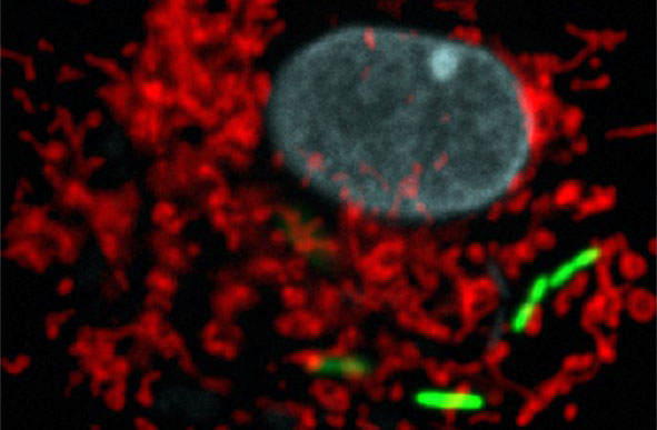
Legionella pneumophila (green), the bacterium responsible for severe acute lung disease inside eukaryotic cells. Mitochondrial network in red, nucleus in blue. © Institut Pasteur.

Scientists at the Institut Pasteur, CNRS and Inserm, together with a team from Switzerland*, have shown that the bacterial pathogen Legionella pneumophila (the causative agent of Legionnaires’ disease or legionellosis) has developed a specific strategy to target the host cell mitochondria, the organelles in charge of cellular bioenergetics. By changing the shape of these host organelles, L. pneumophila impairs mitochondrial respiration leading to metabolic changes in the host cell that are instrumental for the pathogens replication in human cells. This work provides precious information on how a pathogen manipulates the cellular metabolism to replicate intracellularly, and proposes a new concept of protection of host cells from Legionella-induced mitochondrial changes in order to fight infection. This research is published online on August 31, at the Cell Host & Microbe website.

Intracellular pathogens adopt various strategies to circumvent the defences of the host cell and to proliferate intracellularly. One specific strategy is to target host organelles like the mitochondria. Mitochondria are well-defined cytoplasmic organelles, which take part in a variety of cellular metabolic functions. Mitochondria are important as they supply the energy to the cell, thus they are also referred to as the ‘power house’ of the cell. Some bacteria, including Legionella pneumophila, are able to alter mitochondrial functions to the pathogens advantage.

L. pneumophila is a bacterial pathogen that causes Legionellosis – a disease characterized by an acute pulmonary infection, which is often fatal when not treated promptly. In France, between 1200 and 1500 cases are identified each year, with mortality rates ranging from 5 to 15%.

Researchers from the Institut Pasteur, CNRS and Inserm, in collaboration with a team from Switzerland, have discovered a previously unknown mechanism by which L. pneumophila targets mitochondria to modulate mitochondrial dynamics and thereby impairs mitochondrial respiration which in turn leads to a change in the cellular metabolism. These metabolic changes in the host cell favour bacterial replication. Thus the rewiring of cellular bioenergetics to create a replication permissive niche in host cells is a core virulence strategy of L. pneumophila.

The researchers have identified the following mechanism: L. pneumophila establishes transient, highly dynamic contacts with host mitochondria and secretes an enzyme called MitF that modifies the shape of the mitochondria by inducing DNM1L (a host protein that is necessary for fragmenting mitochondria) depended mitochondrial fragmentation. Surprisingly, L. pneumophila induced mitochondrial fragmentation is independent of cell death and ultimately impairs mitochondrial respiration, whereas cellular glycolysis is increased. Thus the bacterial-induced changes in mitochondrial dynamics promote a Warburg-like phenotype (which is characteristic of cancerous cells) in the infected cell that favours bacterial replication.

Researchers also brought the proof of concept that protecting host cells from Legionella-induced mitochondrial changes may help to fight infection. Indeed, pre-treating of human cells with a compound that inhibits changes in mitochondrial morphology allows protecting the host cell from Legionella-induced changes of mitochondria and restricts bacterial infection of human cells.

As Carmen Buchrieser, head of the Biology of the intracellular bacteria research unit at the Institut Pasteur and researcher at CNRS, explains: “this is an important discovery as our results showcase a key strategy used by L. pneumophilia for intracellular replication. By targeting mitochondria, the bacterium ensures that the host cell will be permissive to its replication. It is therefore essential that researchers also focus their studies on metabolic changes caused by pathogenic bacteria, in order to develop new therapeutic strategies against legionellosis and other diseases linked to intracellular bacteria.

This work sheds new light on how a pathogen shapes host metabolic responses during infection of human cells and shows that metabolic changes in the host cell are instrumental for the pathogens replication in human cells and thus to cause disease. It also proposes a new concept, which is to treat infections by inhibiting pathogen induced metabolic changes.


* The researchers and scientific teams involved are: Carmen Buchrieser, head of the Biology of the intracellular bacteria unit at the Institut Pasteur and associated to CNRS, Hubert Hilbi, of the University of Zürich Switzerland, Priscille Brodin, at the Institut Pasteur Lille and associated to CNRS and INSERM, and Jean-Christophe Olivo-Marin, head of the Bioimage analyses unit at the Institut Pasteur.

Follow-up at the age of 2: preterm children have higher survival rates and better health than 20 years ago

©Fotolia

It’s good news. Over the last 20 years there has been a marked improvement in the survival rate of preterm infants, and rates of brain damage at the age of 2 years have halved. These are the latest results of the EPIPAGE-2 study carried out by researchers from the Inserm group EPOPé – Obstetrical, Perinatal, and Pediatric Epidemiology Team at the Center for Research in Epidemiology and Statistics, Sorbonne Paris Cité (CRESS, Unit 1153), AP-HP. These results from follow-up of 5,000 preterm infants were published in the British Medical Journal.

EPIPAGE-2 is a French study that initially included over 5,500 children, born preterm between 22 (5 months) and 34 weeks (7 and a half months) of pregnancy, between April and December 2011. The researchers’ main objective was to achieve a better understanding of the factors associated with children born preterm, in particular their neuromotor and sensory outcomes, and overall development at 2 years. The EPIPAGE-2 results were compared to those collected in 1997 by a similar study carried out in 9 French regions.

The data were collected from doctors who followed up with these children and families.

  • At 2 years, 52% of children born between 22 and 26 weeks of pregnancy, 93% of those born between 27 and 31 weeks, and 99% of those born between 32 and 34 weeks survived.
  • Rates of cerebral palsy (a motor disability often associated with preterm birth) were 7%, 4%, and 1% in these same groups by birth.
  • Less than 1% of children in the cohort had a severe sensory deficit (blindness or deafness).
  • Development, as assessed by parental questionnaires, was at the level expected for their age groups among 50% of children born between 24-26 weeks, 59% of children born between 27-31 weeks, and 64% of those born between 32-34 weeks.
  • Between 1997 and 2011, the rate of cerebral palsy was halved among very preterm babies. The rate of survival without severe motor or sensory impairment has thus increased in all groups by birth, particularly for the most preterm. Before 7 months of pregnancy, it was 74.5% in 1997, and 80.5% in 2011.

“Such studies are needed to better understand the impact of changes in medical practice on outcomes in children, and to adapt the organization of health care based on population-level data,” explains Pierre Yves Ancel. “Parental questionnaires, for example, have allowed children’s development to be evaluated by their own families, and enabled early identification of those requiring further investigation. For us, the challenge is therefore identifying at a very early stage the children most at risk of developmental delay in the future. These questionnaires represent a promising option for providing structured follow-up of babies while focusing resources on those who really need it,” adds Véronique Pierrat, who led the study.

However, the rates of overall and above all disability-free survival have only marginally improved among children born before 5 months of pregnancy. After adjusting for baseline characteristics, rates of survival and rates of survival without severe or moderate neuromotor and sensory disabilities at 2 years appeared to have increased between 1997 and 2011 for children born between 22 and 31 weeks of gestation. However, no change was observed in children born at 24 weeks of gestation or earlier. These statistics are noticeably poorer than in countries where management of the delivery room is more active than in France. Since publication of the initial EPIPAGE-2 results, a working group has been set up in France to consider the management of these extremely preterm children. As in seven other European countries, the recommendation in France is not to provide medical intervention for children born before 24 weeks, rather to provide them with “comfort” care.


Preterm birth

Children born between 22 and 26 weeks of gestation are considered to be extremely preterm.

Children born between 27 and 31 weeks of gestation are considered to be very preterm

Children born between 31 and 34 weeks of gestation are considered to be moderately preterm

Intestinal Viruses Predict the Risk of Graft-Versus-Host Disease

©Fotolia

Teams from the virology, hematology-bone marrow transplant and biostatistics departments of the Saint-Louis Hospital, AP-HP, Université Paris Diderot and INSERM, in collaboration with scientists from the University of California-San Francisco (USA) discovered that a group of viruses in the intestine may trigger the onset of graft-versus-host disease. Researchers demonstrated the role of this intestinal “virome” (the population of viruses found in the intestine) in the onset of graft-versus-host disease by analyzing the intestinal virome of 44 patients receiving a bone marrow transplant.

These results appeared in a letter published in Nature Medicine.

Graft-versus-host disease (GVHD) affects up to 60% of patients who receive a stem cell graft (bone marrow transplant) with a high risk of death. Even though past studies have identified biomarkers associated with the severity of the disease, none of them have been used in practice to date in order to predict the onset of graft-versus-host disease.

The researchers carried out a longitudinal study analyzing the intestinal virome of 44 patients before a bone marrow transplant and six weeks after the transplant. By using high-throughput metagenomic techniques, they sequenced the DNA and RNA in the stool samples, in order to analyze and compare the flux over time in bacterial and viral populations.

The results show that GVHD is associated with significant changes in bacteria, bacteriophage (i.e. the viruses that infect bacteria) and virus populations in the stool.

In patients with intestinal GVHD, the persistent DNA viruses (Anelloviridae, Herpesviridae and Polyomaviridae) are absent from the intestinal tract in the first 2-3 weeks following the graft and then they increase in a significant manner. However, the presence of these viruses is stable as soon as the graft is performed in patients without GVHD.

In particular, the researchers detected the presence of a group of RNA viruses, picobirnavirus (PBV), in more than a third of patients after transplanting stem cells. As opposed to other viruses, the presence of PBV predicts the development of GVHD and is highly correlated to the severity biomarkers of intestinal graft-versus-host disease.

Metagenomic tools have made it possible to identify PBVs that have been little studied to date, since their extreme variability makes developing a test to detect them difficult. These viruses have been described in cases of intestinal gastroenteritis, notably in patients with AIDS, but their pathogenic role has not yet been determined. The potential utility of PBV as a predictive marker of GVHD has rekindled the interest in developing adapted to tools for clinical practice.

These results improve the state of knowledge of this complex disease, notably in terms of the role of viral infections in digestive inflammatory diseases and has opened the door to new therapeutic opportunities.

Testicular macrophages are guardians of fertility

Cross section of newborn mouse’s testis (Ø = 20 µm), where we can see the seminiferous tubules (red) surrounded by macrophages (green).  Confocal micrograph.

© Noushine Mossadegh-Keller and Sébastien Mailfert / CIML

The origin, development, and characteristics of two types of testicular macrophage have been described by a CNRS team at the Centre d’Immunologie de Marseille-Luminy (CNRS / INSERM / Aix-Marseille University). To elucidate the nature of these immune cells, the researchers used a novel cell tracing method. Their findings were published on August 7, 2017, in the Journal of Experimental Medicine, and are of fundamental importance. They may help understand certain kinds of infertility in men and find new treatments for them.


From the start of life, an individual’s immune system learns to distinguish self—that is, native cells—from other, potentially pathogenic cells. But in males, as sperm only appear at puberty, they may be mistaken for foreign cells by certain elements of the immune system.  Testicular macrophages are special immune cells that rush to the defense of sperm. By releasing specific molecules, these guardians of fertility prevent other immune system agents from entering the testes.


Macrophages not only migrate to sites of infection and phagocytose pathogens, but also modulate immune system activity to ensure proper organ function and regeneration. They may arise from either embryonic progenitors or bone marrow cells in adults. Research with mice has enabled the team of Michael Sieweke from the Centre d’Immunologie de Marseille-Luminy (CNRS / INSERM / Aix-Marseille University) to describe both testicular macrophage populations in depth.


The testis is divided into two compartments. One kind of testicular macrophage is found in the interstitial spaces, where testosterone-producing Leydig cells are also located. These interstitial macrophages are of embryonic origin: they are present from the beginning of the individual’s life. The other kind is peritubular—that is, located on the surface of the seminiferous tubules that house sperm cell precursors. Each macrophage population has distinctive cellular markers.


The researchers used a new cell tracing method to follow the movement of peritubular macrophages from the bone marrow to the testes. They discovered that these macrophages only appear two weeks after the mice are born, which corresponds to the pubescent stage in human males. Surprisingly, once they have been established in the testes, macrophages of both populations remain there for the rest of their long lives. Sieweke’s team will next focus their research efforts on the relationships between macrophages, sperm, and testosterone production, which may yield innovative treatments for certain kinds of male infertility.

Microdystrophin restores muscle strength in Duchenne muscular dystrophy

©fotolia

Researchers from Généthon, the AFM-Téléthon laboratory, Inserm (UMR 1089, Nantes) and the University of London (Royal Holloway) demonstrated the efficacy of an innovative gene therapy in the treatment of Duchenne muscular dystrophy. Indeed, after injecting microdystrophin (a “shortened” version of the dystrophin gene) via a drug vector, the researchers managed to restore muscle strength and stabilise the clinical symptoms in dogs naturally affected by Duchenne muscular dystrophy. A first. This work, published today in Nature Communications, has been achieved thanks to donations from the French Téléthon.

Duchenne muscular dystrophy is a rare progressive genetic disorder involving all the muscles of the body, and affects 1 in 5,000 boys. It is the most common neuromuscular disorder in children. It is associated with abnormalities in the DMD gene, which encodes dystrophin, a protein that is essential for proper muscle function. This gene is one of the largest in our genome (2.3 million base pairs, of which over 11,000 are coding). Because of this size, it is technically impossible to insert the entire DNA for dystrophin into a viral vector (or even the 11,000 coding base pairs alone), as is usually done for gene therapy.

To meet this challenge, teams at Genethon developed, in collaboration with a team at Royal Holloway University of London led by Pr. Dickson, and produced, a gene therapy drug combining an AAV-type viral vector with a shortened version of the dystrophin gene (approximately 4,000 base pairs), allowing the production of a functional protein. Dr Le Guiner’s team tested this innovative treatment in 12 dogs naturally affected by Duchenne muscular dystrophy.


By injecting this microdystrophin intravenously, and hence into the whole body of the dogs, the researchers observed that dystrophin expression returned to a high level, and muscle function was significantly restored, with stabilisation of the clinical symptoms observed for over 2 years following injection of the drug (see video). No immunosuppressive treatment was administered beforehand, and no side-effects were observed.

Some Golden Retrievers develop Duchenne muscular dystrophy naturally. The successful treatment of these dogs, which show the same clinical symptoms as children with this disease, and are of a similar weight, is a decisive step toward developing the same treatment in children.

“This preclinical study demonstrates the safety and efficacy of microdystrophin, and makes it possible to consider developing a clinical trial in patients. Indeed, this is the first time that it has been possible to treat the whole body of a large-sized animal with this protein. Moreover, this innovative approach allows treatment of all patients with Duchenne muscular dystrophy, regardless of the genetic mutation responsible,” says Caroline Le Guiner, the main author of this study.

“This is tremendously exciting progress towards a gene therapy for DMD. The studies in GRMD dogs have been spectacular and exceeded our expectations. My team has worked for many years to optimise a gene therapy medicine for DMD, and now the quite outstanding work of colleagues in France, in Genethon, in Nantes and in Paris has taken us close to clinical trials in DMD patients. I pay thanks also to the amazing and steadfast support of this research by AFM-Telethon and MDUK (Muscular Dystrophy UK) which has been essential to this achievement.” commented George Dickson.

For Frédéric Revah, Chief Executive Officer of Généthon: “For the first time, researchers obtained a systemic therapeutic effect on a neuromuscular disease in dogs using microdystrophin, and without immunosuppressive treatment. This highly complex cutting edge technology has been developed as part of an exceptional collaborative effort between Genethon and academic teams from Britain and France. Now our bioproduction experts have the task of producing a sufficient quantity of these new drug vectors, under GMP conditions, for the clinical trial.”
“This new evidence of the efficacy of gene therapy in Duchenne muscular dystrophy strengthens the therapeutic arsenal developed (exon skipping, CRISPR Cas-9, pharmacogenetics, etc.), and the first results are there. We need to forge ahead to complete the final phase and transform these scientific advances into drugs for children,” emphasises Serge Braun, Scientific Director of AFM-Téléthon.

Gene therapy: first results in children with Sanfilippo B syndrome

©Fotolia

On July 13, 2017, the journal Lancet Neurology published the results of a gene therapy trial conducted in four children with Sanfilippo type B syndrome (also known as MPS IIIB). This trial is the achievement of a two-decade partnership with financial support of AFM-Téléthon and the cooperation of the charity “Vaincre les Maladies Lysosomales” (VML). After monitoring of the treated children for 30 months, Dr. Jean-Michel Heard, from the Institut Pasteur and Inserm, and Professors Marc Tardieu and Michel Zérah, from the Paris public hospital administration (AP-HP) and the Paris-Sud and Paris Descartes Universities, conclude that the treatment was well tolerated and associated with neurocognitive benefits for the patients.

Sanfilippo syndrome is a rare genetic disease which affects approximately one in every 100,000 children. It alters brain development after birth and leads to brain degeneration several years later. The first symptoms of the disease are hyperactivity and delayed cognitive acquisition, which are usually noticed when children are around two-years old. A genetic anomaly prevents the production of an enzyme needed to breaking down mucopolysaccharides. Mucopolysaccharides are large macromolecules that help the neurons develop effective connections in young children during learning. Incomplete degradation and accumulation are toxic for brain cells. The disease progressively leads to a state of severe and multiple impairments and to premature death within periods of 5 to 10 years.

The challenge to treat Sanfilippo syndrome lies in the design of a method to supply the missing enzyme to the brain as early as possible after birth.  The therapeutic trial conducted by the Institut Pasteur at the Bicêtre Hospital (AP-HP) used gene therapy for that purpose. A gene therapy vector (AAV2/5) capable of inducing the production of the missing enzyme by brain cells was injected at several sites in the brain and cerebellum of affected children. The specific aim of this phase I/II trial was the assessment of tolerance to the surgical procedure and to the candidate drug delivered by gene therapy.

The clinical study initiated in 2013 was preceded by more than ten years of preclinical studies in animals naturally affected by the disease. The researchers administered the treatment for the first time to four children aged between 1.5 and 4 years (20, 26, 30, and 53 months). No particular clinical, radiological or biological side effects associated with the treatment were observed within 30 months of administration, indicating that it was well tolerated.

Within one month of treatment and throughout the 30 months of the trial, researchers detected the previously missing enzyme in the cerebrospinal fluid of the four treated children. Moreover, very careful and regular neurocognitive monitoring revealed positive impact on cognitive acquisition and behavioral development, which were more pronounced in the youngest treated child.

The encouraging findings of this phase I/II clinical trial suggest that treatment could be proposed to patients with Sanfilippo syndrome in the future.  To reach that goal, the next step would consist in a large and multicentric phase III clinical trial, if an appropriate partner is found.

9th IAS Conference on HIV

© Fotolia

From July 23 to 26, 2017, the Ninth IAS Conference on HIV Science, of which Inserm is a partner, will be held at the Palais des Congrès in Paris. For this edition, the International AIDS Society (IAS) has teamed up with ANRS, the autonomous agency of Inserm, to coordinate and fund research on HIV/AIDS and hepatitis.

The program will cover innovative approaches to HIV prevention, treatment and care. Various speakers, including Inserm researchers, will highlight the various advances in vaccines, new therapeutic approaches and prevention of HIV and hepatitis.

Yves Lévy, Inserm Chairman and CEO, will speak at the inaugural conference on Monday, July 24 on “Exploiting the immune system to prevent and control HIV infection”.

The scientific results of several Inserm researchers, including Dominique Costagliola, Patrizia Carrieri, Guillemette Antoni and Marie Jauffret-Roustide, will be presented at this conference.

Access the program for IAS 2017

Hereditary hearing loss: the ear and auditory brain are both affected

Brain slice showing neurons migrating to the auditory cortex. The nuclei are shown in blue. In green, molecules from the “molecular code” that sends the neurons to this brain region.

©Institut Pasteur

Scientists from the Institut Pasteur, Inserm, the Collège de France and Pierre & Marie Curie University have recently demonstrated that mutations in three genes responsible for Usher syndrome – a hereditary condition that affects both hearing and sight – influence not only the workings of the ear, specifically the function of sensory cells in the cochlea, but also the development of the auditory cortex. Their discovery could explain why some patients, even after being fitted with a cochlear implant (an electro-acoustic device that bypasses the defective cochlea), still have difficulties understanding speech. The findings are reported this week in a paper in the Proceedings of the National Academy of Sciences of the USA.

In most cases of hereditary hearing loss in humans, damage to the auditory sensory organ, the cochlea, is sufficient to account for patients’ hearing impairment. Many forms of hereditary hearing loss affect the hair bundle that acts as the sound receptor for auditory sensory cells. Cochlear implants stimulate the auditory nerve directly, bypassing the need for the sound signal to be processed by the cochlea and restoring a good level of hearing. In some cases, however, patients still have difficulties in understanding speech.

The team led by Prof. Christine Petit[1] from the Genetics & Physiology of Hearing Unit (Institut Pasteur/Inserm/UPMC) – with research by Baptiste Libé-Philippot supervised by group leader Dr. Nicolas Michalski –, working in cooperation with Dr. Christine Métin (Inserm/UPMC), recently identified three genes in mice which, when damaged, affect not only the cochlea but also the auditory cortex, the brain region responsible for analyzing auditory information. These three genes are among the nine currently recognized as causing Usher syndrome (type I and II), the leading hereditary cause of hearing and sight loss. Since damage to the cochlea in these patients prevents the auditory brain from receiving some or all of the acoustic information it normally receives, the cerebral damage had previously gone unnoticed.

The scientists demonstrated that, during embryogenesis, the proteins coded by these three genes are involved in the migration and maturation of some cells destined to become “inhibitory” neurons, which specifically colonize the auditory cortex. These neurons produce parvalbumin and are closely involved in cortical plasticity, which determines the structure and function of neural networks in the cortex based on auditory experience. They also play an important role in the temporal precision of sound detection that is required to understand speech. In mice, a single mutation in one of these three genes prevented this population of neural precursors from entering the developing cortex and reaching the auditory cortex.

The scientists also revealed that these neurons synthesize molecules that act as molecular markers. “These molecules serve as labels that instruct neurons to be sent from their birthplace in the subpallium, in the center of the brain, to the cortical area, their final destination. For the first time, we suggest that inhibitory neuron precursors have a “molecular mailing code”, explains Prof. Christine Petit.

These findings therefore demonstrate that hearing loss genes, previously thought to influence just the cochlea, also have another independent role in the development and formation of neural networks in the auditory cortex. In embryogenesis, this role is performed prior to the one played by the same genes in the cochlea.

Prof. Christine Petit’s team is at the forefront of pioneering research into hereditary hearing loss. It previously demonstrated that the proteins coded by the genes that cause type I and type II Usher syndrome are also responsible for the development and workings of the hair bundle, and went on to unravel the related molecular networks. The scientists hope to use these new findings to develop innovative aural rehabilitation methods designed specifically for patients with damage to the auditory cortex.

This research was funded by the French National Research Agency (LIGHT4DEAF [ANR-15-RHUS-0001] and LIFESENSES LabEx [ANR-10-LABX-65]), the European Commission (ERC-2011-ADG_294570), the BNP Paribas Foundation, the FAUN Stiftung (Suchert Foundation) and the LHW-Stiftung (CP), and the “scientific discovery” award and SEIZEAR grant from the “Agir Pour l’Audition” foundation (NM).

[1] Prof. Christine Petit is a Professor at the Collège de France and at the Institut Pasteur.

Vision restoration by optogenetic therapy within easy reach?

©fotolia

The Vision Institute (Inserm, Université Pierre et Marie Curie (UPMC), National Center for Scientific Research (CNRS)) via the Fondation Voir & Entendre has signed a contract with the United States Defense Advanced Research Projects Agency (DARPA), which could ultimately represent $ 25 million. With the help of an international consortium, researchers from Inserm, CNRS and UPMC, working within the Vision Institute, want to develop a system capable of restoring vision by optogenetic stimulation of the visual cortex. This project is called CorticalSight.

The consortium is coordinated by Professor José-Alain Sahel (Vision Institute and University of Pittsburgh School of Medicine). It is composed of academic partners: Stanford University, Friedrich Miescher Institute for Biomedical Research, the French Alternative Energies and Atomic Energy Commission – Leti and companies GenSight Biologics, Chronocam and Inscopix. Serge Picaud, Inserm research director, will coordinate the research activities at the Vision Institute.

The retinal ganglion cells are neurons which integrate the visual information of the environment in the eye’s photoreceptors and transmit it to the higher visual centers. Impairment of these cells deprives the centers of any visual information coming from the outside, thus causing complete blindness.

The degeneration of retinal ganglion cells is one of the leading causes of blindness in the Western world. It can be the result of various pathological conditions, including ocular trauma, retinal disorders such as glaucoma, diabetic retinopathy or optic neuropathies.

In animals, the restoration of sight after photoreceptor degeneration works thanks to the development of a very recent technique: optogenetic therapy. By this method it becomes possible to optically take control over the activity of very precise areas of the brain to induce behavior in the animal. In this specific case, the visual areas would be directly activated to induce visual perception even though the photoreceptors have not been activated. This first step in animals paves the way for the transfer of this technology to humans.

The CorticalSight project, financed by this contract, thus aims to restore visual perception in people who have become blind, by acting directly on the higher centers of the brain. To do this the researchers will use an intelligent image capture device combined with optogenetic stimulation.

In detail, the system as a whole will consist of several devices operating in series. On the face, a first device attached to glasses will consist of a camera filming the live environment of the patient in high resolution. A second device in the brain will transform the visual information, through complex algorithms, into light signals that the brain can interpret.

And this is where otogenetics comes into play. Using this technique, neurons specific to the visual cortex will be made sensitive to light by the expression within them of a microbial opsin (this algae protein transforms light energy into electrical activity).

It is then sufficient to couple the two external and internal devices so that the light signals coming from the outside are transformed into optical stimulation capable of activating the neurons of the visual cortex.

The human brain then does the rest of the work, as it knows how to, by translating the visual perception into a mental image representing the environment: a face, a tree, etc.

The Consortium

The CorticalSight project is coordinated by the Vision Institute (Inserm/CNRS/UPMC) and brings together international researchers in the field of vision whose individual expertise will be needed at each stage of scientific development.

fermer