Menu

Regenerating Lost Neurons: A Successful Bet for Research

neurones

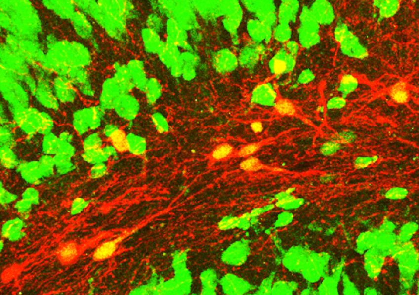
Confocal microscopy image showing induced neurons (red with a yellow nucleus) expressing the NeuN neuronal marker (green) within an epileptic mouse hippocampus. © Extract from: Lentini et al., Cell Stem Cell, 2021.


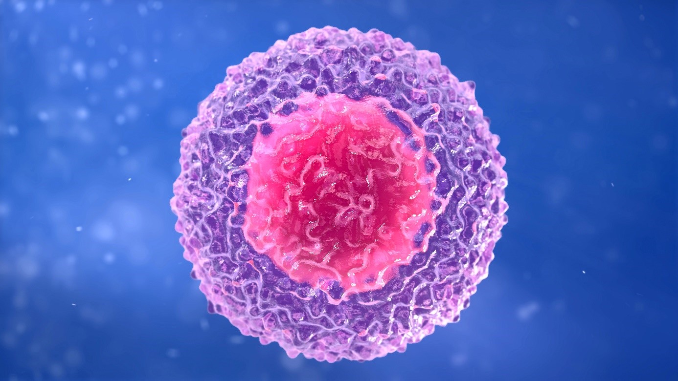
Many central nervous system diseases are associated with the death of neurons without the brain being able to regenerate them. A phenomenon that is observed particularly in Parkinson’s disease, Alzheimer’s disease, following stroke, and in some forms of epilepsy. How can lost neurons be regenerated? This question has been tackled by a team of researchers from Inserm, CNRS and Université Claude Bernard Lyon 1 at the Stem Cell and Brain Research Institute, in collaboration with King’s College London. Using an animal model of epilepsy, the researchers have succeeded in transforming non-neuronal cells in the brain into new inhibitory neurons that reduce chronic epileptic activity by half. This research will in time make it possible to envisage a therapeutic application of this strategy. The findings of this study have been published in Cell Stem Cell.

Our brain generally lacks the regenerative capacity to replace lost or damaged neurons. The goal of regenerative medicine is to replace lost cells in order to correct the functional disorders associated with that loss. Direct cell reprogramming (as opposed to induced pluripotent stem cell reprogramming) has emerged as an innovative strategy that consists of “reprogramming” the identity of certain non-neuronal cells present within the affected brain to transform them into neurons. If this strategy is to be effective, many challenges need to be addressed. The new neurons must be integrated into the networks of surviving neurons and take over from those they replace in order to correct the pathological disorders.

This was the strategy explored in a new study published in the journal Cell Stem Cell. A team of researchers from Inserm, CNRS and Université de Lyon have succeeded in transforming glial cells of the brain into new neurons in a mouse model with mesial temporal lobe epilepsy, the most common form of drug-resistant epilepsy in humans.


Proliferation of glial cells: a cell source from which to generate neurons

In neuronal death, as observed in mesial temporal lobe epilepsy, the most common form of adult focal epilepsy, the glial cells present in the direct environment of the damaged neurons react by multiplying themselves, albeit without resolving the problem.

In the study, the researchers had the idea of taking advantage of this proliferation and using these extra glial cells. First, they had to identify genes making it possible to transform these glial cells into inhibitory neurons, whose loss plays a key role in the onset of seizures, in order to restore the balance of the neuronal activities that had been affected. The researchers therefore selected genes known for being involved in the genesis of these inhibitory neurons during development.

By forcing the expression of these genes, they were able to reprogram the identity of the glial cells to make them so-called “induced neurons” whose properties are comparable to those lost in the disease. Through stereotactic surgery[1], the genes were inserted directly into the brains of the mice at the sites of origin of the epilepsy using deactivated viral vectors that induce reprogramming of the glial cells. Within a few weeks, the vast majority of these gene-treated glial cells had transformed into new neurons.


Functional neurons integrated into the epileptic network

The results of the study show that the induced neurons adopt an identity of inhibitory neurons that present a set of molecular characteristics comparable to those of neurons having degenerated in epilepsy.

Using electrophysiological recordings, the scientists were able to confirm that they were indeed functional neurons, capable of inhibiting the neighboring neurons responsible for seizures, thereby reducing their activity. Then, by tracing connections between the neurons, they were able to determine that the induced neurons were fully integrated into the epileptic network but also more broadly in the brain.

Finally, thanks to electroencephalographic (EEG) recordings in the sites of origin of the seizures, the researchers were able to show, in the reprogrammed mice, that the seizures had reduced by half.

“These findings reveal the therapeutic potential of this cell reprogramming strategy in fighting a pathology such as mesial temporal lobe epilepsy. This represents a blessing in the specific case of this disease at a time when 30% of patients are refractory to pharmacological treatments,” explains Christophe Heinrich, the study’s designer.

Even if much remains to be done before this research can truly be applied to the treatment of patients, this study highlights the reprogramming of glial cells into neurons as a new strategy capable of modifying not just a disease such as epilepsy, but which also could be expanded to include other devastating brain diseases.


[1] Neurosurgery technique that uses a system of 3D coordinates in space for the precision-access of brain regions. 

Inserm Publishes Book on Fake News Making Rigorous and Credible Health Information Accessible to All

chocolat et santé

Is dark chocolate better for you than milk chocolate? Inserm tackles fake news and preconceived ideas in its new book. © JP Courbatze


As the effects of the COVID-19 crisis continue to make themselves felt, how do we answer the many health questions raised by citizens and make space for scientific information that is useful in everyday life? More motivated than ever to provide robust scientific information to all, Inserm has joined forces with publisher Le Cherche-Midi to deconstruct fake news and preconceived ideas regarding our health.

They say that fasting is effective against cancer, that IUDs make you sterile, that HIV is transmitted by mosquito bites… While health misinformation is nothing new, the growing number of communication channels and the rise of social media have given it space to flourish.

We have all seen that the health crisis has been the focus of violent controversy, showcasing individual voices sometimes to the detriment of collective scientific discourse.

This is an issue that Inserm has been fighting since 2018, when it began to publish its series of Canal Détox videos. The aim of these educational scientific videos is to establish reliable scientific information on health topics that are of concern to citizens. With COVID-19, the initiative now includes the publication of informative texts in rapid response to misinformation that has gone viral, continuing to draw on the rigorous and measured scientific discourse of Inserm directors and researchers.

In order to intensify efforts to open up science to as many people as possible, Inserm has joined forces with publisher Le Cherche-Midi to roll out its Canal Détox label in book form. Taking an educational and thematic approach, the collection dissects nearly 80 pieces of health misinformation. COVID (of course!), as well as diet, cancer, mental health, HIV… It uses the latest scientific data and the expertise of Inserm researchers to shatter many preconceived ideas.

“At a time when the health crisis has revealed the need for every citizen to be informed and be able to make informed decisions to protect their health, Inserm’s ‘Science for Health’ motto has never been more relevant. This book illustrates our desire to continue to make Inserm a reference for dialogue with society and make biomedical science accessible to as many people as possible,” emphasizes Dr. Gilles Bloch, CEO of Inserm.

Available in French bookstores from September 30 and written for a general readership, this book maintains the modern, everyday angle and tone of the Canal Détox videos and addresses the most serious and complex scientific issues. In addition to providing responses to the readers’ health questions, its intention is also to share with them the methods of the rigorous, ethical and transparent scientific approach at work in Inserm’s 300 laboratories.

Covid-19 : identification de cellules immunitaires associées à la survie dans les formes sévères de la maladie

lymphocyte immunité

The cellular immune response is based on the T cells recognizing cells that have been infected with the virus. © Adobe stock


A better understanding of the precise mechanisms of immune response to SARS-CoV-2 is essential if we are to improve patient care and continue to develop effective new vaccines. Since the pandemic began, many studies have tried to explain what differentiates the immune response of people with severe forms of COVID-19 resulting in death from that of other patients. In a new study, researchers from Inserm and Sorbonne Université at the Center for Immunology and Infectious Diseases, in collaboration with physician researchers from Paris hospitals group AP-HP, focused on a type of immune response known as the cellular immune response. They show that certain cells involved in this response, CD8+ T cells, are present in smaller quantities in patients who have died from COVID-19. Some of them are considered to constitute predictive markers of survival. This research has been published in JCI Insight.

Since the COVID-19 pandemic began, the immune response triggered following SARS-CoV-2 infection has been the subject of much research. At a time when the Delta variant has spread across the world, understanding the precise mechanisms of the adaptive 1 immune response to the virus appears essential if we are to continue to develop effective vaccines that are less sensitive to the emergence of new variants.

In the majority of cases, infection with SARS-CoV-2 only results in relatively mild symptoms (or even forms that are asymptomatic). However, in some cases, serious forms develop, associated with severe, life-threatening symptoms that sometimes require intensive care.

Numerous publications have already shown that different profiles of immune responses are associated with the severity of symptoms. However, we still do not have enough data to understand the role of the humoral and cellular responses that constitute the adaptive immune response (see box), as well as their involvement in the survival or death of patients with the severest symptoms.

In their study, the scientists from Inserm, Sorbonne Université and Paris hospitals group AP-HP looked at the immune response profiles of 56 patients hospitalized in intensive care, a third of whom died from COVID-19, paying particular attention to the cellular responses, mediated by T cells.

Different types of immune response

There are two broad categories of adaptive immune response: humoral responses, based on the production of antibodies that recognize the virus and can prevent it from infecting its target cells; and cellular responses, based on the T cells recognizing cells that have been infected with the virus. These T cells, through various mechanisms, contribute to the destruction of cells infected with the virus in order to control the viral infection and help eliminate the virus from the body. For this study, the researchers focused on the cellular response.

T cells are a population of heterogeneous cells that can be divided into different subpopulations according to their phenotype (the molecules they express on their surface) and their function (destruction of infected cells, production of molecular messengers, support and activation of other cells…). There are two main types of T cells: CD4+ cells that support the production of antibodies and CD8+ cells, specialized in the destruction of infected cells through the production of cytotoxic molecules.

Numerous publications over the past decades have shown the essential role of cellular responses, in particular that of “cytotoxic effector CD8 cells” (CD8+) in the context of viral infections such as influenza in the elderly or HIV.

Markers of survival

In the study, the team saw significant differences in the amounts of certain T cell subpopulations, between those individuals who survived and those who died from COVID-19.

First, the researchers saw a marked loss of CD8+ T cells capable of recognizing the SARS-CoV-2 nucleocapsid in deceased individuals, compared to individuals who survived the infection. Nucleocapsid is an internal molecule of the virus, highly immunogenic (i.e. capable of inducing an immune response) and well preserved from one SARS-CoV-2 variant to another.

In addition, the individuals who died from COVID-19 had a very low proportion of CD8+ T cells expressing two molecules on their surface (PDL1 and CXCR3), compared to those who survived. Having CD8+ T cells that present PDL1 and CXCR3 could therefore be a molecular signature predictive of survival.

“We have shown that the simple combination of the level of nucleocapsid-specific CD8 T cells and the overall level of CD8+ T cells expressing CXCR3 and PDL1 could predict the survival or death of critical COVID-

19 patients with over 90% accuracy. Both factors are significantly important given the other factors that are potentially important when it comes to severity, such as age and obesity, and more accurately predict the risk of death in patients with severe forms”, emphasizes Inserm researcher Béhazine Combadière, who coordinated the study.

These findings also have important implications for the development of future vaccines. Vaccines capable of targeting nucleocapsid – a highly preserved protein common to the coronaviruses – and of producing a high level of CD8+ T cells specific against this molecule could be useful in preventive vaccination and probably also in therapeutic vaccination in order to promote the survival of patients with severe forms, regardless of which variant they were infected with.

For the team, the next step is to work with people who have already been vaccinated, to see if they produce the immune cells that were predictive of survival in the patients studied in this research.


1 Innate immunity is an immediate response that occurs in any individual in the absence of prior immunization. It is the first barrier of defense against various pathogens and mainly brings into play pre-formed (natural) antibodies and lymphocytes that do not present receptors specific to the antigen. Adaptive immunity is established a few days after contact with the pathogen and is the body’s second line of defense. Unlike innate immunity, adaptive immunity is specific for a given antigen.

Absence d’efficacité du remdesivir chez les patients hospitalisés atteints de Covid-19 et poursuite de l’essai DisCoVeRy pour tester un nouveau médicament antiviral

SARS-CoV-2

Electron microscopy of a cell infected with SARS-CoV-2 © Philippe Roingeard, Anne Bull-Maurer, Sonia Georgeault, Inserm Unit U1259 MAVIVH & University of Tours, France


The Discovery clinical trial was initially launched in France by Inserm in March 2020, to evaluate several possible treatments for COVID-19. Its European expansion was made possible by the EU-RESPONSE[1] project, funded by the European Commission. Interim analysis of the trial data had led to the recommendation to suspend the recruitment of remdesivir group patients for futility – that is to say due to the very low likelihood of this treatment showing a benefit, even if inclusions were continued. In an article published this week in The Lancet Infectious Diseases, the final scientific analysis shows no improvement in patients hospitalized with COVID-19 presenting respiratory symptoms requiring oxygen and treated with remdesivir.

Data from 832 patients hospitalized between March 2020 and January 2021, recruited in 5 European countries (418 patients receiving standard of care and 414 additionally receiving remdesivir), were analyzed. The analysis has shown no difference between the two groups in patient clinical status 15 and 29 days after receipt of the first remdesivir dose, in time to discharge from hospital, and in death rate on Day 28. There was also no demonstrated difference between the groups in terms of speed of elimination of the virus at nasopharyngeal level. Severe side effects were distributed similarly between the two groups. These data support those of the Solidarity trial conducted by the WHO, in particular by providing results on a larger number of endpoints.

In order to continue to analyze the efficacy of the treatments evaluated on a larger number of patients, the data collected during Discovery are being used to perform meta-analyses. The data used to analyze the efficacy of remdesivir are therefore being shared, within the framework of the EU-RESPONSE project, with those of other major international studies, to clarify the results on a larger scale.

“18 months after the launch of Discovery, it can be concluded that 4 different molecules offer no therapeutic benefit in patients hospitalized for COVID-19. This huge undertaking has made it possible to further knowledge on sound scientific foundations. Although, like everyone else, we would have preferred to prove the efficacy of a treatment, we are continuing our research with an approach that specifically targets the virus,” explains Florence Ader, principal investigator of the trial.

With the epidemic still ongoing, it is essential to pursue research efforts to find a treatment for COVID-19 that is effective against the virus and its new variants. The DisCoVeRy trial is thus continuing in 80 hospitals across 14 European countries to assess the efficacy of a combination of two monoclonal antibodies targeting SARS-CoV-2 and currently effective against its variants.

This treatment, developed by AstraZeneca, has appeared effective in a recent preventive treatment trial in which it reduced by 77% the risk of developing symptomatic COVID, thereby reducing hospitalization and potentially harmful outcomes. In addition, another trial has shown decreased mortality in patients hospitalized with COVID-19 who did not develop natural antibodies and who received treatment with monoclonal antibodies.2

Monoclonal antibody treatments are therefore the first antivirals to show efficacy in the prevention and treatment of COVID-19 in non-immunized patients.

Evaluating the efficacy of these monoclonal antibodies in hospitalized patients is therefore important when it comes to identifying curative treatments that reduce mortality and the number of severe forms of the disease.

In addition, within the framework of EU-RESPONSE – now the European platform for clinical trials in response to emerging infectious diseases – the SolidACT trial has started in three European countries with the objective of evaluating baricitinib in patients hospitalized for severe forms of COVID-19.


[1] https://cordis.europa.eu/project/id/101015736

Funded by Europe (European Union Horizon 2020 programme for research and innovation), DisCoVeRy is now the research axis 1 of the EU-RESPONSE project (European Reasearch and Preparedness Network for Pandemics and Emerging Infectious Diseases), which brings together 21 partners (clinics, hospitals, universities…) from 13 countries of the European Union, Norway, Switzerland and Turkey.

Covid-19 : les confinements ont eu un impact délétère sur la sévérité du cancer colorectal métastatique

COVID-19: Discovery of a Molecular Signature of Pediatric Myocarditis

enfant masqué

© Adobe Stock


In very rare cases, children having contracted COVID-19 go on to develop severe inflammation 4 to 6 weeks after infection with SARS-CoV-2. In two-thirds of them, this inflammatory syndrome affects the heart, leading to myocarditis. In a study published in the journal MED, researchers, doctors and teacher-researchers from Inserm, the Paris hospitals group AP-HP and Université de Paris at the Imagine Institute, in collaboration with Institut Pasteur, analyzed blood samples from a cohort of 56 pediatric patients admitted to Hôpital Necker Enfants-Malades AP-HP. What they saw was the abnormal expression of several genes associated with the development of severe forms of myocarditis. A molecular signature that could ultimately help identify those children at risk of developing this rare cardiac inflammation.

Certain children with SARS-CoV-2 develop severe inflammation four to six weeks after infection, with varying symptoms: fever, stomach pain, skin rashes, etc. In around 70% of cases, this so-called “multisystem” inflammatory syndrome affects the myocardium, the muscle responsible for heart contractions. These severe cases of myocarditis were first reported in the UK, in March 2020, before being observed in Italy, France, and then all over the world. What is the explanation behind these rare forms?


Cutting-edge analyses

In a study published in MED – a journal from Cell Press –, conducted by Inserm researchers Frédéric Rieux-Laucat and Mickaël Ménager (*) working in two laboratories at the Imagine Institute (Inserm, Université de Paris, Paris hospitals group AP-HP), in collaboration with doctors from Hôpital Necker-Enfants Malades AP-HP and Institut Pasteur, cutting-edge molecular investigations were conducted in order to find out more. The result of these investigations was the identification of several genes linked to the development of severe forms of myocarditis in these children.

As part of the study, the authors analyzed blood samples from 56 children hospitalized between April 6 and May 30, 2020. A total of 30 had developed multisystem inflammatory syndrome following SARS-CoV-2 infection, 21 of whom with a severe form of myocarditis, and 9 without. “To understand the difference between these two patient groups, we did several analyses using state-of-the-art techniques:  an ultra-sensitive assay of cytokines – the immune system hormones that enable an appropriate response in case of infection –, characterization of blood cell composition, and a cell-by-cell analysis of gene expression“, explains Ménager.


Three molecular abnormalities

In both groups, the researchers found reduced numbers of monocytes and dendritic cells (white blood cells), increased inflammatory cytokine levels, and an overactivation of the so-called “NF-kB pathway” within these cells.

“NF-kB is a molecular pathway that enables a set of genes to be activated, resulting in the production of proteins tasked with orchestrating the immune response,” summarizes Rieux-Laucat. However, it is precisely the overactivation of this system that triggers hyperinflammation in these patients.

Following closer comparison of the dendritic cells and monocytes of the two groups, the authors observed three specific anomalies in patients with myocarditis: lack of inhibition in the NF-kB pathway, overproduction of “TNF-α” (cytokine involved in NF-kB pathway activation), and finally a lack of response to type I and II interferons (cytokines involved in regulating inflammation).

All of these abnormalities can be explained by the abnormal expression of certain genes. In order to identify these genes, the authors carried out a cell-by-cell genetic analysis.

“Like this we were able to identify and validate over one hundred genes overexpressed specifically in the monocytes and dendritic cells of patients with severe forms of myocarditis, explains Ménager. A molecular signature that could ultimately enable the development of tests to identify patients at risk of developing this severe cardiac inflammation.


(*) Frédéric Rieux-Laucat heads up the Immunogenetics of Pediatric Autoimmune Diseases laboratory. Mickaël Ménager heads up the Inflammatory Responses and Transcriptomic Networks in Diseases laboratory and the LabTech Single-Cell@Imagine, a platform dedicated to the cell-by-cell study of gene expression.

Demonstration of the major role of mutations in the PIK3CA gene in sporadic cavernomas

Brain scan, X-ray

Brain scan, X-ray© Adobe Stock


Teams from Inserm, CNRS, AP-HP and Sorbonne University, grouped together within the Brain Institute at Pitié-Salpêtrière AP-HP hospital and coordinated by Dr Matthieu Peyre and Prof. Michel Kalamarides, studied the presence of mutations in the PIK3CA genes in cavernomas. This work was published on September 09, 2021 in the New England Journal of Medicine .

Cavernomas are low-flow cerebrovascular malformations that consist of abnormally enlarged capillary cavities with no visible brain parenchyma between the dilated vascular cavities; this condition affects 1 in 200 to 250 people. Although it is characterized mainly by bleeding visible on MRI but not causing any clinical symptoms, cavernomas can lead to seizures and hemorrhagic strokes with significant neurological complications, especially when localized in the brainstem.

Cavernomas can occur in isolation or as part of a familial genetic disease. Mutations occurring in a family context concern the CCM genes in 80% of cases . The genetics of sporadic cavernomas, which represent up to 90% of cases, are however poorly understood.

In order to study meningeal tumorigenesis and meningiomas (the most common tumor of the central nervous system of which they are experts), Dr Peyre and Pr Kalamarides have generated two new genetically modified murine models of meningiomas by activating mutation of PIK3CA and AKT1 genes in the PI3K-AKT-mTOR pathway.

The unexpected observation of typical cavernomas identical to human lesions prompted them to investigate the possible involvement of PIK3CA and AKT1 mutations in sporadic human cavernomas. They identified 39% mutations in the PIK3CA gene in a series of 88 sporadic cavernomas. Moreover, their results shed new light on the cell of potential origin of the cerebral cavernous malformations which was until now considered to be of endothelial lineage. They have in fact shown that it is in fact the PGDS-positive pericytes which in their models are at the origin of cavernomas by disorganization of the neurovascular unit.

Their results may provide a better understanding of the biology of sporadic cavernous cerebral malformations by highlighting the major role of PIK3CA mutations in them, rather than that of CCM genes , initially considered to be predominant.

This result, which was corroborated by a preclinical model, opens up new perspectives, yet to be validated, for the development of targeted therapies for the treatment of sporadic human PIK3CA mutated cavernomas which are refractory to surgery and radiotherapy or radiosurgery. and lead to frequent complications. PIK3CA inhibitors have indeed shown promising results in patients with CLOVES syndrome (PIK3CA overgrowth syndrome) as well as in patients with a wide range of tumors.

fermer