Menu

COVID-19 Causes 3 Times More Deaths Than Seasonal Flu

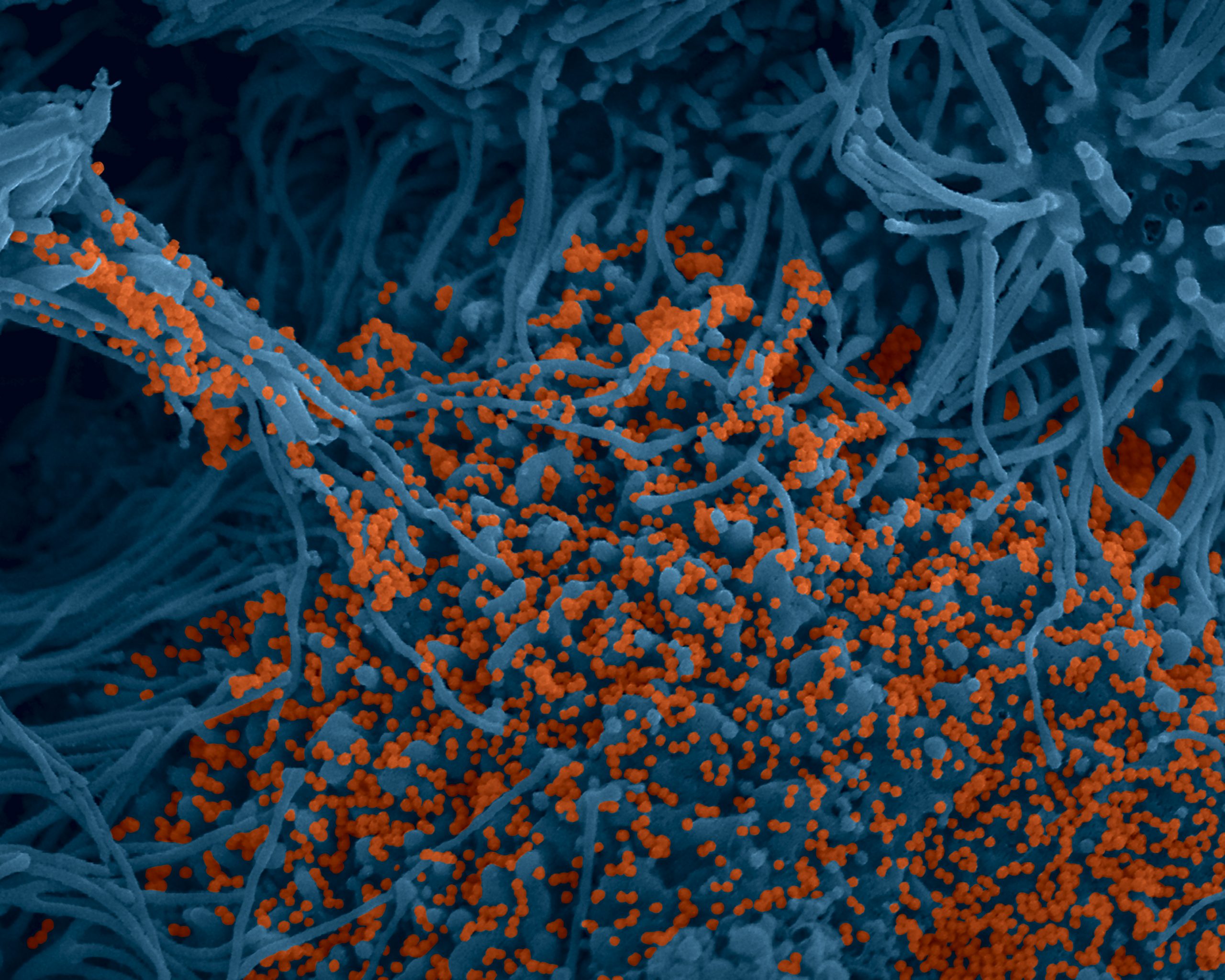
SARS-CoV-2 coronavirus attached to the cilia of human respiratory epithelial cells.© Manuel Rosa-Calatrava, INSERM ; Olivier Terrier, CNRS ; Andrés Pizzorno, Signia Therapeutics ; Elisabeth Errazuriz-Cerda UCBL1 CIQLE. VirPath (Centre International de Recherche en Infectiologie U1111 Inserm – UMR 5308 CNRS – ENS Lyon – UCBL1). Colorized par Noa Rosa C.


A study by Inserm and Dijon University Hospital based on French nationwide data on around 130,000 patients hospitalized for either COVID-19 or seasonal influenza shows that the mortality rate among those admitted for COVID is three times higher than that of seasonal influenza. These findings have been published in The Lancet Respiratory Medicine

This study uses data from the French national administrative database (medicalized information system program – PMSI), which contains information on all patients admitted to public and private hospitals in France, such as the reasons for their admission and the treatment they received. The researchers compared the admissions for COVID-19 (between March 1 and April 30, 2020) with those for seasonal influenza (between December 1, 2018 and February 28, 2019).


The results reveal:


  • A mortality rate among the patients hospitalized for COVID-19 three times higher than that of those admitted for seasonal influenza. Of the 89,530 patients admitted for COVID, 15,104 [16.9%] died versus 2,640 [5.8%] of the 45,819 patients hospitalized for influenza.


  • More COVID-19 patients required admission to intensive care with an average stay that was almost twice as long (15 days versus 8 days).


  • Fewer children under 18 years of age were hospitalized for COVID-19 than for seasonal influenza, but a larger proportion of the patients younger than 5 years needed intensive care support for COVID (14 out of 613) than for influenza (65 out of 6973). The mortality rate for the children under 5 years of age was similar for both groups and very low (less than 0.5%).


  • Almost twice as many people were hospitalized for COVID-19 at the peak of the pandemic compared to those who had been hospitalized for influenza at the peak of its 2018/2019 season.


  • In addition, a larger proportion of the COVID-19 patients had a severe form of the illness requiring intensive care than those with influenza. Looking at the number of admissions to intensive care: out of the 89,530 COVID-19 patients, 14,585 [16.3%] were admitted to intensive care versus 4,926 [10.8%] of the 45,819 influenza patients.


  • More than one in four COVID-19 patients suffered from acute respiratory failure, versus fewer than one in five with influenza.


  • Consistent with previous reports, the most common underlying medical conditions among the patients admitted with COVID-19 were hypertension (33.1%), overweight or obesity (11.3%), and diabetes (19.0%).

The researchers point out that their study has several limitations. In particular, testing practices for influenza are likely to be highly variable across hospitals, whereas practices for COVID-19 may be more standardized. This may partly explain the increased number of admissions for COVID-19 compared to seasonal influenza. In addition, the difference in hospitalization rates may be partly due to existing immunity to influenza in the population, either from previous infection or vaccination.

Nevertheless, the conclusions of this research confirm the importance of measures to prevent the spread of both diseases. Measures that are particularly relevant at a time when several countries are preparing for the COVID-19 pandemic to continue in parallel with seasonal influenza outbreaks during the winter months.

“This study is the largest to date comparing the two diseases and confirms that COVID-19 is much more serious than influenza. The finding that the COVID-19 death rate was three times higher than that of seasonal influenza is particularly striking when one recalls that in the last five years the 2018/2019 influenza season has been the worst in France in terms of number of deaths,” declares Catherine Quantin, researcher at Inserm and Professor at Dijon University Hospital.

“Taken together, these findings clearly indicate that COVID-19 is much more severe than seasonal flu. While no treatment has yet been shown to be effective in preventing serious illness in COVID-19 patients, this study underlines the importance of the various prevention measures (barrier measures) and highlights the need for access to effective vaccines,” concludes Pascale Tubert-Bitter, research director at Inserm.

Discovery of a Mechanism Explaining the Beneficial Action of Cancer Drug Tamoxifen on the Cardiovascular System

Image showing the endothelial healing process in mice 3 days following carotid artery injury. © Coralie Fontaine.

A commonly used treatment in some forms of breast cancer, tamoxifen acts on the cancer cells by blocking the estrogen receptor (ER)a and thereby inhibiting their proliferation. However, the action of this drug appears to be more complex than that, with the addition of protective effects on the arteries that could reduce the risk of developing cardiovascular disease. Researchers from Inserm and Université Toulouse III – Paul Sabatier at the Institute of Cardiovascular and Metabolic Diseases have studied the effects of tamoxifen on the arteries in animal models in order to better understand its mechanism of action and refine its clinical use. Their findings have been published in the journal Circulation Research.

Following breast cancer, women are at increased risk of developing cardiovascular disease. Several studies have confirmed this association, highlighting risk factors common to both disease types as well as the toxicity of certain cancer treatments, such as chemotherapies, to the cardiovascular system. However, experimental and clinical data suggest that tamoxifen – a hormone therapy that reduces the risk of recurrence of certain forms of breast cancer[1] – also has protective effects against cardiovascular disease.

In the cancer cells, tamoxifen acts as an anti-estrogen: without eradicating the production of this hormone, it takes its place in its receptors (the ERa receptors), thereby blocking the proliferation of these cells.

However, the action of this drug is not limited to blocking the estrogen receptors of the cancer cells. In other cell types it could have the opposite effect of activating these same receptors, as estrogens do.

Different mechanisms of action

In their study, the researchers have shown that tamoxifen accelerates arterial healing by promoting the renewal of the endothelial cell layer that protects the arteries, thereby revealing a novel beneficial effect of this drug in terms of cardiovascular risk.

In order to explain this novel beneficial action of tamoxifen, the team shows that contrary to its inhibiting effect on the cancer cells, the drug mimics the action of the estrogens in the arteries, bringing about their healing.

However, the cellular mechanisms involved are thought to be different.

Whereas estradiol (the main estrogen) induces this effect by directly activating the estrogen receptors in the arterial endothelial cells, the researchers show that tamoxifen produces this same effect on the arteries by also activating the estrogen receptors, but in another cell type (the underlying smooth muscle cells).

This research therefore shines a new light on the action of tamoxifen, showing that this molecule can mimic the action of estrogens by targeting the different functions of their receptors in different cell types.

These findings could have various clinical implications, particularly because they enable a deeper understanding of the spectrum of action of this drug that is prescribed to thousands of patients in oncology.

They are also expected to generate further research on this molecule that has been in use for 40 years.

“The vision we have of tamoxifen at present is that of a hormone therapy that blocks the receptors present on the cancer cells, but this only partially explains its action. Our study emphasizes that this drug mimics estrogens by targeting pathways that are not always the ones we expect. We have revealed a protective effect on the arteries through an indirect action on the endothelial cells, but this action could also affect the immune system cells, which play a key role in the immune surveillance of tumors”, emphasize Jean-François Arnal, professor at Université Toulouse III – Paul Sabatier, and Coralie Fontaine, Inserm researcher, who have coordinated this research.


[1]So-called “hormone-dependent” cancers, for which the cancer cells express the estrogen receptor

Study – Particularly active antibodies to act as a barrier to SARS-CoV-2

Colorized image of human bronchial cells (blue) infected with SARS-CoV-2 virus (orange). © Institut Pasteur. Image by Rémy Robinot, Mathieu Hubert, Vincent Michel, Olivier Schwartz & Lisa Chakrabarti, colors by Jean Marc Panaud

Teams from the Pitié-Salpêtrière AP-HP hospital, Sorbonne University, Inserm and the Pasteur Institute have carried out work to study the role that IgA-type antibodies play in the protection of body against Covid-19 in the mucous membranes, in particular respiratory. This work in press in Science Translational Medicine , and which is the subject of a pre-publication on Monday, December 7, 2020 on the website of the journal Science Translational Medicine , shows that the IgA antibody response plays a key role in neutralizing the early and particularly effective SARS-CoV-2 virus.

IgA-type antibodies play an essential role in the protection of the organism at the level of the mucous membranes, in particular respiratory. It therefore made sense to study this particular antibody response in patients infected with the SARS-CoV-2 virus. Researchers and clinicians affiliated with the CIMI Research Center (Sorbonne University and Inserm) in collaboration with several clinical departments of APHP-Sorbonne University and teams from the Institut Pasteur, show that the IgA response plays a key role in neutralizing the early and particularly effective SARS-CoV-2 virus.

Somewhat surprisingly, IgA antibodies are often even the first detectable virus specific antibodies. An unusual profile since immunological dogma wants the IgM response to be dominant when encountered with an unknown pathogen. The stimulation induced by the virus induces a very large expansion of young cells secreting IgA antibodies (plasmablasts) which circulate between the blood and the mucous membranes in which they are able to reside. However, this IgA antibody response is slowly declining, including in the saliva.

The teams in charge of this work show that in the first weeks following infection, IgA type antibodies do most of the work of neutralizing the virus.

The level of these antibodies decreases rapidly in the blood, becoming weakly detectable in most people 30 days after the onset of symptoms. However, these antibodies remain detectable and active in saliva longer (up to 73 days after the onset of symptoms) even though this level of local protection also seems to decline slowly. In a subgroup of patients having presented a mild ambulatory form, it is indeed reported that approximately 6 months after the counting, saliva no longer exerts a neutralizing power on the virus.

In conclusion, this work highlights the powerful protective nature of IgA. It raises the question of the possible role of secretory IgA in limiting the transmission of the virus.

It will therefore be interesting to assess to what extent the various vaccines which will soon be widely available could induce, or not, a systemic IgA response, or even mucosal.

If necessary, it may be considered to eventually include local stimulation in our vaccine strategies, for example in the form of local nebulizations.

Published Now in the New England Journal of Medicine: The Initial Results of the Solidarity/Discovery Clinical Trial

©Adobe Stock

Back at the start of the pandemic, Inserm, through its REACTing consortium, set up Discovery: a European clinical trial to evaluate the efficacy of four antiviral drugs repurposed for the treatment of patients hospitalized with COVID-19 (remdesivir, hydroxychloroquine, lopinavir and interferon beta-1a). In parallel, the World Health Organization (WHO) set up Solidarity, a major consortium of clinical trials also aimed at testing the efficacy of these four treatments. Discovery then joined forces with Solidarity to help supply it with robust and rigorous data. The initial results of Solidarity have now been published in the New England Journal of Medicine.

Launched in March 2020 under the aegis of Solidarity – the World Health Organization (WHO) global clinical trials – Discovery is a clinical trial to study efficacy and safety. It is the only large-scale European academic trial of COVID-19 treatments.

Focusing on patients hospitalized with severe COVID-19 in France and other European countries, this trial is both randomized (the treatments are randomly assigned to the participants) and open-label (the patients and their caregivers know which treatment they have been allocated). The data obtained in Discovery form part of the data analyzed within the framework of Solidarity and have been presented in the study by the New England Journal of Medicine.

The analysis covers 11,330 adult patients in 405 hospitals across 30 countries and includes Solidarity’s “daughter trials” (with one of the main contributors being Discovery). The patients were assigned to different groups in order to receive one of the following regimens:

  • Remdesivir + standard of care
  • Hydroxychloroquine + standard of care
  • Lopinavir + standard of care
  • Interferon (or interferon and lopinavir) + standard of care
  • Standard of care that is given to all patients hospitalized with severe COVID-19

The results suggest that none of these treatments have an effect on the clinical improvement of patients. None of them significantly reduce overall mortality, the risk of having to initiate mechanical ventilation, or the duration of hospitalization.

However, within the framework of Solidarity and Discovery, it has nevertheless been decided to continue enrolments in the remdesivir group. Indeed, the meta-analysis presented in the study suggests that although mortality is not reduced in the mechanically ventilated patients in intensive care having received remdesivir, it does show that remdesivir may slightly reduce mortality in the subgroup of hospitalized patients who do not require mechanical ventilation. In addition, while a scientific consensus is emerging as to the lack of efficacy of the other therapeutic combinations tested in Solidarity, data published in other studies remain contradictory on the subject of remdesivir.

The decision by the Discovery investigators to continue enrolments in this group has therefore been made with the aim of obtaining new data in order to decide one way or the other on the utility of remdesivir in severe COVID-19.

Discovery has received funding from the European Commission and is currently enrolling patients hospitalized for severe COVID-19 in five European countries (other European countries will soon follow and are awaiting regulatory approvals).

A Disruption of the Microbiota Is Linked to the Formation of a Molecule Promoting Type 2 Diabetes

Diet plays a major role in the composition of our gut microbiota. From what we consume, the gut bacteria produce organic compounds known as metabolites, which can affect our health. © Adobe Stock

An imbalanced diet has been linked to a disruption of the gut microbiota, which promotes metabolic diseases such as diabetes. Researchers from Inserm, Sorbonne Université, Paris hospitals group AP-HP and the French National Research Institute for Agriculture, Food and Environment (INRAE) in collaboration with a Swedish team have shown, in a large European cohort, that changes in the composition of the gut microbiota lead to increased blood levels of the molecule imidazole propionate. A molecule known to render the body’s cells resistant to insulin, thereby increasing the risk of developing type 2 diabetes. Their findings have been published in Nature Communications.

Diet plays a major role in the composition of our gut microbiota. From what we consume, the gut bacteria produce organic compounds known as metabolites, which can affect our health if they are present in too large or too small a quantity in our body.

Previous studies have shown that changes in the makeup of the gut microbiota and the production of certain metabolites can directly influence the development of type 2 diabetes.

For example, they have revealed that lower levels of the bacteria that produce butyrate, a fatty acid known to improve insulin sensitivity, are linked to an increased risk of diabetes.

Other recent research suggests that an alteration of the gut microbiota disrupts the metabolism of histidine, an amino acid found in many foods, leading to increased levels of the metabolite imidazole propionate. This molecule blocks the action of insulin, preventing it from lowering blood glucose levels.

The present study published in Nature Communications confirms these initial findings in a large European cohort of 1990 participants from France, Germany and Denmark, called METACARDIS. Coordinated by Inserm, the objective of this cohort is to study the impact of changes in the gut microbiota on the onset and progression of cardiometabolic diseases and associated pathologies. “METACARDIS is a unique and valuable database in that it allows us to access very detailed characteristics of each person enrolled in the cohort with large amounts of phenotypic, metabolic, and bacterial genetic data,” emphasizes the project’s coordinator, physician Karine Clément, teacher-researcher in nutrition at Sorbonne Université.

She and her colleagues show that in the cohort, subjects with prediabetes[1] or type 2 diabetes do indeed have higher levels of imidazole propionate in their blood. The gut microbiota of these subjects is also characterized by a significant depletion of bacteria.

The researchers suggest that these alterations in the bacterial composition of the microbiota are linked to an imbalanced diet. They cause a disruption in the metabolism of histidine, which in turn leads to an increase in imidazole propionate and problems regulating blood glucose. The risk of developing type 2 diabetes then becomes higher.

“Our study suggests that people with poor diets have increased levels of imidazole propionate and that there is a clear link between the depleted composition of the microbiota, diet and type 2 diabetes. Its aim is to convey a message of prevention, emphasizing that a more varied diet can enrich the microbiota. This study also has therapeutic implications since we could envisage the future development of drugs to modify the synthesis of certain metabolites such as imidazole propionate,” explains Clément.

A number of questions continue to be raised and are expected to be elucidated in future research based on METACARDIS. In particular, the researchers want to understand how the elevation of one or more metabolites can predict, in people with diabetes, the risk of developing other complications, such as those affecting the cardiovascular system. They also want to study how increased imidazole propionate levels in people with prediabetes could increase their risk of developing type 2 diabetes earlier on in their clinical course.


This large-scale research project, based on close collaboration between several European scientific teams, has received support from the European Community (7th Framework Programme FP7-Metacardis), as well as from the Leducq Foundation.

[1] Prediabetes is a blood glucose disorder at a less advanced stage than diabetes itself. It is characterized by fasting blood glucose levels of between 1.10 g/L and 1.25 g/L (normal fasting blood glucose is below 1.10 g/L). The risk of going on to develop type 2 diabetes is increased.

Deciphering the energetic code of cells for better anticancer therapies


© Olivier Cabaud

A procedure that may help personalise anticancer therapies has just been developed by the CNRS, Inserm, and Aix-Marseille University scientists at the Centre d’Immunologie de Marseille-Luminy, in association with colleagues from the University of California San Francisco and the Marseille Public University Hospital System (AP-HM), with support from Canceropôle Provence–Alpes–Côte d’Azur. Their patented technique1 reveals the energy status of cells, an indicator of their activity. It is presented in Cell Metabolism (1 December 2020).

Immunotherapies are a promising anticancer arsenal and work by mobilizing the immune system to recognize and destroy cancer cells.2 Currently, however, only a third of patients respond to immunotherapies: the tumour environment can be hostile to immune cells, depriving them of their source of energy, which diminishes treatment efficacy. The energy status of the various types of immune cells is a marker of their activity, and particularly of their pro- or antitumour action. To boost the effectiveness of immunotherapies, it is thus essential to have a simple method for characterising the energy profiles of immune cells from tumour samples.

SCENITH1 is just such a method. Developed by scientists working in Marseille and San Francisco, it identifies energy sources on which the different cells in the tumour are dependent and, most importantly, the specific needs of immune cells in this hostile environment.

It uses the level of protein synthesis, a process responsible for half of cellular energy consumption, as an indicator of a cell’s energy status. The biopsy sample is separated into subsamples that are each treated with an inhibitor of a metabolic pathway through which cells produce energy. Levels of protein synthesis are then measured using a flow cytometer,3 which also makes it possible to differentiate types of cells in the sample and identify cell surface markers targeted by therapies. The SCENITH method thus identifies the energy status of each immune or cancer cell within the tumour, its energy sources, and the metabolic pathways it relies upon.

The scientists behind SCENITH have already begun working with clinical research teams to better understand how it might be used to predict patient treatment response.4 They seek further collaborations of this kind to determine profiles associated with different responses to immuno- and chemotherapy. SCENITH seeks to enable personalised treatment for each patient that exploits the strengths of the immune response and the weaknesses of the tumour.


  1. SCENITH: Single Cell ENergetIc metabolism by profilIng Translation inhibition. Patent: PCT/EP2020/060486
  2. The 2018 Nobel Prize in Physiology or Medicine was awarded for the discovery of immunotherapeutic mechanisms. Find out more about immunotherapy: https://news.cnrs.fr/articles/cancer-the-immunotherapy-revolution.
  3. A flow cytometer is an apparatus that can be used to evaluate each sample cell’s size or shape, or any other cell component or function affecting the intensity of the fluorescent stains employed.
  4. Lopes N, et al. Metabolism and function of γδ T cell subsets in the tumour microenvironment. Nature Immunology (In press).

The SCENITH project received support from Canceropôle Provence–Alpes–Côte d’Azur, the French National Cancer Institute (INCa), France’s Sud regional authority, and INSERM Transfert.

Find out more:

Neuroscience Provides New Ways to Understand the Origin of Our Emotions

Emotions have long been considered as innate and universal biological experiences, quite distinct from each other. ©Adobe Stock

Are our emotions innate or are they the product of our culture and environment? This question has long been the subject of debate in the field of neuroscience. Researchers from Inserm, Université de Caen Normandie, Ecole Pratique des Hautes Etudes and the University Hospitals of Caen and Rennes provide robust clinical data in favor of the second hypothesis. Their work suggests that our ability to know and recognize emotions is built up gradually and depends on our knowledge of language. Their findings have been published in the journal Brain.

Throughout the history of neuroscience, the question of the origin of emotions has always intrigued scientists. Based on Charles Darwin’s theories, they have long considered emotional states as innate and universal biological experiences, quite distinct from each other.

Faced with the observation that emotions are not defined in the same way in all cultures, and that the boundaries between the categories (joy, sadness, anger, etc.) are not the same throughout the world, this viewpoint has nevertheless continued to evolve. A so-called “constructionist” hypothesis of emotions has thus developed over recent decades, postulating that emotions are not innate but rather concepts learned in childhood and associated with our physical sensations.

These concepts would be enriched throughout life, according to our experiences and environment. However, robust data from brain imaging and clinical practice were lacking to confirm this theory.

To decide between the two schools of thought, Inserm researcher Maxime Bertoux and the team of the Neuropsychology and Imaging of Human Memory laboratory (Inserm/Université de Caen Normandie/École Pratique des Hautes Etudes) in association with the University Hospitals of Caen and Rennes and GIP Cyceron studied 16 patients suffering from a rare neurodegenerative disease, known as “semantic dementia”.

This disease is characterized by a degradation of conceptual memory, i.e. a loss of knowledge about the world and language. “Patients have difficulty mobilizing what they have learned throughout their lives, for example, remembering that Paris is the capital of France. They are also unable to identify everyday objects and remember how they work or what they are used for, or understand the meaning of words. However, the degradation of conceptual knowledge associated with this disease should not impact patients’ ability to know and recognize emotions, if they are truly innate,” explains Bertoux.

Brain network identified

The participants were tested on their conceptual knowledge of four emotions: anger, pride, surprise and embarrassment. As part of the first step, they were asked to give a synonym for each of these emotions and then choose another similar word from a list. They were then asked to give an example of a context in which this emotion could be felt and then, from a list of situations, choose the one most likely to cause the emotional state in question. As part of the second step, the participants watched photos and videos of actors expressing emotions. They then had to recognize which emotion was represented.

Compared to the healthy participants, the conceptual memory of emotions was more impaired in the participants with semantic dementia. On average, these patients were, for example, less able to give or choose the correct synonym for a particular emotion, and also less able to select the appropriate context in which to expect to feel it. They also had greater difficulty recognizing emotional states expressed by others, whether positive or negative, presented in photographs or videos.

Based on these findings, the researchers reveal a strong correlation between the loss of conceptual knowledge memory and the difficulty in recognizing emotions and their positive or negative nature.

The researchers also used brain imaging techniques to identify the brain networks mobilized during the performance of these various exercises. Their results suggest that the same network is at work in both the tasks of recognizing facial emotions and those of mobilizing conceptual knowledge about emotions.

“Our study highlights the strong link between “affective” neurocognitive processes, linked to the recognition of emotions, and “conceptual” processes that were supposedly distinct. We show that our conceptual and linguistic knowledge play a decisive role in how we perceive emotions. This enables us to supply new elements to confirm the constructionist theory of emotions: that we culturally construct our emotions from childhood,” emphasizes Bertoux.

This work is also of interest in the clinical field. Indeed, many psychiatric and neurodegenerative diseases cause emotional disturbances.

“Our study supports the utility of cognitive, behavioral and emotional approaches in mental illness and neuroatypical states. Recognizing an emotion in others but also regulating our own emotions depends on our ability to have learned to name them and distinguish them conceptually,” concludes Bertoux.

Links between nutrition and the brain: how a maternal omega-3 deficiency can influence the behavioural development of offspring in animals

Animal and vegetable sources of omega-3 such as salmon, avocado, flax seeds, eggs, butter, nuts, almonds, pumpkin seeds, parsley leaves and colzal oil. © Fotolia


Omega-3 fatty acids* are essential, necessarily supplied by the diet and indispensable to brain development. Scientists from INRAE and University of Bordeaux, working in collaboration with INSERM, Laval and Toronto Universities in Canada and other partners (Harvard, Fondation Basque, etc.) have focused in particular on the impact of the maternal diet during gestation and lactation on the brain development of their offspring. They have thus shown for the first time in mice how an insufficient intake of omega-3 in the mother can alter the development of neuronal networks in the offspring, causing memory deficits. They have also deciphered the molecular mechanisms underpinning these effects. This unprecedented work, which is the result of several years of research, is published on 30 November 2020 in Nature Communications.

Essential fatty acids (omega 3 and 6) are massively incorporated in the brain of offspring via the maternal diet during gestation and lactation. Patchy scientific findings indicated that an insufficient consumption of these fatty acids by the mother during the perinatal period constitutes a risk factor for cognitive deficits in children (language, memory, learning, etc.). But what is the causal mechanism?

INRAE scientists from the Nouvelle-Aquitaine Research Centre and University of Bordeaux, and their colleagues, focused on a particular cell type in the brain: the microglial cells (or microglia) that participate in the shaping of the neuronal networks sustaining memory abilities. These brain macrophages lie at the interface between the environment and neurons.

During brain development, the microglia “sculpt” neuronal networks by “engulfing” useless synapses – the connectors between neurons – and only retaining those that are essential for satisfactory brain functioning.


The scientists focused their studies on a mouse model to determine whether maternal omega 3 status – and hence that of offspring – could exert an effect on microglia activity.

Omega 3 deficiency impacts the activity of a particular cell type in the mouse brain

The results showed for the first time that an insufficient intake of omega 3 via the maternal diet affects the activity of microglia in the developing brain; these cells adopt abnormal functioning and become hyperphagic; i.e. they lose the ability to recognise the synapses that needed to be deleted, hence “engulfing” too many of them. The neuronal network is thus poorly formed, causing deficits in the offspring memory capacities. The scientists were also able to decipher the molecular mechanisms responsible for this abnormal microglial activity.

To study this link between omega 3 intake and brain development, the scientists also developed several innovative technologies to evaluate the modification of microglial behaviour towards synapses, to analyse their lipid content, and to test different molecules in order to identify those responsible for this dysfunction and determine how it could be restored.

This work offers new perspectives for research, and studies will continue in humans in order to better understand the links between omega 3 and brain development.

In the general population, many pregnant women are deficient in omega 3, and the early identification of individuals at risk could enable preventive measures in order to counterbalance this deficiency.

* Omega 3 fatty acids are a family of essential fatty acids. This contains the fatty acids that are essential to developing satisfactory functioning of the body, but they can only be supplied by diet. They are found in numerous vegetable oils (walnut, rapeseed, linseed, etc.) and in the flesh of fatty fish.

A new cerebral canalopathy associating intellectual disability and abnormal movements

Brain scan, X-ray © Fotolia


Dysfunctions of ion channels – or channelopathies – in the brain are today associated with more than 30 neurological diseases such as epilepsy or cerebellar ataxias. Structures located on the membrane of cells allowing the passage of ions (for example sodium and potassium ions) between the interior of a cell and its external environment (extracellular environment), these channels make it possible in particular to generate and control d potentials. action in neurons. A study conducted at the Brain Institute (Sorbonne University / Inserm / AP-HP / CNRS) identified a new cerebral channelopathy originating from dominant mutations in the KCNN2 gene, encoding the SK2 ion channel. The results were published in Brain on November 27, 2020.


Pathogenic variants of the KCNN2 gene identified in patients and their location on the protein structure of the SK2 channel.

The variants in red are pathogenic variants truncating (introducing a stop codon into the protein sequence). Variants in black are pathogenic missense variants associated with loss of function. The variant in gray was classified of unknown significance because the channel with this variant did not show any particular deficit in electrophysiology.


Dr Fanny Mochel, geneticist in the genetics department of the Pitié-Salpêtrière hospital AP-HP and researcher at the Brain Institute (Sorbonne University / Inserm / AP-HP / CNRS) and Professor Christel Depienne, A geneticist at the Institute of Human Genetics at the University Hospital of Essen (Germany) and also a researcher at the Brain Institute have identified a new syndrome associated with mutations in the SK2 channel. The study published in the scientific journal Brainconcerns 10 patients, 6 men and 4 women aged 2 to 60 years with more or less severe intellectual delays associated, for some, with autism spectrum disorders or psychotic episodes. These cognitive disorders are in all cases associated with tremors, symptoms of cerebellar ataxia or even abnormal movements.

Thanks to a collaboration with Agnes Rastetter from the genotyping / sequencing platform of the Brain Institute (Sorbonne University / Inserm / AP-HP / CNRS), the genome of a first patient recruited at Pitié-Salpêtrière was analyzed at the search for genetic mutations at the origin of this syndrome. This analysis revealed a mutation in the KCNN2 gene interrupting its coding sequence, absent from the patient’s parents ( de novo mutation ). Brain imaging by MRI (magnetic resonance imaging) in this patient showed abnormalities in the structure and integrity of the white matter of the brain, that is, the cerebral sheath that protects the axons of neurons.

In addition, an international collaboration has enabled researchers to identify 9 other patients with mutations in the KCNN2 gene . The majority of these mutations had arisen de novo while a mutation was transmitted in a familial form of the same syndrome.

Finally, by working jointly with Carine Dalle from the electrophysiology cell exploration platform of the Brain Institute, the teams of Dr Mochel and Depienne have shown a deleterious role of these mutations on the function of the SK2 channel, i.e. that is to say a loss of function leading to a dysfunction of the ion channel SK2 and therefore a loss of regulation of the action potential, support of the nervous message.

The results of this new study have identified a new cerebral channelopathy originating from dominant mutations in the KCNN2 gene , encoding the SK2 ion channel. This new syndrome is characterized by the presence, on the one hand, of cognitive symptoms, in particular an intellectual disability, and, on the other hand, of motor symptoms such as abnormal movements.

This new pathology, the cause of which is now known, is very heterogeneous from a point of view of symptoms and requires multidisciplinary management at the border between genetics, for the search for mutations in the KCNN2 gene, pediatric neurology and neurology. for the management of cognitive and motor manifestations of patients.

Food Emulsifiers Increase Pathogenicity of Certain Bacteria and Risk of Intestinal Inflammation

Certain bacteria of the intestinal microbiota (shown in red) are able to penetrate the normally sterile mucus layer (shown in green). © Benoit Chassaing

Diet is believed to play a role in triggering intestinal inflammation that can lead to the development of certain conditions, such as Crohn’s disease. Researchers from Inserm, CNRS and Université de Paris have shown that the emulsifiers present in many processed foods could have a harmful impact on specific bacteria in the gut microbiota, leading to chronic inflammation. Their findings have been published in Cell Reports.

The prevalence of chronic inflammatory bowel disease is increasing in all countries of the world and is thought to affect nearly 20 million people. Characterized by inflammation of the wall of part of the digestive tract, these conditions include Crohn’s disease and ulcerative colitis.

Several factors, both genetic and environmental, have been implicated in explaining the intestinal inflammation associated with these diseases. For several years, Inserm researcher Benoît Chassaing and his team at Institut Cochin (Inserm/CNRS/Université de Paris) have studied the role of diet, particularly the impact of certain additives such as emulsifiers.

Widely used by the food industry in many processed products, the purpose of emulsifiers[1] is to improve texture and extend shelf life. For example, lecithin and polysorbates ensure the smooth texture of mass-produced ice cream and prevent it from melting too quickly once served.

In previous studies based on animal models, the researchers had already shown that the consumption of dietary emulsifiers negatively alters the microbiota in such a way as to promote inflammation.

Moreover, in mouse models where the microbiota had been comprised of a low diversity of bacteria, they observed that the animals were protected against the negative effects of certain emulsifiers.

This led to their hypothesis that the emulsifiers would impact only specific bacteria, which are harmless under “normal” conditions but have the potential to cause disease. It is only in the presence of emulsifiers that these bacteria would be able to promote the development of chronic intestinal inflammation and its associated diseases.

E. coli as a model

As part of their study published in Cell Reports, the researchers used two mouse models: one without a microbiota and the other with a simple microbiota containing only eight species of bacteria. They colonized them with a strain of Escherichia coli (“AIEC bacteria”) associated with Crohn’s disease.

The researchers were interested in the effects of two emulsifiers administered following the colonization of the mice by the AIEC bacteria. Although the consumption of the emulsifiers had no harmful effects on the animals in the absence of these bacteria, they observed the development of chronic intestinal inflammation and metabolic deregulation when they were present. The presence of both the AIEC bacteria and the emulsifier was necessary and sufficient to induce chronic intestinal inflammation.

Further analysis revealed that when these bacteria were in contact with the emulsifiers, they over-expressed groups of genes that increased their virulence and propensity to induce inflammation. “We were able to identify a mechanism by which dietary emulsifiers can promote chronic intestinal inflammation in people who harbor certain bacteria, such as AIEC bacteria, in their digestive tract,” says Benoît Chassaing , who coordinated the study.

The next step is to list all the bacteria that have the same effects in contact with these food additives.

In the longer term, studies to identify and stratify patients according to the composition of their microbiota and risk of inflammation could be set up with the aims of taking a preventive approach and implementing personalized nutritional recommendations. People with specific microbiotas, sensitive to emulsifiers, could benefit from such recommendations.

“And while it is illusory to think that we can banish emulsifiers from our diet, the models and methodologies we have developed here will also allow us to test the action of several types of emulsifiers on the microbiota in order to identify those without harmful effects, and thus encourage their use,” concludes Chassaing.


[1] An emulsifier is a compound that has an affinity for both water and oil and allows the different phases of a compound to remain mixed together.

An Immune “Signature” to Identify Diabetic Patients at Risk of Developing Severe Covid-19

Researchers have identified biomarkers in blood samples taken from diabetic patients. © Inserm/Latron, Patrice

Type 2 diabetes is a risk factor for the development of a severe form of Covid-19. Identifying the immune- and inflammatory markers associated with these severe forms of the disease in this patient population would enable earlier and more appropriate care. Researchers from Inserm, the Paris hospitals group AP-HP and Université de Paris have identified an immune signature in hospitalized diabetic patients that would make it possible to predict the risk of admission to intensive care. Their findings have been published in EMBO Molecular Medicine and supplement those of other studies published in recent months on the identification of biomarkers predictive of severe forms of Covid-19.

In the early months of the Covid-19 pandemic, type 2 diabetes was identified as a risk factor for developing a severe form of the disease and has been linked to higher mortality. Therefore, understanding why this is and identifying biomarkers to predict which diabetic patients will progress to a severe form of Covid-19 requiring intensive care constitutes a research priority in order to improve their care and increase their chances of survival.

Type 2 diabetes is characterized by chronic inflammation, related to the disruption of adipose tissue which produces lipids recognized as “danger signals” by certain cells of the immune system. The immune response is then deregulated, leading to local and then systemic inflammation.

As part of the team led by Inserm Research Director Nicolas Venteclef at Cordeliers Research Center (Inserm/Université de Paris/Sorbonne Université), researchers Fawaz Alzaid and Jean-Baptiste Julla prepared an observational study in a hospital setting. It was conducted at the University Center for the Study of Diabetes and its Complications led by Jean-François Gautier, a diabetologist researcher at Lariboisière Hospital AP-HP. The objective was to better understand the link between pre-existing inflammation in diabetes and the risk of developing a severe form of Covid-19. The scientists sought to characterize the immune and inflammatory “signatures” of diabetic patients hospitalized following infection with SARS-CoV-2 and who presented severe symptoms of the disease.

They looked at the immune response of 45 patients hospitalized with Covid-19, thirty of whom had type 2 diabetes. Among the study participants, 35% of the diabetic patients developed a severe form of the disease requiring a stay in intensive care, compared to 25% of the non-diabetic hospitalized patients.

The researchers analyzed blood samples from all of the patients. They found that those most severely affected had fewer lymphocytes (a type of white blood cell) than those who had not been in intensive care. The team observed particularly low levels of cytotoxic CD8+ lymphocytes, immune cells particularly involved in the antiviral response with important functions of recognizing and eliminating infected cells. This was observed in all of the intensive care patients, regardless of diabetic status.

However, the diabetic patients having required intensive care differed from non-diabetic patients in the same case because they also had fewer monocytes (another type of white blood cell) in their blood. Changes in the morphology of these monocytes were also observed, as these immune cells in patients with type 2 diabetes had a larger average size than those found in blood samples from non-diabetic patients.

Finally, the researchers noted an increased presence of inflammatory markers associated with the type 1 interferon pathway, powerful antiviral molecules.

“These findings have major clinical implications as they suggest that there is an immune- and inflammatory signature specific to diabetic patients at risk of developing severe Covid-19. If physicians notice a decrease in monocyte frequency and a change in their morphology, they have the possibility to identify patients who will require further follow-up and potentially a place in intensive care. This will make it possible to refine and improve care,” explains Inserm researcher Fawaz Alzaid.

This research also provides data to support ongoing clinical studies that suggest the importance of a disruption of the type 1 interferon pathway in the development of severe forms of the disease, and the potential therapeutic value of anti-interferon drugs, already highlighted in recent research involving Inserm, published in Science.

fermer