Menu

Stopping the vicious circle of tumour progression in children with bone cancer

Primary bone cancer develops when the cells which are continually forming our bones get out of control. If cancer takes hold, these cells can degenerate and form bone haphazardly without any specific organisation. Researchers in Inserm Unit 957 “Bone Resorption Physiopathology and Primary Bone Tumour Therapy” in Nantes have recently developed an innovative treatment which stops the vicious circle that allows bone cancer to progress.

Published in today’s issue of Nature Communications, their study shows an inhibition of tumour progression and a reduction in bone degradation combined with extended lifespan in animals.

Survival rates for primary bone cancer, which mainly affects children and teenagers and whose incidence peaks around the age of 15, are 50 to 70% in the best cases for localised forms and 20 to 30% in the event of metastasis, relapse or treatment resistance. This prognosis has not changed over the last 30 years. Although these forms of cancer, which include osteosarcoma, Ewing’s sarcoma and chrondrosarcoma, are caused by a diverse range of factors of which little is yet known, they seem to involve similar types of cell dysfunction. However, there have been no major advances in the treatment of these types of cancer for about fifteen years.

Bone is living tissue

Bone tissue in physiological condition is constantly reforming, a process which entails phases of bone destruction and bone formation. Bone consists mainly of two types of cells: osteoclasts and osteoblasts which are constantly interacting to maintain a balance between bone destruction and formation. Osteoblasts are the cells responsible for bone formation. Osteoclasts are responsible for bone resorption. Any disruption of the balance in bone formation/destruction can cause cancer.

Thanks to work carried out by the Inserm research team, it has been clearly established that an imbalance between the action of osteoblasts and osteoclasts is involved in the development of primary bone tumours. Indeed, when a tumour cell grows on a bone site, major resorption occurs causing bones to weaken (with lesions, fractures etc.).

Stopping the vicious circle: a challenge overcome

Primary bone tumours ‘use’ the bones’ microenvironment in order to spread. Tumour cells disrupt the system’s natural balance by releasing proteins known as ‘growth factors’. These molecules are capable of activating osteoclasts/osteoblasts, not only causing major bone degradation but also releasing other growth factors normally trapped inside the bone. Once released these stimulate tumour growth. The larger the quantities of growth factors present in the tumour’s microenvironment, the more the tumour spreads. This is what we refer to as the ‘vicious circle’.

No treatment is yet capable of inhibiting these three components of the vicious circle, namely the tumour, osteoblasts and osteoclasts.

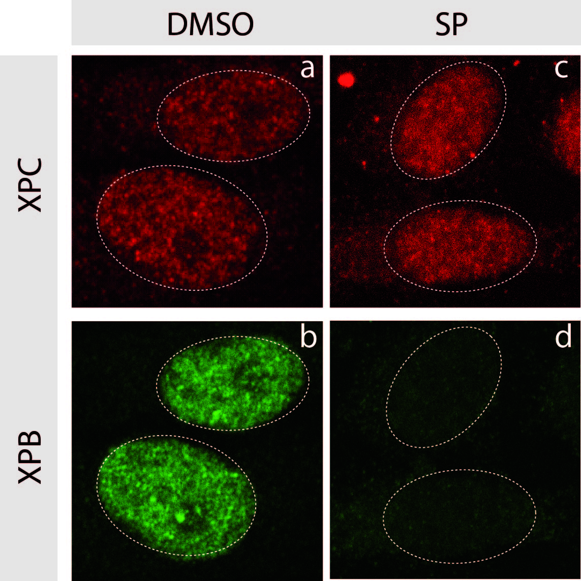
The researchers decided to focus on the fact that cells can turn tumorous if the expression of certain genes known as ‘tumour facilitators’ gets out of control. A number of proteins are involved in controlling the expression of these genes, in particular the BRD protein family. The researchers have proven for the first time that an innovative treatment targeting these transcription-controlling BRD proteins inhibits the three components of the vicious circle, namely the tumour cells and the differentiation of osteoclasts and osteoblasts. By chemically inhibiting the BRD4 protein (which belongs to the BRD family), researchers limited the spread of primary bone tumours while maintaining bone architecture.

Further tests were performed on patient biopsies to illustrate and confirm the results achieved with animals.

Tibias3D

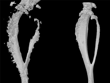
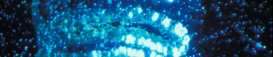
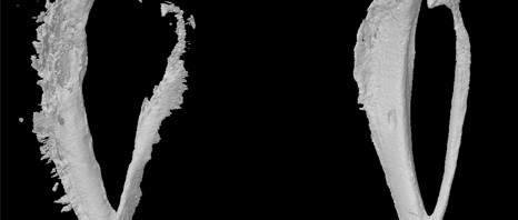
3D reconstruction of a mouse tibia with a primary bone tumour (left) and post-treatment (right)©Inserm/F Lamoureux 

“Our study clearly demonstrates inhibition of tumour progression and bone degradation combined with extended lifespan in animals”, comments Inserm researcher François Lamoureux. At 32 days, all the mice in the control group had died while the treated mice were still alive after 40 days. “On the basis of work, we can now seriously consider the possibility of developing a new treatment for primary bone tumour patients which tackles both the tumour and the associated bone degradation.” Moreover, since bone architecture is preserved, we could envisage a wider range of indications including bone metastases in patients with prostate or breast cancer and also non-tumorous bone diseases (e.g. osteoporosis)”.

Schéma

Diagram: treatment tackles the 3 components of the vicious circle

Non-coding genomic regions ameliorate the severity of beta-thalassemia and sickle cell anemia

Beta-thalassaemia and sickle cell anaemia are genetic disorders caused by mutations in a single gene but non-coding genomic regions seem to have a strong influence on disease severity. The teams of Eric Soler, researcher at Inserm unit 967, Fontenay-aux-Roses, France (Inserm / CEA), Swee Lay Thein, Clinical Director of the Red Cell Centre in King’s college London and King’s College Hospital, London, UK, and Frank Grosveld, professor of Cell Biology at the Erasmus Medical center, Rotterdam, unraveled the molecular mechanisms explaining how non-coding genomic variants, located far away from genes, were able to modify the clinical severity of beta thalassaemia and sickle cell anaemia. To reach this goal, the researchers have combined different expertise, including the study of spatial chromosome architecture. This work will be published in The Journal of Clinical Investigation and will be accessible online starting from March 10th 2014.

It is now clear that the vast majority of mutations and genetic variations uncovered by Genome-Wide Association Studies (GWAS), associated with common and rare diseases (diabetes, cardiovascular diseases, cancers) lie within non-coding sequences in the genome, i.e. do not directly hit the structural part of genes. Surprisingly, these genomic variants, found in the human population may be located at considerable distances from genes. This fact strongly complicates their functional analysis, and illustrates the complexity of the human genome organization and its relationship with diseases.

Etude de la drépanocytose. © C Feo/Inserm

Beta-thalassaemias and sickle cell anaemia are among the most common inherited disorders affecting red blood cells. In particular, sickle cell anaemia which affects 300,000 newborns annually, is fast becoming the most common serious genetic disease in UK, France, and the rest of Europe. These disorders are caused by mutations affecting a single gene – the beta globin gene- leading to alterations of the adult haemoglobin. Despite being single gene mutations, both disorders display an extremely variable range of disease severity, many factors modify the disease severity including the ability to produce fetal haemoglobin. Fetal haemoglobin is normally ‘silenced’ in adults, but some adults are able to escape this silencing and continue to produce fetal haemoglobin, and although harmless in healthy adults, fetal haemoglobin  can compensate for the altered adult haemoglobin in patients with beta thalassaemia and sickle cell anaemia, reducing the severity of the anaemia. In 2007, Swee Lay Thein’s lab identified a number of variants in a “gene desert” on chromosome 6q23, at tens of thousands of base-pairs from the closest genes, as affecting the ability to produce fetal haemoglobin in adults. How these genetic variants and the mechanism involved in ‘reactivating’ fetal haemoglobin were the focus of the three labs.

The researchers used a combination of chromosome conformation technologies and high throughput DNA analyses on thalassemia patient samples to elucidate the molecular mechanisms of how non-coding variants exert their action and improve the symptoms of thalassaemia and sickle cell anaemia.

The researchers showed that these variants, in a normal context, physically interact with the MYB gene, located at a distance of more than 80 000 base-pairs, via chromosome folding. In individuals carrying the variants, chromosome folding is diminished leading to a decrease in expression of the MYB gene. 

The decrease of MYB expression in patients carrying the variants leads to a reactivation of fœtal haemoglobin which compensates for the defect of adult haemoglobin and significantly ameliorates sickle cell and beta thalassaemia severity “, the authors say.

These data identify directly and for the first time the MYB gene as the target of chromosome 6q23 non-coding variants. Thus, the MYB gene represents a major therapeutic target for the induction of fœtal haemoglobin, a potential therapeutic approach for beta thalassaemia and sickle cell anaemia, Eric Soler and Swee Lay Thein suggest.

Enhancement of chemotherapy by prevention of tumour cell repair

Chemotherapies are cancer treatments that work by inducing lesions in the DNA of tumour cells in order to inhibit their proliferation. However, the body naturally tries to repair these lesions, and thus reduces the efficacy of chemotherapy. Blocking the mechanisms for DNA repair would help to potentiate chemotherapy by reducing the resistance of cells to treatment. A team of researchers directed by Frédéric Coin, Inserm Research Director at the Institute of Genetics and Molecular and Cellular Biology (IGBMC) in Strasbourg (a Joint Inserm/CNRS/University of Strasbourg Research Unit), has discovered a new drug that inhibits repair: spironolactone, which seems likely to be used in the very short term as an adjuvant to chemotherapy.
Their results are published in
Chemistry & Biology.

UV rays, physical or chemical agents—the human body is constantly subject to environmental insults that cause more or less damage to our DNA. The body has therefore developed a whole system for proofreading and repair. Among these mechanisms, NER (Nucleotide Excision Repair) has been studied for several years by the researchers in a team led by Frédéric Coin and Jean-Marc Egly at IGBMC. This mechanism can thus detect a lesion, and then replace the damaged DNA fragment with an intact fragment.

Cytotoxic chemotherapy is aimed at blocking the division of malignant cells in order to prevent tumour growth. Included among the drugs used to treat many cancers such as colorectal, face and neck, testicular, bladder, ovarian and lung cancers are medications based on platinum. These drugs bind to cellular DNA, induce damage in the latter, and thus prevent its replication. Blocking DNA repair mechanisms, specifically NER activity, would help to potentiate chemotherapy by reducing the resistance of cells to the treatment.

The researchers at IGBMC therefore sought a drug that would inhibit NER activity. They thus tested over 1,200 therapeutic drugs and demonstrated the action of spironolactone, a drug already used for the treatment of hypertension, on NER activity. Specifically, the researchers showed that its action, when combined with that of platinum derivatives, caused a substantial increase in cytotoxicity for malignant colonic and ovarian cells.

Since spironolactone is already in use for other purposes, it does not require a new application for marketing authorisation, and its side-effects are already known. This result thus bodes very well for the rapid development of new chemotherapy protocols that include spironolactone.

illustration coin

© Inserm/ Frédéric Coin

Immunofluorescence labelling, 1 h after treatment, of XPC proteins (in red) and XPB proteins (in green) involved in NER activity. On the right, treatment with spironolactone induces rapid degradation of XPB, which explains its inhibition of NER.

Leukemia: mode of action of a targeted treatment clarified

The mechanism of senescence – or premature cell ageing – can have an anticancer effect. This new work, conducted by Hugues de Thé and his team (Paris Diderot University/ Inserm/ CNRS/ AP-HP), was published in Nature Medicine on 12 January 2014. It reveals that targeted treatments for acute promyelocytic leukaemia, a rare form of blood cancer, cause a cascade of molecular events leading to cellular senescence and recovery. This action model could be activated in other types of cancers. 

The PML/RARA* protein causes the proliferation of cancer cells in patients affected by acute promyelocytic leukaemia. Existing targeted treatments combining a hormone – retinoic acid – and a poison – arsenic – result in permanent recovery for the majority of patients, without us having a precise understanding of their action on cancer cells. Previous work by Prof Hugues de Thé’s team has shown that the combination of arsenic and retinoic acid causes destruction of the PML/RARA protein and the elimination of leukaemic stem cells. It remained to understand the link between these two events.

This new research contributes the factors needed to understand the recovery. It demonstrates the unexpected involvement of a cascade of events leading to senescence. The aim of the treatment is to reach this final ageing stage of the cells in order to render them incapable of multiplying.

During this targeted treatment researchers showed that the p53** protein, arbiter between cell death and survival, triggers senescence through the involvement of PML nuclear bodies. These spherical structures are present in normal cells but are disorganised by PML/RARA in leukaemia. The treatment reorganises them (see figure below), activating p53 and triggering senescence. In this cascade of events (treatment, PML/RARA degradation, reformation of nuclear bodies, p53 activation), only one link has to be missing to block all the therapeutic effects.

2_cellules

Leukaemic cells before (left) and after treatment (right). The blue represents DNA in the nucleus; the red is nuclear corpus PML. These one are reorganised by the treatment targeting PML/RARA. © Photos provided by Prof Hugues de Thé

It is this phenomenon that enables the elimination of diseased cells and leads to total recovery of the patient, using only combined retinoic acid/arsenic treatment. The absence of chemotherapy avoids many severe side effects.

This understanding of the cellular and molecular mechanism of recovery from acute promyelocytic leukaemia opens prospects for activating this same PML/p53 pathway in other types of cancers.

This work was financed by the French Ligue contre le cancer [cancer research charity], the French Fondation ARC pour la recherche sur le cancer [ARC Foundation for Cancer Research] and the European Research Council (ERC).

* Acute promyelocytic leukaemia is caused by the modification of two genes, RAR and PML, leading to the development of cancer cells;
** the gene coding for p53 protein plays an essential role in cell proliferation under normal conditions and in maintaining the integrity of the cell genome.


Mechanics and genetics: an indispensable cocktail for embryonic development

In the fruit fly Drosophila and zebrafish, mechanical strain may activate the genetic cascade that initiates the formation of the future organs during embryogenesis. A discovery made by Emmanuel Farge (Inserm Research Director at Institut Curie) and his staff might explain the emergence of the first complex organisms more than 570 million years ago.
The results of this work are published in the journal
Nature Communications.


embryon

signal de phosphorylation de la béta-caténine dans le tissu ventral qui invagine (mésoderme) dans l’embryon de Drosophile en vue ventrale  de haut © E Farge

Living things exist in a multiplicity of forms. At the very beginning—whether they are early multicellular or embryonic forms of life—they are all nothing more than a mass of cells. A long series of morphological changes causes all existing life forms to develop from this one form.

The embryo adopts a particular form at each stage of its development. These successive deformations, which are genetically regulated, in turn create mechanical strain within the embryo. This strain also seems to influence or even regulate the expression of genes involved in development.

From Drosophila to zebrafish

“Whether in zebrafish or Drosophila, we have found that activation of the β-catenin protein at the beginning of embryonic development follows mechanical compression developed during the very first change in the form of the embryo,” explains the researcher.


At the very start of development, a morphological change—known as invagination in Drosophila and epiboly in the zebrafish—allows expression of the genes that specify the mesoderm1[1], in response to the mechanical activation of β-catenin in tissues that are particularly deformed by these movements. Complex organs such as the muscles, heart, or gonads are derived from the mesoderm.

In their publication in Nature Communications, Emmanuel Farge and his team show in detail that the mechanical strain occurring during this morphological transition induces a modification of β-catenin (phosphorylation), which induces its relocation from the surface of the cell to its centre.

This protein may take on several roles: on the cell surface, it is responsible for cell-cell adhesion and may thus undergo mechanical strain, become phosphorylated and then released into the cell; within the cell, it can activate certain genes and thus modify the fate of the cells. Thus mechanical compression can lead to the acquisition of an identity similar to that of mesodermal cells following relocation of β-catenin to the interior of the cell. “To reproduce the mechanical strain naturally undergone by the embryo, we introduced liposome-encapsulated magnetic nanoparticles into the embryo, which we then exposed to a micromagnet.

An answer to the origins of evolution into complex organisms?

“The main point is that the mechanosensitivity of gene expression has been conserved by Drosophila and zebrafish during evolution,” explains the researcher. “Its origin therefore goes back to the last common ancestor shared by the two species, i.e. over 570 million years.” Moreover, specialists in evolution associate this same period with a major transition in evolution: the emergence of the mesoderm in ancestral life forms, possibly related to the jellyfish, for example, which do not have a mesoderm. The origin of this transition, which led to the development of complex organisms, such as the vertebrates, has not been well understood until now. Researchers are therefore only now on-track to answer this long-standing question.

Going back still further in time, mechanosensitivity might have even contributed to the emergence of the very first organisms. And what if compression, triggered, for example, in a mass of cells because it was resting on the substratum, gave rise to local deformation of the cell mass and thus activated the first invagination of the very first primitive gastric organ, as suggested by the experiments previously done by the team?

Cancer genes, reactivation of sensitivity to compression

Since the genes for embryonic development are implicated in the process of tumour progression, the mechanical induction of genes constitutes a new avenue for studying the development of cancers. The β-catenin protein is not unknown to cancer specialists. Thus during development of colon cancer, dysregulation of the β-catenin pathway is often described as one of the events correlated with loss of the APC gene. Furthermore, the development of cancer leads to the generation of physical strain affecting the neighbouring tissues.
It is a little as if the mechanism necessary for the development of the embryo were reawakened at the wrong moment. “Indeed”, underlines Emmanuel Farge, “when all is going well, APC protein degrades the β-catenin released into the cytoplasm by abnormal mechanical cues. When APC is mutated (which happens in 80% of colon cancers correlated with genome modifications), the β-catenin released into the cytoplasm is no longer degraded effectively, and is free to enter the nucleus and stimulate the expression of genes that promote tumour development.”


[1]  The mesoderm is one of the three embryonic germ layers, and is formed between the endoderm and ectoderm at the time of gastrulation. During development, it gives rise to most of the internal organs. Its existence is a feature of the most highly evolved organisms.

Chemotherapy: when our intestinal bacteria provide reinforcement

Research jointly conducted by investigators at Institut Gustave Roussy, Inserm, Institut Pasteur and INRA (French National Agronomic Research Institute) has led to a rather surprising discovery on the manner in which cancer chemotherapy treatments act more effectively with the help of the intestinal flora (also known as the intestinal microbiota). Indeed, the researchers have just shown that the efficacy of one of the molecules most often used in chemotherapy relies to an extent on its capacity to mobilise certain bacteria from the intestinal flora toward the bloodstream and lymph nodes. Once inside the lymph nodes, these bacteria stimulate fresh immune defences which then enhance the body’s ability to fight the malignant tumour.

Results of this work are published in the journal Science on 22 November 2013

bacteria - blue version

©Fotolia

The intestinal microbiota is made up of 100,000 billion bacteria. It is a genuine organ, since the bacterial species that comprise it carry out functions crucial to our health, such as the elimination of substances that are foreign to the body (and potentially toxic), or keeping the pathogens that contaminate us at bay. They also ensure the degradation of ingested food, for better intestinal absorption and optimal metabolism. These millions of bacteria colonise the intestine from birth, and play a key role in the maturation of the immune defences.

However, the bacterial species that make up the intestinal microbiota vary from one individual to another, and the presence or absence of one or another bacterial species seems to influence the occurrence of some diseases, or, conversely, may protect us.

In the cancer area, the French team directed by Prof Laurence Zitvogel, Director of Inserm Unit 1015,Tumour Immunology and Immunotherapy,” at Institut Gustave Roussy, in close collaboration with Institut Pasteur (Dr Ivo Gomperts Boneca, “Biology and Genetics of the Bacterial Cell Wall” Unit) and researchers at INRA (Drs Patricia Lepage and Joël Doré, Micalis Unit, “Food Microbiology in the Service of Health”), has just provided evidence that the intestinal flora stimulates an individual’s immune responses to combat cancer during chemotherapy.

Cyclophosphamide is one of the most widely used drugs in chemotherapy. However, like any treatment, it involves side effects (inflammation of the mucosa etc.), and disrupts the normal balance of the intestinal microbiota. Certain bacteria (of the Gram+ group of bacteria) can pass the intestinal barrier and enter the bloodstream and lymph nodes.

These bacteria, once in the general circulation of the body, may be considered harmful, and the body generates an immune response.

“This chain reaction, a side effect of the treatment, actually turns out to be very useful,” explains Laurence Zitvogel. “Surprisingly, the immune response directed against these bacteria helps the patient to better fight his/her tumour, by stimulating fresh immune defence mechanisms.”

More specifically, immunisation against bacteria leads to the recruitment of effector lymphocytes different to those mobilised by chemotherapy. Their role consists of helping anti-tumour lymphocytes to stem the growth of tumours.

To verify these observations in mice, researchers suppressed all Gram+ bacteria from their intestinal microbiota. Results showed that the efficacy of the chemotherapy was reduced. The researchers also suggest that some antibiotics used during chemotherapy may destroy these Gram+ bacteria, and thus negate their beneficial effect.

“Now that these “beneficial” bacteria that potentiate the anti-tumour immune response have been identified, we should soon succeed in supplying more to the body, especially via pro- or prebiotics and/or a specific diet,” the researcher concludes.

This work has received support from the French National Cancer League, the French National Cancer Institute (lNCa; SIRIC SOCRATES) and from LABEX Onco-Immunology

Cancer treatment: a step towards personalized chronotherapy

Cancer chronotherapy consists in administering treatment at an optimal time. Because the body is governed by precise biological rhythms, the efficacy of anti-cancer drugs can be doubled and their toxicity reduced five-fold depending on the exact timing of their administration. However, important differences in biorhythms exist between individuals, which chronotherapy has not been able to take into account until now. An international study conducted on mice and coordinated by researchers from Inserm, CNRS and Université Paris-Sud[1] has paved the way towards personalized chronotherapy treatments. In an article published in the journal Cancer Research, the team has shown that the timing of optimal tolerance to irinotecan, a widely used anti-cancer drug, varies by 8 hours depending on the sex and genetic background of mice. They then developed a mathematical model that makes it possible to predict, for each animal, the optimal timing for administering the drug. They now hope to test this model on other drugs used in chemotherapy.

The body’s metabolism follows a 24 hour rhythm, driven by the circadian clock. Consequently, at certain precise times of the day or night, a given drug may prove to be more toxic to cancer cells and less aggressive to healthy cells. Cancer chronotherapy, discovered some twenty years ago by Francis Lévi, seeks to improve the efficacy of chemotherapy treatments. His research has shown that this efficacy can be doubled, depending on the time at which they are administered. Furthermore, it is precisely at this optimal time that the drugs prove to be five times less toxic to the body.

However, research points to the need for personalizing chronotherapy. Indeed, biorhythms can change from one person to the next. For example, although the optimal timing is the same for 50% of patients, the remaining 50% are either ahead of or behind this time. The team headed by Lévi wanted to elucidate the factors that affect these differences in biorhythms.

To do this, the researchers studied the toxicity of irinotecan, an anti-cancer drug widely used in the treatment of cancer of the colon and pancreas, as a function of the timing of its administration in four strains of male and female mice. For the first time, they were thus able to observe that the time of best tolerance to treatment varied by up to eight hours from one group of rodents to the next, depending on their sex and genetic background.

The researchers then worked on developing a method able to predict this optimal drug timing independently of sex and genetic background. To do this, they measured the expression of 27 genes in the liver and colon over 24 hours and then analyzed these measurements using a methodology derived from systems biology. In this way, the researchers were able to construct and validate a mathematical model to precisely predict the timing at which irinotecan is less toxic to the body using the expression curve of two genes, known as Rev-erbα and Bmal1, which regulate the metabolism and proliferation of cells.

The researchers are now aiming to validate this model on other drugs used in chemotherapy. In addition to gene expression, they would also like to find other physiological parameters related to the biological clock that could help predict the optimal timing of treatments for each patient. This work should make it possible to enhance the efficacy and tolerance of such treatments as well as considerably improve the quality of life of patients.

This project was funded in particular by the European Union (7th Framework Programme for Research and Technological Development) and ERASYSBIO+, the European consortium of funding bodies, ministries and project management agencies


[1] Coordinated by the Unité Rythmes Biologiques et Cancers (Inserm/Université Paris-Sud), this work also involved the Institut de Biologie de Valrose (CNRS/Inserm/Université de Nice Sophia Antipolis), the Laboratoire des Signaux et Systèmes (CNRS/Supélec/Université Paris-Sud) and the Milan Institute of Pharmacology.

Identification of a new mechanism in the most commonly used immunotherapy for lymphoma

Using innovative dynamic imaging technique, scientists at the Institut Pasteur, Inserm and the VU Medical Center in Amsterdam have uncovered the mode of action of anti-CD20, an antibody therapy frequently used in the treatment of lymphomas (cancers of the immune system) as well as some auto-immune diseases. In a lymphoma model, the scientists have been able to carry out real time in vivo imaging of the cellular events activated by the treatment and resulting in the destruction of tumor cells. These discoveries should help optimize the efficacy of future therapies involving anti-CD20 antibodies. This work is the subject of an article published online November 1 on the Journal of Clinical Investigation website.

A lymphoma usually develops as a result of abnormal proliferation of one of two types of immune cells:  B lymphocytes (in the vast majority of cases) or T lymphocytes. For the last fifteen years or so, anti-CD20 antibody therapy has frequently been used in the treatment of B-cell lymphomas (in particular those known as non-Hodgkin lymphomas), in combination with conventional chemotherapy. These antibodies are directed against B lymphocytes, bind to cancer cells and mark them for depletion by other immune cells. Anti-CD20 antibody therapy also triggers a decrease in the normal B lymphocyte population, dampening immune responses. For this reason it is also used to treat autoimmune diseases. However, how anti-CD20 antibody therapy works in vivo was not fully understood.

A study led by Philippe Bousso, head of the Dynamics of Immune Responses Unit (Institut Pasteur / Inserm U668), along with researchers at Inserm and the VU Medical Center in Amsterdam, has provided the first conclusive answers. Using dynamic imaging techniques developed at the Institut Pasteur, the scientists have carried out real time in vivo imaging of the destruction of cancerous and normal B lymphocytes during anti-CD20 antibody treatment. The scientists noticed that the phenomenon of B lymphocyte depletion resulting from anti-CD20 antibody therapy primarily takes place in the liver and involves a specific cell type, known as Kupffer cells. The images produced by the scientists clearly show Kupffer cells (in green) capturing cancerous B lymphocytes (in orange) and preventing their circulation before destroying them.

These discoveries provide important insight for optimizing the efficacy of future treatments using anti-CD20 antibodies. Non-Hodgkin lymphomas affect 10,000 people per year in France, and account for 10% of pediatric cancers.

A better evaluation of the body’s ability to fight tumours

Some individuals are better able to fight cancer for many years compared with others. This ability to fight tumours depends on the immune response, as observed for colorectal cancers by Jérôme Galon, Research Director at Inserm, and his team, the Laboratory of Integrative Cancer Immunology, at the Cordelier Research Centre (Inserm/UPMC/Paris Descartes University). The investigators show that the proportions of immune system cells in and around the tumour change with the stage of progression of the cancer, and demonstrate the importance of an increased concentration of some cell types to the survival of patients, namely, follicular T-helper cells (Tfh) and B lymphocytes. A better understanding of the dynamics of these cells will help to identify new strategies for developing targeted immunotherapies.

The results of this study are published in the 17 October issue of the journal Immunity.

The immune system is able to fight some tumours before they can affect health. Once the tumour has been identified, immune system cells are mobilised to kill and eliminate the tumour cells. However, tumour cells can sometimes manage to survive the response of the immune cells, and become established. The tumour becomes malignant when it develops in an uncontrolled manner. The investigators at the Cordelier Research Centre (Inserm/UPMC/Paris Descartes University) study the manner in which the immune system fights tumours, in an effort to effectively unleash the body’s intrinsic potential for fighting cancer.

Two factors indicate the body’s potential for “fighting or defeating” a tumour: the intensity of the immune response, and the mechanisms adopted by tumours to escape recognition by the immune system. The complex interactions between tumours and their microenvironment were poorly known until now. In the present study, the investigators examined the spatiotemporal dynamics of 28 different types of immune system cells that infiltrate colorectal tumours. By combining the study of cellular interactions with bioinformatics, they observed that the proportions of immune system cells infiltrating tumours change with the stage of progression of the tumour.

The research team has demonstrated the importance of an increased concentration of some types of immune system cells to patient survival, i.e. the T follicular-helper (Tfh) cells and B lymphocytes. These results obtained for human tumours were also demonstrated in three mouse models of colon cancer.

The investigators also studied more specifically in patients the instability of the gene for the chemokine CXCL13, which modulates the infiltration of Tfh and B lymphocytes. CXCL13 and IL-21 have proven to be additional factors that promote the death of tumour cells: high levels of these molecules are correlated with patient survival.

These observations indicate that T, Tfh and B lymphocytes form a network of cells that communicate inside tumours. High levels of Tfh and B lymphocytes prevent tumour progression and recurrence in colorectal cancer. As in patients, T, Tfh and B lymphocytes control tumour development in murine models of colon cancer.

“The immune response develops during cancer progression. The immune landscape that we describe in relation to colorectal tumours helps us to understand this development in order to intervene in the right place at the right time,” explains Jérôme Galon, Research Director at Inserm and last author of the study. The clinical outcome is highly variable among patients with the same stage cancer. Understanding why some individuals are able to defend themselves against cancer for many years is crucial in combating the disease,” concludes the main author of the study.

The investigators have also developed a test, known as “Immunoscore,” which predicts the ability of an individual’s immune system to fight tumour cells. Using Immunoscore as part of routine prognostic evaluation may provide critical new information on prognosis, and facilitate clinical decision-making (including guidance for therapeutic decisions). In order to promote Immunoscore for routine use in hospitals, an international consortium directed by Jérôme Galon has been launched in association with the US Society for Immunotherapy of Cancer (SITC) and a large number of international institutions from 17 countries throughout the world.

(French) : Cancers du rein, mélanomes de l’œil et mésothéliomes : un gène de prédisposition en commun

Vesicular transport: a requirement for T immune response

T-lymphocyte (TL) activation requires recognition by the T-cell receptor (TCR) of the ligands present on the antigen-presenting cell (APC). The ligand–TCR link bound to the TL plasma membrane  introduces a signalling cascade that converts a signal received from the outside into a suitable response, for example cytokine secretion. Claire Hivroz, INSERM Research Director (INSERM Unit 932, Institut Curie, Paris) and her colleagues recently showed in an article published in Nature immunology how a specific protein known as VAMP7 is essential for transporting the signal produced by the ligand–TCR link. 

The setting up of this activation cascade has been the subject of numerous research papers and yet remains little known. In particular, the location of the cell in which the signalling occurs and the mechanisms governing the formation of the protein complexes needed for such activation remain the subject of controversy.

Claire Hivroz’s team showed in 2004 that a protein that played a key role in TCR signalling, the LAT molecule, acts as a framework on which the other signalling proteins are assembled. It is present on the plasma membrane and in each of the intra-cellular vesicles. In response to TCR stimulation, these vesicles are attracted to the area in which the stimulation takes place, namely, the contact area between the TL and the APC, known as the immunological synapse (IS).

ImageJ=1.46i

Crédit photo : © Inserm-Institut Curie/C. Hivroz

Evanescent wave microscopy image showing recruitment into the immunological synapse of LAT-containing vesicles (magenta), VAMP7 (green) or the two proteins (white) in a T-lymphocyte interacting with a glass slide covered in activating antibodies (INSERM–Institut Curie/C. Hivroz)


Researchers wanted to discover the role played by this intracellular LAT ‘pool’ and the mechanisms that lie behind its recruitment by the IS. The study being published this month in Nature Immunology was performed in collaboration with Thierry Galli, a specialist in the proteins involved in the transport of vesicles to the neurological synapse, known as SNARE proteins, involved in membrane fusion reactions during vesicular transport.

By using genetically modified mice, RNA interference mechanisms and high-resolution microscopy, Claire Hirvoz’s team showed that VAMP7, a SNARE protein, is necessary for transporting LAT-containing vesicles to the IS. This transport controls LAT activation and the formation of the protein complex that sets up the signalling cascade. Consequently, T-lymphocytes deprived of VAMP7 will not respond normally to stimulation of their TCRs.

In conclusion, this new data will contribute to understanding the way in which information is propagated over time and space after TCR stimulation and shows for the first time how the transport of vesicles is involved in setting up a response to T-lymphocytes.

These results also show that molecules present in the neuronal synapses and involved in their functions are also involved in the functions of the immunological synapse.

fermer