Menu

The efficacy of certain anti-cancer vaccines depends on how they are administered

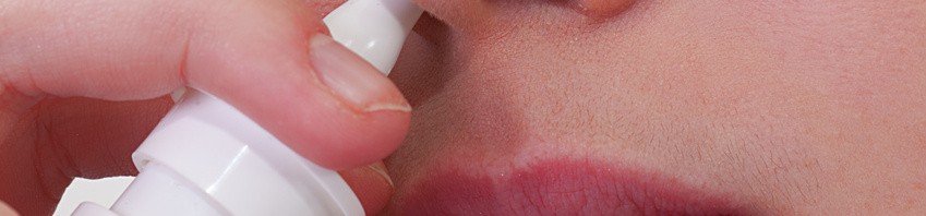
The therapeutic efficacy of certain anti-cancer vaccines depends on how they are administered. This is what the team of researchers headed by Eric Tartour at the Paris-Centre de recherche Cardiovasculaire (Université Paris Descartes, INSERM Unit 970 PARCC, AP-HP), in collaboration with researchers from the CNRS[1], have just demonstrated. A paper on the subject was published on 13 February 2013 in Science Translational Medicine. In the case of so-called mucosal cancers of the lungs or ENT area, the vaccine should be administered directly via the mucosa if it is going to be effective (intra-nasally for example). The same vaccine administered in the usual way that immunisation is performed, i.e. intramuscular or sub-cutaneous injection, will be ineffective. In this research, the investigators also identified an important method by which the vaccine acts against tumours. These results will have a major impact on the creation and effectiveness of anti-cancer vaccines targeting mucous tumours.


© Fotolia

Vaccines to treat cancer represent a new and promising avenue of therapy. The aim is to stimulate certain cells in the immune system, such as the T-CD8 lymphocytes, in order to cause the tumour to shrink. Many hopes are being pinned on these types of vaccines. Numerous trials are currently in progress, but some have suffered setbacks in the clinical phase even though they proved successful on animal models.  

So as to understand why this happens, Eric Tartour and his team (Université Paris Descartes, INSERM  Unit 970 PARCC, AP-HP) used mice to test the efficacy of two different routes of administration, the intramuscular route, on the one hand, and the intranasal route on the other. The tests were performed on a candidate vaccine against mucous, oropharyngeal cancers developed in collaboration with Ludger Johannes at the Institut Curie.

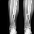
They proved that only vaccination via the mucosa will shrink a tumour in the lung or the ear, nose and throat (ENT) area. The same anti-cancer vaccine administered in the usual immunisation way, i.e., intramuscularly or subcutaneously, is ineffective.

Our results may explain the failure of anti-tumoural vaccines designed to treat tumours of the mucosa in humans, and should lead to a change in how anti-cancer vaccines are used to target mucosal tumours”, explained Eric Tartour, Professor at Paris Descartes University and hospital doctor at the Hôpital Européen Georges Pompidou (AP-HP).

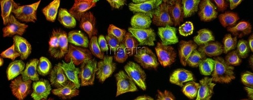
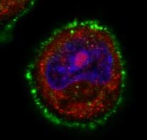
To try and explain the difference in efficacy, researchers sought to identify the mechanisms that operated differently depending on the method of administration. They showed that vaccination through the mucosa caused a certain protein (the mucous integrin CD49a) to be produced that enabled the T-CD8 lymphocytes (whose production is stimulated by the administration of the vaccine) to migrate into the tumour. Once they are at the site of the tumour, the T-CD8 lymphocytes destroy the cancerous cells.

In the opposite case, if this molecule is blocked it prevents the penetration of T lymphocytes into the mucosal tumour and prevents their anti-tumoural activity. The integrin CD49a is essential in the promotion of migration of the anti-tumoural lymphocytes produced through vaccination in the case of pulmonary tumours and ENT tumours and thus the efficacy of the vaccination in combatting mucosal cancers.

To check the applicability of these results in humans, the researchers analysed samples from patients suffering from lung cancer. Here again, they demonstrated the elevated expression of mucosal integrin CD49a on T-CD8 lymphocytes present in patients with lung tumours[1].

These results are encouraging for the future extrapolation of the study to humans.


[1] Travaux menés par Laurence Zitvogel, Unité immunologie des tumeurs et immunothérapie (Université Paris-Sud/Inserm) ; Institut Gustave Roussy, Villejuif 


[1] Laboratories involved: Unité de pharmacologie chimique et génétique et d’imagerie (CNRS/Université Paris Descartes/Inserm/Chimie ParisTech); Compartimentation et dynamique cellulaires (CNRS/Institut Curie/UPMC). 

Cancer du col de l’utérus : première résolution 3D d’une oncoprotéine du virus à papillome humain

Regenerative medicine: clinical trials launched for the treatment of delayed union fractures

50% of fractures do not heal alone and require surgical bone grafting; this figure equates to around one million patients in Europe. The REBORNE project (Regenerating Bone Defects using New biomedical Engineering approaches), funded by the European Commission and coordinated by Inserm, has just received the green light from the French Medicinal Agency ANSM to begin orthopaedic surgery clinical trials. The clinical trials aim to repair bones using adult stem cells combined with biomaterials. It will be conducted in France in the Créteil University Hospital Centre and the Regional University Hospital Centre in Tours, in collaboration with the French Blood Establishment.

The REBORNE project began three years ago and its originality lies in using mesenchymal stem cells (MSC), found in bone marrow, to help heal fractures. Adult stem cells are amplified in culture, then are associated with biomaterial before being implanted on the fracture. Promising pre-clinical trials have now paved the way for the launch of these new clinical trials.

Following trauma (fall or road accident), there is a high incidence of delayed union or non-union in tibia, femur or humerus bones requiring a surgical apposition of an autologous bone graft (patient’s own bone). However, the bone graft available is limited and complications are often observed at the second surgical site.

The partners of the European project REBORNE propose an alternative treatment using autologous mesenchymal stem cells associated with a synthetic bone substitute.

© P. Layrolle, Inserm

Thanks to a bone marrow aspiration under local anaesthesia, the mesenchymal stem cells are isolated and amplified in culture for 21 days. During the surgical intervention stem cells are associated with biphasic calcium phosphate ceramic granules and implanted at the site of the fracture. In this way, the biomaterial is used as “scaffolding” and encourages stem cell proliferation. The stem cells are then differentiated into bone cells and regenerate the bone tissue around the fracture.

© P. Layrolle, Inserm

Bone formation (in green) and bone barrow formation (in red) further to a mixture of human stem cells and biomaterial granules (grey/white) being implanted at a subcutaneous site in nude mice. This histological section shows osteoinduction by the cell/biomaterial mix with the formation of around 41% of bone tissue four weeks after implantation.

The French Medicinal Agency ANSM gave its green light on 3 January 2013 so that these Inserm-sponsored trials can now begin in France, with seven patients receiving treatment in the Créteil University Hospital Centre and the Regional University Hospital Centre in Tours. Ultimately, thirty patients will be included in France, Spain, Germany and Italy to be part of this multi-centre European study.

“The objective of this trial is to show that biomaterials and stem cells are safe and are at least equivalent to standard treatments, without their disadvantages. This surgery is less invasive and preserves the patients’ bone stock. For these reasons, it is preferable to bone grafts in terms of triggering bone healing”.

 explains Pierre Layrolle, director of Research, Inserm and coordinator of REBORNE.

For further information:

REBORNE – Regenerating Bone Defects using New biomedical Engineering approaches

The objective of REBORNE is to develop new biomaterials that stimulate bone tissue formation with a view to correcting bone regeneration defects in orthopaedic and maxillofacial surgery. Biomaterials, combined with the use of stem cells, are interesting alternatives to biological grafts.

The REBORNE project kicked-off in January 2010 and will run for five years using an overall budget of roughly 12 million Euros provided by the European Union (PC7). The project involves 24 partners, based in eight European countries: https://www.reborne.org/presentation.html

La protéine à l’origine de la maladie de Huntington impliquée dans les tumeurs mammaires

Improving chemotherapy effectiveness by acting on the immune system

An Inserm team in Dijon directed by François Ghiringhelli (Inserm unit 866 ‘Lipids, nutrition and cancer’) is to publish an article this week in the Nature Medicine review.  The article suggests that two chemotherapy drugs frequently used to treat digestive and breast cancers may encourage the development of tumours by modulating the anti-tumoural immune response. These results reveal how the immune system can then limit the effectiveness of some cancer chemotherapies. The researchers now intend to block the molecules responsible for negative immune system activation to increase the efficiency of chemotherapy. A clinical trial to test this hypothesis should begin very soon.

Chemotherapy is one of the most frequently used treatments to eliminate cancerous cells. These drugs kill all cells that are multiplying, or block their proliferation (for example, cells responsible for hair growth, explaining the hair loss of treated patients).  In addition to their direct toxic effects, the chemotherapeutic agents also seem to act on the immune system and could make it possible for the body to trigger a direct antitumor immune response in a second phase.

© fotolia

However, this last point is still the subject of hot debate, since some studies suggest, conversely, that chemotherapy eliminates all immune defences.

What now?

The Inserm team directed by Professor François Ghiringhelli (Inserm unit 866 “Lipids, nutrition and cancer”) from the Georges François Leclerc Cancer Research Centre in Dijon observed that two chemotherapeutic agents, 5-fluorouracile and gemcitabine, used to treat colon, breast and pancreas cancers activate a protein complex “inflammasome NLRP3” within some cells in the immune system.

To be more specific, this activation leads to releasing proinflammatory cytokine (interleukin IL-1beta) through these cells. This cytokine “distorts” the immune response related to lymphocytes T and causes the production of another cytokine (cytokine IL-17), which has protumoral properties by encouraging tumour angiogenesis, i.e. vascular irrigation of tumours.

Our results have made it possible to ascertain that the activation of inflammasome limits the effectiveness of chemotherapy. The challenge was then to see whether we could prevent the activation of inflammasome”

explains François Ghiringhelli. The researchers then tested two different strategies:

The first was to test the two drugs on inflammasome NLRP3- or cytokine IL-17-deficient mice.  In these cases, the researchers showed that antitumor activity was not only present, but it actually increased, demonstrating that these two elements (NLRP3 and IL-17) slow down the chemotherapy action.

The second strategy was to treat the mice using an IL-1beta inhibitor. Here again, the effectiveness of chemotherapy was again increased.

These results suggest that targeting the inflammasome and IL-1beta channels, combined with the use of these two chemotherapy agents, can improve the effectiveness of the latter. These tumour cells are eliminated and, in parallel, the damaging immune responses are deleted.

A therapeutic trial combining 5-fluorouracil and IL-1 beta is currently being prepared and should begin soon at the Georges François Leclerc Cancer Research in Dijon.

Predicting the age at menopause of women having suffered from childhood cancers

Researchers from Inserm, the French public hospital organization (AP-HP), the Institut Gustave Roussy and the Université Paris-Sud studied the age at menopause of a cohort of 706 women who had suffered from childhood cancer. The data revealed that these women, in particular those who had undergone unilateral oophorectomy or who had been treated with alkylating agents and who had received a radiation dose to the ovaries, were menopaused on average 4 to 7 years earlier than average. This study provided important data about the fertility window of women who had suffered from childhood cancer and information concerning the associated risk factors, but did not confirm the greater risk of premature menopause (before the age of 40) that was reported by the American studies.

The results were published in the review Human Reproduction of November 15.

Women who have suffered from childhood cancer are known to run a greater risk of premature menopause. However, data about the associated risk factors is limited. Researchers from unit 1018 “Centre for Research in Epidemiology and Population Health (CESP)” (Inserm/Université Paris-Sud/Institut Gustave Roussy) and from the AP-HP analyzed the data from a French cohort, named Euro2k, concerning 1522 survivors of childhood cancer diagnosed between 1945 and 1986 when they were under 18, initially in order to study the mortality rate. The study estimated the radiation doses received at the ovaries by the women in this cohort who had been treated by radiotherapy. 706 of these women filled in a detailed questionnaire about their state of health. 32% of these women had already reached the age of 40 years; 7% were over 50 years of age. The research team studied the age at menopause of these women and the potentially associated risk factors. The researchers based this study on self-reported questionnaires sent to the women in order to obtain information about the menopause, without confirming by measuring FSH levels.

Analysis of this data showed that 97 women (13,7%) were menopaused at a median age of 44 years, in other words, 7 years earlier than the general population. For a third of these women (36%), menopause was surgically induced.

The researchers concentrated on the risk factors of menopause in these women, who had been subjected to various cancer treatments during childhood. It appeared that being treated during puberty was associated with a risk of non-surgical menopause. At a given age, the maximum risk of early menopause was observed in women who had been treated after the onset of puberty with alkylating agents (either alone or along with even a minimum dose of radiation to the ovaries, for example 0.01 Gray). Menopause occurred on average 4 years earlier in women who had been exposed to these agents. Having undergone unilateral oophorectomy is also associated with a 7-year earlier age at menopause.

The results showed that women who had suffered from a childhood cancer were more likely to suffer from early menopause, but did not conform the high risk of premature menopause (in other words, before 40 years of age), such as was reported in the American studies. This is probably due to the difference between the populations studied (there were no cases of leukemia or lymphoma in the Euro2k cohort population).

“In this cohort , very few women had received high doses of radiotherapy in order to receive bone marrow transplants, and only 21% of them (i.e. twice the occurrence of the general population) suffered premature menopause before the age of 40”, explained Cécile Teinturier, the principal writer of the study. The main risk factors associated to these cases of premature menopause are: the older the women is when being treated for cancer, the dose of alkyating agents such as Cyclophosphamide or Melphalan received during bone marrow transplants, and the radiation dose received at the ovaries.

“This study provided information about the risk factors affecting the fertility window of women who have suffered from childhood cancer. This new data should help both to inform patients who are at risk of premature menopause, by advising them not to delay their first pregnancy until after the age of thirty, and to reassure women who present a low risk” concluded Cécile Teinturier.

It is planned to extend Cohort Euro2k to cover all patients treated for solid cancers who were under 18 years of age before the year 2000 inFrance. The aim is to study the impact of high-dose chemotherapy on the occurrence of premature menopause.

New developments reveal a molecule with a promising function in terms of cancer treatment

©Inserm

One of the current challenges in terms of cancer treatment is how it can be best adapted to patients: today the emphasis is on personalised treatment (factoring in genetic and metabolic profiles). In response to this growing need for personalisation, there is an increasing demand for fundamental research to develop adapted future treatments. Researchers from Inserm and CNRS from the Institute for genetics and molecular and cellular biology (IGBMC) and from the Research Institute at the Strasbourg school of biotechnology (Irebs) have focussed their efforts on PARG, currently thought to be a promising new therapeutic target in the treatment of cancer. Their work has revealed the role of this molecule in regulating gene expression. The results were published on 25 October 2012 in the on-line Molecular Cell review.

Cells are subjected to various stresses throughout their life. Some of this stress can damage DNA. Fortunately, cells have several mechanisms used to repair these lesions. Breaking two DNA strands is one of the most serious afflictions a chromosome can suffer. The cell must repair this break if it is to continue dividing. Repair actions are either performed in an optimal manner, and the cell resumes its normal division cycle, or the lesion is not repaired correctly, causing the cell to die or the appearance of an anomaly that may trigger cancer.

One of the repair mechanisms used is poly(ADP-ribosyl)ation. In this mechanism, some molecules (PARPs) detect DNA breaks and cause poly(ADP-riboses) synthesis that binds with proteins, thus initialising the lesion repair system. As such, this system can act as a ‘saviour’ if the repairs are correctly completed, but, it can be equally damaging in the event of incorrect repair.

In the case of cancer, to ensure the cells are destroyed, PARP inhibitors are currently undergoing clinical tests as therapeutic adjuvant used to increase the receptivity of cancer cells to specific chemotherapies.

In terms of fundamental research, researchers know that the poly(ADP-ribosyl)ation mechanism is reversible and is closely regulated by Poly(ADP-ribose) glycohydrolase (PARG). PARP and PARG thus seem to make up a tandem of molecules dedicated to maintaining genome integrity. PARG targeting has proved to potentiate the action of genotoxic agents, making this molecule a promising new therapeutic target in the treatment of some cancers, as is the case for PARP.

However, little research has been conducted into PARG, but in light of its close links with PARP, researchers are now taking a very close look at its functions.

In this new work, the researchers have demonstrated that in addition to its genome repair role, PARG was involved in modulating the cell’s transcriptional activity.

Given the keen current interest on PARP and PARG inhibitors in the treatment of cancer, it is essential to gain accurate knowledge of the functions and action modes of these promising therapeutic targets, as well as the consequences of their invalidation. This study is the first to highlight how PARG action mechanism regulates gene expression.

TIM and TAM: 2 paths used by the Dengue virus to penetrate cells

A study carried out by Ali Amara’s team at the combined Inserm/CNRS- Université Paris Diderot “Molecular pathology and virology” unit in the Saint-Louis hospital in Paris, working jointly with the team from the Pasteur Institute in Paris and the team from the Salk Institute in San Diego, has identified two families of receptors that play an important part in the penetration of the Dengue virus into cells. By demonstrating that it is possible to inhibit the viral infection in vitro by blocking the bonding between the virus and these receptors, the researchers have opened the way to a new antiviral strategy. These works were published on line in the review “Cell Host & Microbe” of October 18, 2012.

The Dengue virus circulates in four different forms (four serotypes). It is transmitted to humans by mosquitoes. It is a major public health problem. Two billion people throughout the world are exposed to the risk of infection and 50 million cases of Dengue fever are recorded by the WHO every year. The infection is often asymptomatic, or resembles influenza symptoms, but its most serious forms can lead to fatal haemorrhagic fevers. At present, there is no preventive vaccine or efficient antiviral treatment for these four Dengue serotypes. So it is of vital importance that we develop new therapeutic strategies.

Ali Amara’s team performed genetic screening in order to identify cell receptors used by the virus to penetrate target cells[1]. The researchers have determined the important function played by the TIM receptors (TIM-1, 3, 4) and TAM receptors (AXL and TYRO-3) in the penetration process of the four Dengue serotypes. Mr. Amara’s team has succeeded in demonstrating that the expression of these 2 receptor families makes cells easier to infect. In addition, the researchers observed that interfering RNA or antibodies that target the TIM and TAM molecules considerably reduced the infection of the cells targeted by the Dengue virus. The TIM and TAM molecules belong to two distinct families of transmembrane receptors that interact either directly (TIM) or indirectly (TAM) with phosphatidylserine, an “eat-me” signal that allows the phagocytosis and the elimination of these apoptopic cells. Unexpectedly, the work of the Inserm researchers discovered that phosphatidylserine is abundantly expressed at the surface of virions and that it was essential that the TIM and TAM receptors recognize the phosphatidylserine to allow infection of target cells.

These results have helped to understand the first key stage in the Dengue virus infectious cycle, by discovering a new method of virus entry that works by mimicking the biological functions involved in the elimination of the apoptotic cells. The discovery of these new receptors has also opened the way for new antiviral strategies aimed at blocking bonding of the Dengue virus with the TIM and TAM molecules.

This research has been patent-protected by Inserrm Transfert.


[1] Up till present, only DC-SIGN receptors and L-SIGN receptors were known to play an active role in the penetration of the Dengue virus into target cells

(French)Un test automatisé pour évaluer la migration cellulaire et l’efficacité de nouvelles molécules anticancéreuses

un Un dispositif qui réduit la carcinogénèse et détermine la réponse thérapeutique des cancers

A white mouse …

Mice with black fur that turns white? Specialist cancer researchers from Inserm, CNRS, the Institut Curie and the Université Paris-Sud have taken steps to better understand the development of skin cells responsible for pigmentation (known as melanocytes). By genetically manipulating mice with black fur, the researchers identified two key players: B-Raf and C-Raf proteins. These proteins are required for melanocyte stem cell self-maintenance and, as such, correct pigmentation throughout the mice’s life span. Without these two proteins, the mice’s fur turns white.

Their research is published in the review ‘Cell Report’ and paves the way for serious possibilities in terms of stopping the formation of melanomas, tumours that originate from melanocyte cells.

Melanocytes are cells in the organism used for skin, fur and hair pigment. This pigmentation function provides protection from the sun and lends organisms their colour. Malfunctions in these cells may lead to skin cancer known as melanoma. Melanomas are highly aggressive cancers that become very difficult to treat as they develop and metastases occur.

A few years ago, researchers discovered that, in humans, the B-Raf gene (coding gene for protein of the same name) is mutated in more than 50% of melanoma. Spectacular progress has been made in recent years in the treatment of this cancer, thanks to the development of pharmacological inhibitors that target an enzyme: the B-Raf kinase.

However, despite this treatment, cancer returns in several patients, indicating that not all cancerous cells have been eliminated.

This led researchers to believe that B-Raf is not the only element driving the cancer process.

In this new research, scientists have tried to understand how melanocytes function normally, to then understand their specific role in cancer. To this end B-Raf protein expression, then, in turn, C-Raf protein expression, were removed from mice with black fur (ideal to clearly see any changes in pigmentation).

Black mice that become white with age

No changes in pigmentation were observed for mice that only had their B-Raf or C-Raf expression removed by researchers from the line of cells producing melanocytes.

Mice that had both coding genes for B-Raf and C-Raf removed simultaneously had a normal colour at birth. However, they progressively lost their pigmentation as they grew. They turned grey from black, before becoming increasingly white.

souris blanches et noires

A. Eychène / F. Bertrand (Institut Curie)

For Alain Eychène, the research team leader, “these observations represent a fault in melanocyte renewal. Since the colour black is present at birth, the pigment cells clearly exist. However, the progressive whitening of the fur, once B-Raf and C-Raf have been removed from the cell line, proves that both these proteins are required for melanocyte renewal”.

As is the case for all cells, melanocytes originate from stem cells; the latter are responsible for renewal during moulting.  This research shows that it is specifically this population of stem cells alone that disappears progressively in mutant mice.

For Alain Eychène, “This is the first in vivo demonstration of the role of RAF proteins in the self-renewal of stem cells”.

The fact that B-Raf and C-Raf are both involved in controlling and renewing pigment stem cells represents another step towards understanding and treating melanoma. By blocking these proteins (using inhibitors) in patients undergoing treatment, it is possible that in time researchers will succeed in eliminating all cancerous stem cells, i.e. the likely cause behind cases of cancer reoccurrence.

fermer