Menu

Restoring natural immunity against cancers

Scientists at the Institut Pasteur and Inserm have successfully increased the infiltration of immune cells into tumors, thus inducing the immune system to block tumor growth. In an article published in Nature Immunology, the scientists show that, in combination with existing immunotherapies, this process efficiently destroys cancer cells.

Chemokines are small molecules that can attract immune cells towards inflammatory tissues, acting for example during tumor development or upon infection, in order to support migration of lymphocytes into diseased tissues. However, these molecules may be degraded by enzymes, a process that limits the influx of immune cells. For example, the chemokine CXCL10, which induces the recruitment of T lymphocytes into pathological tissues, is rapidly degraded by the enzyme dipeptidylpeptidase 4 (DPP4).  

The Dendritic Cell Immunobiology Unit, led by Matthew Albert (Institut Pasteur and Inserm), had previously shown that increased levels of DPP4 and the degraded form of CXCL10 in hepatitis C patients correlate with patients’ inability to respond to interferon treatment. Following these results, the scientists predicted and have now confirmed that inhibiting this enzyme could improve the efficacy of immune responses, in particular antitumor responses.

In a recently published study, Rosa Barreira da Silva, Matthew Albert and their colleagues showed that oral administration of a specific DPP4 inhibitor (sitagliptin) slows the development of several types of cancer in mice. In addition, the authors demonstrated that DPP4 inhibition increased the infiltration of T lymphocytes into tumors, and that the combination of this innovative treatment with existing immunotherapies eradicated the tumor.  
tumeur

DPP4 inhibition blocks tumor growth. Histological section of two mouse melanomas – (a) untreated and (b) treated with sitagliptin, a specific DPP4 inhibitor. Stain: hematoxylin and eosin; scale bar: 500 mm. © Institut Pasteur


Since health authorities have already approved DPP4 inhibitors for the treatment of type 2 diabetes, the conclusions drawn from these studies may quickly translate into clinical studies in humans. In fact, Matthew Albert’s team, in collaboration with clinical colleagues, has already submitted a proposal for a phase I clinical trial, to evaluate the impact of sitagliptin treatment in patients with hepatocellular carcinoma.   The cross-disciplinary nature of the projects undertaken by the teams at the Institut Pasteur and Inserm, along with collaboration between scientists and clinicians, allows clinical observations and scientific discoveries to be rapidly applied for the management of human disease.  

This project has received funding from the Pasteur-Roux grant, the French Cancer League (Ligue Contre le Cancer), the Fondation ARC cancer research organization and the French National Research Agency (ANR) as part of the “Immuno-Onco” LabEx (Laboratories of Excellence) program. 

How a tumour mechanically transforms its healthy neighbouring cells into tumour cells and amplifies its own development

A growing tumour exerts considerable ongoing abnormal pressure on the healthy neighbouring cells. The CNRS/UPMC/Institut Curie team directed by Emmanuel Farge, Inserm Research Director at Institut Curie, has just discovered that this force can induce tumour gene expression. The physical stress induced by tumour growth might even trigger the initial phases of malignant transformation in the adjacent tissues. This major discovery is published in the 11 May 2015 issue of Nature.

While a tumour is actively growing, it gradually induces an abnormal and permanent pressure on the healthy neighbouring cells. Could this stress transform compressed healthy cells into tumour cells and affect the development of the tumour? This is the novel approach adopted by the Mechanics and Genetics of Embryonic and Tumoral Development[1] team led Emmanuel Farge, Inserm Research Director.

First, using experimental models, the researchers measured the pressure exerted by the growth of a colon tumour on the adjacent tissues. In doing so, they demonstrated that this mechanical stress activates the beta-catenin signalling pathway in the healthy tissues adjacent to the tumour, leading to the activation of tumour genes. “Beta-catenin is well known to activate tumorigenesis in many cancers,” notes Emmanuel Farge.

The mechanics of cancer: propagation strategies

Using magnets and healthy tissues loaded with magnetic liposomes, the team then mimicked the mechanical forces induced by a tumour in the surrounding tissues, and observed the consequences. “After two weeks of such mechanical stress, we observe an increase in phosphorylation (i.e. activation) of beta-catenin, together with its translocation to the cell nucleus,” observe the scientists. Under the effect of the stress, the beta-catenin protein detaches from the cell membrane to enter the nucleus, where it then activates oncogenes that promote tumour growth.

After a month, overexpression of the c-Myc oncogene, a target for beta-catenin, is then detected, which causes uncontrolled division of healthy cells, as well as overexpression of the target gene Zeb-1, which is responsible for the loss of cell adhesion that leads to invasiveness and metastasis.

After 2-3 months, aberrant crypt foci form in the colon (enlargement of the crypts and alteration of their structure), which corresponds to the initial steps in malignant transformation. “Activation by mechanical stress of the beta-catenin signalling pathway in the healthy tissues surrounding the tumour indicates a new way for a tumour to spread,” says Emmanuel Farge. “It creates an amplificatory autoregulation loop, a chain reaction, a real ‘domino effect:’ malignant changes mechanically induced by the tumour in the genetically healthy neighbouring cells cause abnormal growth of these cells, which itself applies abnormal stress to the as yet non-malignant neighbouring cells, and so on, in  a process likely to considerably amplify tumour growth and spread.”

In addition, it might contribute to tumour heterogeneity: the tumorigenic processes triggered in the adjacent cells might generate tumour cells with characteristics that are distinct from those at the core of the tumour and different in their type of response to treatment. This method of proliferation might thus constitute a factor in resistance to therapeutic treatments. 

Everything is therefore not purely biochemical in the development of colon cancer. Abnormal mechanical stresses, activating tumour biomolecules, thus appear to be a potential new engine for tumour progression and invasion.

This discovery reveals that mechanical stresses caused by growth of the tumour are likely to modify the healthy cells adjacent to it, activating the malignant transformations that boost its development.


These data should therefore be incorporated into therapeutic approaches, since the complete elimination of tumours should include action on all mechanisms used by the tumour in order to grow.

[1] “PhysicalChemistry Curie” Unit, CNRS/UPMC/Institut Curie Joint Research Unit 168

Targeting a host receptor instead of the virus: a new experimental approach against hepatitis C virus.

An international collaboration led by Professor Thomas Baumert (Inserm/University of Strasbourg Joint Research Unit 1110, “Institute for Viral and Liver Disease”) has shown that a monoclonal antibody directed specifically against claudin-1, a liver protein essential for infection by the hepatitis C virus (HCV), enables the prevention and treatment of chronic infection by this virus in an animal model. It turns out that this antibody, which was known to inhibit HCV entry and thereby prevent the initiation of infection, can also eliminate infected cells. This discovery, published in a letter in the Nature Biotechnology issue of 23 March 2015, opens the way to developing an approach to hepatitis C that is not only preventive, but therapeutic as well.

Infection with hepatitis C virus (HCV) leads to cirrhosis of the liver and liver cancer, the second leading cause of cancer death in the world. These complications are major indications for liver transplantation, but HCV reinfection of the transplant is a challenge. To date there is no vaccine, and the new treatments developed recently can be accessed by only a minority of patients worldwide because of their high cost. The development of new preventive and therapeutic strategies therefore continues.

The team directed by Prof. Thomas Baumert (Inserm/University of Strasbourg Joint Research Unit 1110, “Institute for Viral and Liver Disease”), in collaboration with international teams, decided to target a liver protein essential for viral infection instead of targeting the virus. They chose claudin-1, a molecule that is important in the initial steps of HCV infection, and involved in cell-cell contacts.

Using mouse models with humanised liver, the researchers show that a monoclonal antibody directed against claudin-1 can prevent HCV infection by blocking the entry of the virus into liver cells. Surprisingly, the researchers also observed that this antibody enables the treatment of chronic HCV infection by inhibiting the activation of intracellular signalling pathways needed by the virus for survival. As a result, the infected cells disappear and are gradually replaced by uninfected cells.

The advantage of this strategy is that it does not need to be combined with an antiviral agent.

 Moreover, by using different viral strains, the researchers show that it is difficult for the virus to escape from this antibody and develop resistance.

“Claudin-1” is a protein that is usually localised in the tight junctions that are the points of contact between adjacent cells. It is interesting to note that tight junction proteins constitute receptors for other pathogens, such as dengue virus and Shigella species. This innovative approach, employing injection of a monoclonal antibody directed against a protein on the host cell, makes it possible to foresee the development of a vaccine strategy and new therapeutic approaches against HCV, and also against other pathogens that use similar infection mechanisms.

This study received support from the European Union (ERC, INTERREG-IV-Upper Rhine [ERDF], FP7), ANRS (French National Agency for Research on AIDS and Viral Hepatitis), the HepSYS and netRNA Laboratories of Excellence of the French National Research Agency (ANR), ARC Foundation for Cancer Research, IHU Strasbourg MIX-Surg, Wilhelm Sander Foundation, Alsace Region, French National Cancer Institute (INCa), French National Institute of Health and Medical Research (Inserm), French National Scientific Research Centre (CNRS), University of Strasbourg, Ghent University (GOA 01G01712), Flanders Research Foundation (FWO) and Cardiex (Nantes).

Predicting the end of fertility for women after paediatric cancer

Researchers from the Paris Public Hospitals (AP-HP), Inserm, the Gustave Roussy and Curie Institutes, and Oscar Lambret Cancer Centre, coordinated by Dr Cécile Thomas-Teinturier of the Paediatric Endocrinology Service at Bicêtre Hospital, have studied the impact of certain therapeutic agents on the fertility of women who have been cured of a paediatric cancer. This research, carried out with support from the French National Cancer League, is published in the journal Human Reproduction on 23 March 2015.

Now that survival is increasing, the impact of therapeutic agents on the future fertility of girls cured of cancer in childhood can affect their quality of life. In all women, the length of reproductive life is related to the number of follicles present in their ovaries, a reserve that cannot be renewed, and that declines over time. Menopause occurs when this number declines below a certain threshold, 5-10 years after the end of fertility.

In this study, the researchers proposed the hypothesis that women who have undergone chemotherapy with a class of drugs known as alkylating agents—cyclophosphamide, ifosfamide, procarbazine—to treat a childhood cancer can have reduced follicular reserve, even though their apparent ovarian function may still be intact (regular cycles).

They evaluated the ovarian reserve in 105 women cured of cancer during childhood, who had received alkylating agents during their childhood, but no radiation therapy in the pelvic area. Investigations included measurement of hormone levels, particularly anti-Müllerian hormone (a reliable marker of ovarian reserve), ultrasound measurement of ovarian size, and follicle count. Results were compared to those for 20 women of the same age who had not received chemotherapy.

The team observed that the 105 women cured of paediatric cancer had smaller ovaries than the untreated women, and a significantly lower level of anti-Müllerian hormone. This reduction was more marked in patients who had received procarbazine for Hodgkin’s lymphoma, or chemotherapy with high doses of alkylating agents prior to a bone marrow transplant. Neither the cyclophosphamide nor ifosfamide dose seemed to be associated with reduced ovarian reserve.

“These results seem to confirm our hypothesis,” explains Dr Thomas-Teinturier. “From a theoretical point of view, the end of fertility is likely to occur earlier in these women who have been cured of a paediatric cancer. This fact, when combined with increasing age at first pregnancy, is likely to increase problems with reproduction.”

However, although assessment of ovarian reserve seems to be a good predictor of the pregnancy rate in infertile women undergoing medically assisted reproduction techniques, there are few data regarding its real use when advising young women cured of paediatric cancer about the potential effects on their fertility, and their risk of early menopause.

“It therefore seems necessary to monitor these markers in this patient cohort in order to define the thresholds that may make it possible to predict the window of fertility and onset of menopause in the following years,” continues Dr Thomas-Teinturier. “The ultimate objective of our study is to be able, in future, to advise these young women individually on their potential for reproduction in the following five years, based on the results of their review at a given moment.

Anne Dejean-Assémat, is awarded the Inserm Grand Prix for 2014

The annual Inserm awards ceremony for medical research will take place on 3 December 2014 at the Collège de France, in the presence of Marisol Touraine, Minister for Social Affairs and Health, Geneviève Fioraso, Secretary of State for Higher Education and Research, and Yves Lévy, Chairman and Chief Executive Officer of Inserm. Eight prizes will be awarded to eminent scientists who contribute through their work to advances in research and to the excellence of the Institute. The ceremony will conclude an exceptional year, marked by over a hundred events celebrating the 50th anniversary of Inserm.

The event will be streamed live on the Inserm website.

The Inserm Grand Prix is awarded to Anne Dejean-Assémat, director of Inserm/Institut Pasteur Joint Unit 993 “Nuclear Organization and Oncogenesis,” for her entire body of research on the molecular and cellular mechanisms involved in the development of human cancers. PhotoCP webAnne Dejean-Assémat ©Inserm

This researcher established the mutagenic role of hepatitis B virus in liver cancer. She identified one of the main receptors for retinoic acid (RAR), the active form of vitamin A, and subsequently demonstrated its role in some human cancers.

By discovering the systematic alteration of the receptor for this acid in patients with acute promyelocytic leukaemias, and the associated cellular alteration, Anne Dejean-Assémat and her colleagues clarified the molecular and cellular basis for leukaemogenesis, and made it possible to shed light on the mechanisms involved in the effective treatment of this type of leukaemia. These observations are a most illustrative example of targeted cancer therapy.


Since 2004, an Honorary Prize and an International Prize have been awarded, as a tribute to the careers of particularly eminent scientists. The 2014 Honorary Prize will be awarded to William Vainchenker (Inserm/ Institut Gustave-Roussy/University Paris-Sud Unit 1009), and the International Prize will be awarded to Sir Leszek Borysiewicz (University of Cambridge, UK).

Research Awards distinguish researchers, lecturer-researchers and clinician-researchers whose work has marked the area of basic research, clinical and therapeutic research, and public health research. The 2014 laureates are Nadine Cerf-Bensussan (Inserm/ Paris-Descartes University Unit 1163) and Hélène Dollfus (Inserm/University of Strasbourg Unit 1112).

Innovation prizes reward engineers, technicians or administrators for original achievements in the service of supporting research. The 2014 laureates are Frédéric De Bock (Inserm/CNRS/Montpellier Universities 1 and 2 Unit 661) and Mathieu Ducros (Inserm/Paris-Descartes University Unit 1128).

Since 2013, an Opecst-Inserm Award has been jointly awarded by the French Parliamentary Office for the Evaluation of Scientific and Technological Choices and Inserm. It is aimed at rewarding a researcher for his/her involvement in developing the results of research. The 2014 laureate is Mickaël Tanter (Inserm/CNRS/ESPCI de Paris Unit 979).

Further information:

The detailed biography of Anne Dejean-Assémat can be consulted on the French Académie des Sciences website

Photos of the laureates can be downloaded from Serimedis, the Inserm image gallery

Films devoted to the work of the laureates will be available from 4 December on Serimedis


Lung cancer diagnosed before it is detected by imaging

A team of researchers from Inserm led by Paul Hofman (Inserm Unit 1081/University of Nice) has just made a significant advance in the area of early diagnosis of invasive cancers. In a study which has just been published in the journal Plos One, the team shows that it is possible to detect, in patients at risk of developing lung cancer, early signs, in the form of circulating cancer cells, several months, and in some cases several years, before the cancer becomes detectable by CT scanning. This warning could play a key role in early surgical intervention, thereby making it possible to attempt the early eradication of the primary cancer site.

PhotoCP web cancer poumon

© Fotolia

Studies carried out in animals have clearly shown that invasive tumours shed cancer cells into the bloodstream from the very earliest stages of their formation, even before the tumours are detectable by diagnostic imaging. The possibility of identifying these “sentinel” cells is considered a major asset in the race against time for the early detection, and hence treatment, of cancer. Circulating cancer cells are extremely rare in the bloodstream, are very heterogeneous and fragile, and are difficult to isolate without bias or loss.

The team of researchers led by Paul Hofman used a blood test developed during French research[1], which isolates all types of tumour cells from the bloodstream, without any loss, leaving them intact. The team studied a group of 245 people without cancer, including 168 patients at risk of later developing lung cancer because they had Chronic Obstructive Pulmonary Disease (COPD). Participants systematically underwent the blood test and standard diagnostic imaging tests. Using the blood test, circulating cancer cells were identified in 5 patients (3%), whereas imaging did not show any nodules in the lungs.

In these 5 patients, a nodule became detectable 1-4 years after detection of circulating cancer cells by the blood test. They immediately underwent surgery, and analysis of the nodule confirmed the diagnosis of lung cancer. Monitoring of the patients for a minimum of one year after surgery showed no sign of recurrence in the 5 patients, leading one to hope that the cancer had been eradicated. At the same time, no nodules were detected during monitoring of subjects who did not have circulating cancer cells, and no cancer cells were detected in the bloodstream of “control” subjects without COPD.

Detection of these circulating cells via this blood test could play a key role in early surgical intervention, thus making it possible to aim for early eradication of the primary cancer site.

Lung cancer is one of the most lethal cancers. According to the American Cancer Society (ACS), one-year survival among these patients is 44%, and 5-year survival only 16%. Only 15% of these cancers are presently diagnosed at a stage where the disease is localised. Early detection could both improve patient survival and help to improve health economics. COPD is the 3rd leading cause of deaths in the USA, and is mainly caused by smoking.

[1] known as ISET (Isolation by SizE of Tumour cells), and developed by Rarecells Diagnostics.

Colon cancer: Two genetic alterations that generate metastases

With approximately 42,000 estimated new cases in France in 2012, colon cancer is, for both sexes combined, the third most common type of cancer, and the second most common cause of cancer deaths after lung cancer. One of the challenges is to succeed in treating the cancer when metastases are present. Researchers at Institut Curie, Inserm and CNRS describe—theoretically and experimentally—in the journal Nature Communications, the combination of two genetic alterations responsible for tumour dissemination. As well as contributing knowledge on tumour progression, the cancer colon model thus developed offers the possibility of testing new therapies for eliminating metastases.


At the time colon cancer is diagnosed, 25% of patients already show metastases, and 25-35% of patients will develop metastases as their disease progresses. If the starting point of any tumour process is the alteration of the DNA in one cell, metastases occur as a result of a series of genetic accidents. Not all alterations are associated with the same risk. So what are the essential steps in the occurrence of metastases? This question is crucial for researchers and caregivers in oncology, since, as long as the cancer remains localised, a combination of surgery and radiotherapy can eliminate it. Conversely, once it has begun to spread, its treatment becomes more difficult. The development of effective therapies, targeting the flaws in the cell, calls for an understanding of tumour biology.

In order to better understand this long and extremely complex process, and to discover new therapeutic possibilities, it is crucial to elucidate all stages of tumour progression, from the initial mutation until the development of metastases.

Kcôlon

From tumour to metastases, a multitude of events
© Eléonore Lamoglia/Institut Curie

A blend of theory and experiment
With the help of a mathematical model compiling the data from over 200 scientific publications, we first identified two vital components in the epithelial-mesenchymal transition of intestinal cells,” explains Inna Kuperstein, a researcher from Emmanuel Barillot’s team. This transition converts the epithelial cells into a form known as mesenchymal[1]. These less specialised and more plastic cells lose their capacity to adhere to one another, and acquire properties that enable them to migrate and “blend” into their immediate environment. The epithelial-mesenchymal transition may represent the first step for tumour cells towards dissemination.

For this to happen, our model shows that two events must occur in the intestinal cells: the Notch receptor must be activated, and the p53 gene must be lost,” comments Andrei Zinovyev, coordinator of the mathematical study at Institute Curie.

The researchers subsequently developed an animal model carrying these two alterations in the intestinal tissue. “This model makes it possible to study tumour cells throughout their development and thus better understand the modifications required for metastases to form,” explains Sylvie Robine, an Inserm Research Director at Institut Curie.

First observation: these mice developed numerous metastases, in several organs. The combination of alterations in Notch and p53 provides the starting ground needed for the development of metastatic colon cancer.

When colon cancer cells begin to disseminate, they progressively lose the features of epithelial cells, the tissue from which they originate, to acquire the specific features of mesenchymal cells,” she continues.

In addition, the hallmarks of mesenchymal tissue are uniquely present in the cells of the invasive front of the tumour, cells that are moving towards the “exit” from the intestinal tissue (the stroma). The cells that escape from the original tissue are therefore cells that have begun the epithelio-mesenchymal transition. “This result has been corroborated by analysing samples from invasive colon cancers and metastases taken from patients,” states Prof. Daniel Louvard[2], CNRS Research Director at Institut Curie. “The cells present in these specimens have the characteristic features of mesenchyme, not of epithelia.

Thanks to research carried out jointly by bioinformaticians from Emmanuel Barillot’s team and biologists from Prof. Daniel Louvard’s team, the stages in tumour progression and the various pathways leading to colon cancer are gradually yielding up their secrets.

The combination of alterations in p53 and Notch creates the most conducive conditions for the development of metastases in colon cancers” explains Prof. Daniel Louvard.

The mice developed constitute an excellent preclinical model, and as such they may provide the basis of a search for new therapeutic targets. By knowing the specific alterations of the tumour from an individual, better targeted and more effective personalised treatments can be created.

[1] Mesenchyme is an embryonic connective tissue that gives rise to various types of connective tissue in the adult.

[2] Prof. Daniel Louvard is the Honorary Director of the Institut Curie Research Centre, and is currently advisor to the President on international relations of Institut Curie. Maia Chanrion, a member of Daniel Louvard’s team, contributed to the experimental work, Inna Kuperstein and David Cohen, members of Emmanuel Barrillot’s team, participated in the bioinformatics study.

New resistance mechanisms to melanoma targeted therapies : contribution of the translation of RNAs into proteins

French investigators have discovered new resistance mechanisms to targeted therapies used for less than three years in the treatment of melanoma. This discovery enables us not only to better understand why these treatments become ineffective but also to reveal new avenues for the management of these aggressive tumours. These studies have been published in the review Nature and have the benefit of an early on-line publication.


The treatment of metastatic melanoma remains a major problem in oncology. Half of the patients suffering from this disorder have a mutation of a protein called BRAF. Medicines targeting this mutated protein, vemurafenib (Zelboraf®) and dabrafenib (Tafinlar), enable the progression of this type of skin cancer to be significantly delayed. Unfortunately, over time, these anti-BRAF compounds loose their efficacy.

Investigators from the Predictive Biomarkers and new molecular strategies in anti-cancer therapy laboratory (Inserm/Gustave Roussy/Paris-Sud University) have shown that the mechanisms used by tumours to resist these treatments involves a protein complex called eIF4F which regulates the synthesis of proteins from RNA. 

 From the biopsies of tumours from patients, investigators also showed that the formation of this complex was diminished in tumours which responded to anti-BRAF and was increased in resistant metastases.

They have also shown that compounds developed by a pharmacochemistry team of the CNRS and by the University of Strasbourg which inhibit the elF4F complex bring about an improvement in the efficacy of vemurafenib in cellular and murine models.

Mélanome

Inserm/Dantchev, Dimitri

These results offer new prospects for the prediction of the efficacy of melanoma treatments using medicines targeting the BRAF protein.

Moreover, over the long term they may result in more effective new treatments emerging to treat not only this fearsome type of cancer, but also certain types of thyroid, colon, lung and brain cancers.

These studies have been conducted by Stéphan Vagner (Inserm U981/Gustave Roussy/Université Paris-Sud, Villejuif; Current address: CNRS UMR3348/Institut Curie, Orsay) and by Caroline Robert (Inserm U981/Gustave Roussy, Dermatology Department/Paris-Sud University, Villejuif) in collaboration with Laurent Désaubry (Therapeutic Innovation Laboratory, CNRS UMR 7200/University of Strasbourg, Illkirch).

The team of Drs Caroline Robert and Stéphan Vagner was supported by PAIR melanoma (Fondation ARC, The league Against Cancer and by INCa), Cancéropôle Ile de France and the group “Ensemble contre le mélanome” (Together against Cancer). This study has also benefited from the support of AAREC Filia Research, from the Wenner-Gren Foundation and from the Swedish Society of Medicine.


Radiotherapy ‘flashes’ to reduce side effects

Treating hard and fast seems to be a good way to limit the side effects of radiotherapy. This is the discovery made by researchers at the Curie Institute, Inserm and the Vaud University Hospital, published in Science Translational Medicine on 16 July.
Radiotherapy remains one of the benchmark local treatments for cancer patients: increasingly accurate, it consists of irradiating cancer cells to destroy them while preserving neighbouring healthy tissues and organs as much as possible. By increasing the intensity of the irradiation 1,000 times over a very short time, the researchers have shown that the efficacy remains the same, but healthy tissues are better protected.


“Eradicating the tumour, while limiting side effects, has always been the aim of radiotherapists”
, emphasises Vincent Favaudon, researcher at the Curie Institute. Radiotherapy is still one of the most effective approaches for treating cancers. It is offered to more than half of patients, combined with surgery and/or chemotherapy. For more than 20 years, developments in imaging, data processing, dosimetry and accelerators have made it possible to ‘sculpt’ the irradiation volume increasingly accurately, depending on the location and shape of the tumour. Despite everything, side effects due to irradiation of healthy tissues remains a crucial problem.

An effect for each mode of administration

In collaboration with Marie-Catherine Vozenin (Inserm and Vaud University Hospital, Lausanne, Switzerland), the radiobiologist Vincent Favaudon, eminent Inserm Research Director, studied the effects of radiotherapy on healthy and tumorous tissues depending on its mode of administration. “The Curie Institute laboratories on the Orsay site has an experimental electron linear accelerator that can deliver high radiation doses in a very short time, as a flash”, he explains. “To give an idea of scale, this accelerator delivers a radiation dose-rate 1,000 to 10,000 times more intense than conventional radiotherapy”.

The researchers wondered if this modified the effects on the tissues. “In our tumour models, a conventionally-administered 15 Gy dose to treat a lung tumour certainly leads to the occurrence of a pulmonary fibrosis between 8 weeks and 6 months after irradiation, while when using a ‘flash’ irradiation no fibrosis appears below 20 Gy”, explains the radiobiologist. This protective effect was also observed on apoptosis (programmed cell death produced following unrepaired damage to the DNA), blood capillaries and skin lesions.

“On the other hand, anti-tumour efficacy remained the same on all the tumour models we tested”, notes Marie-Catherine Vozenin, Inserm researcher and Head of the radiobiology laboratory in the Radio-oncology Department Vaud University Hospital. ‘Flash’ irradiation therefore protects healthy tissues very selectively from side effects arising.

The equipment currently used in most radiotherapy departments, which operate using X-rays, is not efficient enough to generate the dose-rates needed for ‘flash’ irradiation. A major technological advance would be required to achieve it”, continues Vincent Favaudon. “However, the ‘Pencil Beam Scanning’ system currently being installed at the Curie Institute Protontherapy Centre will be capable of such performance and the medical team, assisted by the researchers, is planning to undertake a pre-clinical trial very quickly”.

Pencil Beam soon at the Curie Institute Protontherapy Centre

Since spring 2013, the Curie Institute Protontherapy Centre (Orsay) has been preparing to commission the technology known as ‘Pencil Beam Scanning (PBS)’, which will enable a proton beam to sweep over the tumour.

Institut de Recherche en Cancerologie de Montpellier (IRCM)

Plateforme de radiothérapie clinique du (CRLC) centre régional de lutte contre le cancer. Val d’Aurelle-Paul Lamarque, Montpellier. Inserm/ P Latron

Installed in the treatment room with an isocentric arm – with which it is possible to orientate the beam around the patient depending on all effects – this cutting edge technology will enable the indications for protontherapy to be extended even further.

In this way we will be able to treat new locations, particularly extra-cranial tumours with complex volumes, by ensuring very good conformation to the volume of the tumour while improving the protection of neighbouring healthy tissues and organs“, rejoices Dr Remi Dendale, the centre’s medical director.

 “This is what we call Intensity-Modulated Protontherapy or IMPT, which will enable us to simplify preparation of treatments, by avoiding the need to manufacture compensators (used to adjust dose distribution at depth) and some of the collimators (used to modulate the outline of energy distribution in the lateral plane)“, says physicist Nathalie Fournier-Bidoz.

Images of tissue sections

Effect of irradiation with 17 Gy administered on 0.28 s on healthy lung tissue, equivalent to a dose-rate of 60 Gy/s (middle picture) and in 548 s, equivalent to a dose-rate of 0.031 Gy/s (right picture). Tissue irradiated with a very high dose-rate looks the same as non-irradiated tissue, while tissue irradiated at low dose-rate is completely altered.

Tissu pulmonaire


Cancer: life two years after diagnosis

On 10 June, at a special colloquium, the French National Cancer Institute (INCa) and Inserm will present and debate the results of a large survey of 4349 people with cancer conducted two years after diagnosis. Known as VICAN2 for “Vie après le Cancer à deux ans du diagnostic” (Life after Cancer two years post-diagnosis), this survey conducted in 2012 represents the only national study that reports on the living conditions of people with cancer.
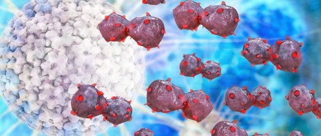
Fotolia_37438608_XS

 ©fotolia

Background to the survey “Cancer: life two years after diagnosis”

The incidence of cancer has been increasing for several decades, but advances in treatment have significantly contributed to reducing the mortality associated with this disease. Although the prognosis remains poor for some cancer locations, the prospects for recovery and long-term survival are showing favourable trends in France for many cancers. Thus 3 million people in France have been or are currently affected by cancer. Cancer remains a hard trial both physically and psychologically. For several years after their diagnosis, people have to cope with the risk of a recurrence, and the side-effects of the disease and treatment, as well as resuming their daily lives.

In order to better know and understand these everyday difficulties, the French National Cancer Institute (INCa) wished to repeat the survey, which was first conducted in 2004 under the aegis of the (French Government) Directorate for Research, Studies, Evaluation and Statistics, on the lives of people two years following their cancer diagnosis. The National Cancer Institute entrusted the survey to Inserm Unit 913, SEESSTIM[1] . This research was conducted through a partnership with the French National Health Insurance Fund for Salaried Workers (CNAMTS), the French Agricultural Social Insurance Fund (MSA), and the Independent Workers’ Health Insurance Scheme (RSI).

Main results of the survey “Cancer: Life Two years After Diagnosis”

The VICAN2 survey addresses the different aspects of life for people with cancer from the initial care for their illness, and their relationship with the care system, to the state of their health two years after diagnosis, the impact of the illness on resources and employment, and problems encountered in their everyday and social lives. It emphasises:

The burden of inequalities

The results of this survey illustrate the scale of the impact of cancer on the existence of those affected, and indicate the burden of health inequalities, all along the person’s pathway. These inequalities sometimes reflect missed opportunities suffered by younger and older people in accessing care; however, they are mostly associated with socioeconomic problems, which sometimes carry as much if not more weight than the location of the cancer, the treatments received or the sequelae experienced. Cancer thus seems to be a factor that aggravates the person’s preexisting social inequalities.

News of the diagnosis is usually given under the right conditions

The survey explores the circumstances of diagnosis. Although the manner in which the diagnosis is given to the patient has advanced since 2004, it is still considered too inhumane by 18% of those surveyed. People with less formal education and lower incomes express this opinion even more often. However, the most obvious variations are seen to be associated with the age and gender of those surveyed: women and younger people most often consider that their diagnosis was given to them in an abrupt manner (This is the case for 28% of women aged from 18 to 40 years).

An apparent improvement in the sharing of information with caregivers

The proportion of people satisfied with their involvement in the choice of treatments has progressed.


Moreover, the proportions of those surveyed who believe that the information provided by caregivers was excessive or over-complicated, or that they themselves were poorly able to formulate their questions, have all considerably decreased in this new survey compared with 2004.

A close association between the impact of the disease and location of the cancer

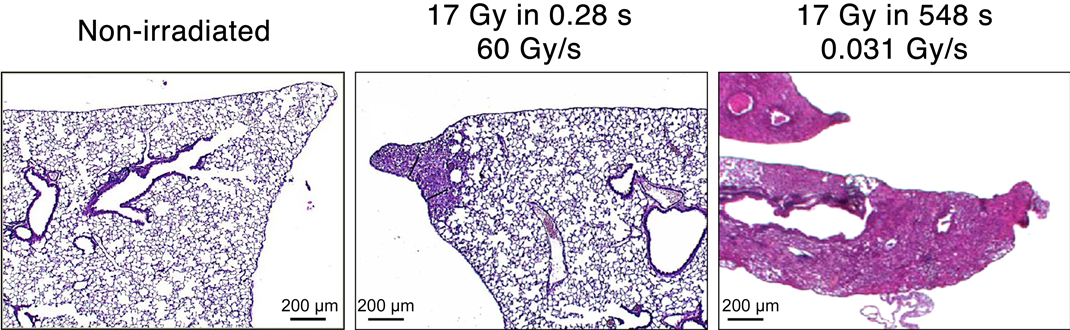
Results show a general deterioration in quality of life caused by cancer; however, this deterioration greatly depends on the location (more frequent for lung cancer, and more rare for prostate cancer), the treatment received and the sequelae experienced.

Apart from than these medical factors, the quality of life measured also depends on possible situations of social fragility (low income, unemployment).

A very marked impact on work situation also

Cancer has an impact on work situation: at the time of diagnosis, eight out of ten people were employed, compared with six out of ten two years later.

The loss of employment more strongly affects the less well educated, younger people and older people, those working as operatives (manual and non-manual workers), who have an insecure work contract or are employed by SMEs.

Moreover, the severity of the cancer underscores the inequalities—the worse the initial prognosis, the greater the difference observed between those working as operatives and those working in management. Thus for a cancer with “a good prognosis,” the rate of staying in employment two years after diagnosis is 89%, for people working in management and 74% for operatives, as against respectively 48% and 28% for cancers with a poor prognosis.

Discrimination infrequent but still present

One out of ten of those surveyed claim that they have already experienced prejudice or discrimination directly related to their disease from their close associates. Women and younger people surveyed are the most likely to report such experiences.

Discrimination is experienced by up to 25% of people who declare that their household has financial problems (compared with 4% of those who claim to be “comfortable”). Social inequalities are thus reflected in the experience of discrimination at the hands of close associates.

These results will be discussed at a colloquium organised on Tuesday 10 June by the National Cancer Institute, bringing together all stakeholders, particularly patients and representatives of patient associations. This reflection will help to support the actions of the 2014-2019 Cancer Plan, which is especially aimed at limiting the social and economic consequences of the disease, facilitating the consideration of cancer in the workplace, continuation of schooling and study, and authorises a “right to be forgotten” for those seeking loans.

[1] Sciences Économiques et Sociales de la Santé et Traitement de l’Information Médicale (Economics and Social Sciences, Health Care Systems and Societies)

Metastases: Tumour Cell Dissemination

When tumour cells acquire the capacity to move around and invade other tissues, there is a risk of metastases and cancer treatment becomes more difficult. Carine Rossé, INSERM research fellow and Philippe Chavrier, Research Director at CNRS, working alongside Dr. Anne Vincent-Salomon, medical researcher at the Institut Curie, have recently discovered one of the mechanisms that allow triple negative breast cancer cells to exit the mammary gland. These results were published on-line by PNAS on 21st April 2014.

Understanding how primary tumours infiltrate tissues and how certain cells detach and migrate to form metastases, is a major challenge in current cancer research. As such, the Institut Curie launched the incentive and cooperative research programme (PIC) “Breast Cancer: Invasion and Motility” in 2011 to give teams battling in this field access to important resources. Both programme coordinators Philippe Chavrier, Research Director at CNRS1 and medical researcher2 Anne Vincent-Salomon have joined forces in order to better understand how breast cancer cells move away and invade other tissues.

Furthermore, their latest discovery focuses on one of the most aggressive forms of cancer today – triple negative breast cancer. Dr. Anne Vincent-Salomon explains “this type of breast cancer is devoid of oestrogen and progesterone receptors with no overexpression of HER2. Women who have this type of cancer can neither benefit from hormonal therapy nor specific anti-HER2 therapy such as Herceptin”

A Tunnel in the Basement Membrane

Carine Rossé3 and Philippe Chavrier have discovered how these breast cancer cells break away from their original tissue. According to Philippe Chavrier “the tumour cells escape by digging a tunnel in the basement membrane that acts as a barrier around the mammary gland”.

His team has shown that the PKCλ protein and protease MT1-MMP are driving this cell “invasion”.  If PKCλ is “depleted” in stem cells derived from aggressive breast cancer, the supply of MT1-MMP to the cell surface is inhibited and cell invasion is no longer possible.

Thanks to the Biological Research Centre4 at the Institut Curie, who keep close to 60,000 tumour samples, researchers have also directly studied these proteins in tumour samples. “We have seen expressive correlation between both proteins associated with unfavourable prognosis in breast cancer” explains a researcher. “We have also identified a mechanism that allows both proteins work together to promote breast tumour cell invasion.”

Researchers have found an essential step in the quest for the early identification of highly invasive tumours, or even blocking the formation of metastases.

“We have identified some interesting potential therapeutic targets, but they remain to be validated” clarified a biologist. Drugs capable of halting these mechanisms will follow in time with the help of chemists.

Chain Reaction

interaction proteines

Zoom-in of the inside of a cell is used to see the reactions between various proteins triggered by PKCλ. This latest protease MT1-MMP traffic “control” (red) enables the association of cortactin (green) with dynamin 2 (blue). This reaction sequence is necessary to allow the cell separate from its neighbours and invade other tissues.
© Carine Rossé – Philippe Chavrier/ Institut Curie


1 1st class Research Director at CNRS, Philippe Chavrier is head of the “The Membrane and Cytoskeleton Dynamics” group “Subcellular Structure and Cellular Dynamics – Institut Curie/CNRS” laboratory, run by Bruno Goud.
2 Anne Vincent-Salomon is a medical pathologist in the Department of Biopathology at the Institut Curie Hospital Complex and has been a researcher in the “Genetics and Developmental Biology – Institut Curie/Inserm/CNRS” laboratory since January 2014, run by Prof. Edith Heard.
3 Carine Rossé is an INSERM research fellow within the “The Membrane and Cytoskeleton Dynamics” group, run by Philippe Chavrier.
4 Department of Biopathology at the Institut Curie Hospital Complex.
fermer