Menu

Beginning of life: how does symmetry come into play?

The first embryonic division, which follows gamete fusion (oocyte and spermatozoon), starts the development of a new individual, the genesis of a functional adult body. This division is symmetric in the one-cell embryo stage (also known as the zygote); it leads to the formation of two daughter cells of identical size. Conversely, it is asymmetric in the oocyte, which has the same size and shape as the zygote. Why? What directs the zygote to divide symmetrically, while the oocyte divides asymmetrically during meiosis? Such are the questions pondered by Marie-Emilie Terret, a researcher at Inserm, and Marie-Hélène Verlhac, a researcher at CNRS and director of the Asymmetric Divisions in Oocytes team at the Center for Interdisciplinary Research in Biology (CIRB; Inserm/CNRS/Collège de France)[1]. By combining biology, physics and mathematics, the researchers succeeded in demonstrating, in mice, the regulatory mechanics that determine, in a very short time, the geometry and hence the fate (symmetric or asymmetric division) of the cell. In future, the elements arising from this work may contribute to improving the efficacy of in vitro fertilisation.

Details of these results are published today in the journal Nature communications.


At the one-cell stage, the embryo very closely resembles an oocyte: a round, isolated cell, similar in size to an oocyte. The geometry of a cell’s division is determined by the position of the microtubule spindle, the machinery that carries and separates the chromosomes. In most animal cells, the centrosomes organise the microtubule network, which is essential to the formation and positioning of the division spindle. But oocytes and zygotes lack centrosomes. A major difference between these two types of cells, however, lies in the geometry of their divisions. Indeed, during meiosis oocytes divide in an extremely asymmetric manner in terms of size, allowing the formation of a single enormous oocyte and the expulsion of “polar bodies” containing the excess genetic material. Conversely, the zygote divides in a perfectly symmetric manner, leading to the formation of two daughter cells of identical size.

CP Terret

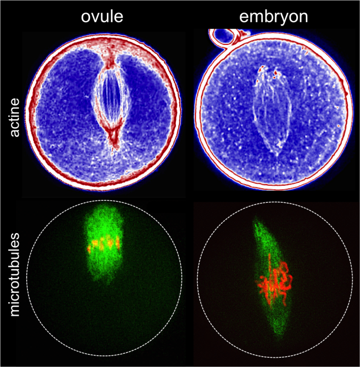
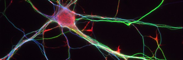
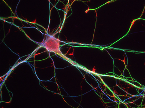
Mouse oocyte and embryo at the one-cell stage. The top images show the actin networks; the bottom images show the microtubule spindles with aligned chromosomes.

(c) Marie-Emilie Terret

The geometry of division is determined by the position of the spindle: eccentric in oocytes, centred in zygotes. The Asymmetric Divisions in Oocytes team had previously shown that the eccentric position of the division spindle in the oocyte depends on the mechanics of the actin networks. In the work published today, the team of researchers shows that the centred position of the division spindle in the zygote is also due to the mechanics of the actin networks, but controlled differently.

Three steps are required for this symmetric division:

  1. The coarse centring of the male and female pronuclei, requiring a network of actin and myosin-Vb.
  2. The fine centring of the division spindle, requiring strong rigidity in the oocyte.
  3. The passive maintenance of the spindle in the centre of the cell.

The mechanics of actin/myosin networks therefore make it possible to change from an asymmetric division to a symmetric division, a change in geometry needed for the transition from oocyte to embryo.


The research team is already formulating hypotheses regarding the mode of action of actin, which plays a role in the physical characteristics of the cell membrane (rigid or soft), which in turn influence the geometry of division. “Our next work will involve the more detailed study of interactions between actin and microtubules in an attempt to understand their respective roles in the architecture of the cell at the moment of division, and the potential involvement of other intermediary proteins,” explains Marie-Emilie Terret.

A better understanding of the physical characteristics and behaviour of the oocyte during division, whether fertilised or not, has the potential to contribute useful new elements to medically assisted conception. During in vitro fertilisation (IVF), for instance, the storage temperature of the oocytes can have an impact on the quality of the actin networks, and consequently affect division, and hence the formation of a zygote.

Increasing the efficacy of IVF might therefore represent a long-term objective for this research team, one of the few in France working on this theme.

[1] In collaboration with researchers from the Laboratory for Analysis and Modelling for Biology and Environment (LAMBE; CNRS/CEA/University of Évry Val d’Essonne/Cergy Pontoise University), the Theoretical Physics of Condensed Matter Laboratory (LPTMC; CNRS/UPMC) and the Curie Physical Chemistry Laboratory (CNRS/Institut Curie/UPMC).

From pluripotency to totipotency

While it is already possible to obtain in vitro pluripotent cells (ie, cells capable of generating all tissues of an embryo) from any cell type, researchers from Maria-Elena Torres-Padilla’s team have pushed the limits of science even further. They managed to obtain totipotent cells with the same characteristics as those of the earliest embryonic stages and with even more interesting properties. Obtained in collaboration with Juanma Vaquerizas from the Max Planck Institute for Molecular Biomedicine (Münster, Germany), these results are published on 3rd of August in the journal Nature Structural & Molecular Biology.
Cellules Souches embryonnaires

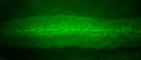
Human embryonic stem cells have the potential to form in vitro neural tube -like structures of the embryo. ©Inserm/Benchoua Alexandra
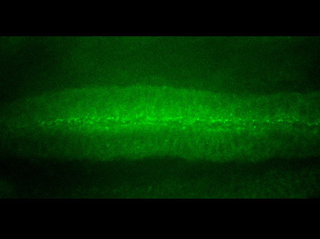
Just after fertilization, when the embryo is comprised of only 1 or 2 cells, cells are “totipotent“, that is to say, capable of producing an entire embryo as well as the placenta and umbilical cord that accompany it. During the subsequent rounds of cell division, cells rapidly lose this plasticity and become “pluripotent”. At the blastocyst stage (about thirty cells), the so-called “embryonic stem cells” can differentiate into any tissue, although they alone cannot give birth to a foetus anymore. Pluripotent cells then continue to specialise and form the various tissues of the body through a process called cellular differentiation.

For some years, it has been possible to re-programme differentiated cells into pluripotent ones, but not into totipotent cells. Now, the team of Maria-Elena Torres-Padilla has studied the characteristics of totipotent cells of the embryo and found factors capable of inducing a totipotent-like state.

When culturing pluripotent stem cells in vitro, a small amount of totipotent cells appear spontaneously; these are called “2C-like cells” (named after their resemblance to the 2-cell stage embryo). The researchers compared these cells to those present in early embryos in order to find their common characteristics and those that make them different from pluripotent cells. In particular, the teams found that the DNA was less condensed in totipotent cells and that the amount of the protein complex CAF1 was diminished. A closer look revealed that CAF1 -already known for its role in the assembly of chromatin (the organised state of DNA)- is responsible for maintaining the pluripotent state by ensuring that the DNA is wrapped around histones.

Based on this hypothesis, the Torres-Padilla team were able to induce a totipotent state by inactivating the expression of the CAF1 complex, which led to chromatin reprogramming into a less condensed state.

These results provide new elements for the understanding of pluripotency and could increase the efficiency of reprogramming somatic cells to be used for applications in regenerative medicine.

Natural reparative capacity of teeth elucidated

Researchers at Inserm and Paris Descartes University have just taken an important step in research on stem cells and dental repair. They have managed to isolate dental stem cell lines and to describe the natural mechanism by which they repair lesions in the teeth. This fundamental discovery will make it possible to initiate unprecedented therapeutic strategies to mobilise the resident dental stem cells and magnify their natural capacity for repair.

These results are published in the journal Stem Cells.

The tooth is a mineralised organ, implanted in the mouth by a root. The “living” part of the tooth or dental cavity is the dental pulp (in yellow in the photograph shown opposite) composed of vessels and nerves. Around it is a hard substance, the dentine or ivory, which is in turn covered by an even harder tissue, the enamel. When a dental lesion appears, the dormant stem cells in the pulp awaken and try to repair the tooth by an unknown process.dent1

3D modelling of a teeth. The dental pulp is in yellow. ©Inserm/ Chappard, Daniel

In this study, the researchers from Inserm and Paris Descartes University at Unit 1124, “Toxicology, Pharmacology and Cellular Signaling,” have succeeded in extracting and isolating tooth stem cells by working on the pulp from the mouse molar.

The researchers were thus able to analyse the cells in detail, and identify 5 specific receptors for dopamine and serotonin on their surface, two neurotransmitters that are essential to the body (see schema on page 2).

The presence of these receptors on the surface of these stem cells indicated that they had the ability to respond to the presence of dopamine and serotonin in the event of a lesion. The researchers naturally wondered what cells might be the source of these neurotransmitters, a warning signal. It turns out that the blood platelets, activated by the dental lesion, are responsible for releasing a large quantity of serotonin and dopamine. Once released, these neurotransmitters then recruit the stem cells to repair the tooth by binding to their receptors (see schema on page 2). The research team was able to confirm this result by observing that dental repair was absent in rats with modified platelets that do not produce serotonin or dopamine, i.e. in the absence of the signal.

“In stem cell research, it is unusual to be simultaneously able to isolate cell lines, identify the markers that allow them to be recognised (here the 5 receptors), discover the signal that recruits them (serotonin and dopamine), and discover the source of that signal (blood platelets). In this work, we have been able, unexpectedly, to explore the entire mechanism,” explains Odile Kellermann, leader of the team from Inserm and Paris Descartes University, and the main author of this work.

To take things a stage further, the researchers tried to characterise the different receptors they found. One of the 5 receptors does not seem to affect the repair process. On the other hand, the other 4 turn out to be strongly involved in the repair process. In vivo blocking of just one of them is enough to prevent dental repair.

“Currently, dentists use pulp capping materials (calcium hydroxide) and tricalcium phosphate-based biomaterials to repair the tooth and fill lesions. Our results lead us to imagine unprecedented therapeutic strategies aimed at mobilising the resident pulpal stem cells in order to magnify the natural reparative capacity of teeth without use of replacement materials,” concludes Odile Kellermann.

The foundations have been laid for extending this research done in rodents to stem cells of the human tooth in order to initiate new strategies for repairing teeth.

schéma mécanisme dent EN

© Inserm / Odile Kellermann, Anne Baudry

A mechanism for eliminating proteins accidentally localised to the cell nucleus

An international collaboration coordinated by the German Cancer Research Center (DKFZ) (University of Heidelberg), including French researchers from the Institute of Genetics and Development of Rennes (IGDR) (CNRS/University of Rennes 1) under the leadership of Gwenaël Rabut, Inserm Researcher, and teams from Sweden and Canada, has just demonstrated a new molecular mechanism that may allow cells to destroy proteins accidentally localised to the nucleus.

This research is published in the journal Nature.

Biological processes are far from perfect. Despite millions of years of refinement, the molecular mechanisms that help living beings to function make many errors, which can have serious consequences unless they are detected and corrected. For example, many cancers are caused by errors that occur while our genetic material is being copied. Similarly, incorrect folding of some neuronal proteins leads to the formation of toxic aggregates that disrupt nervous system function and cause neurodegenerative diseases, such as Alzheimer’s disease or Parkinson’s disease.

To prevent this happening, cells have established complex molecular mechanisms that control the quality of proteins and eliminate those that are defective. These mechanisms are localised and implemented mainly in the cytoplasm (the cellular compartment where the proteins are synthesised).

While working on several factors involved in protein quality control, researchers discovered that some of them are also localised in the cell nucleus (the compartment that contains the genetic material), and that they enable the degradation of proteins that are abnormally present in this compartment.

During this study, researchers from the Institute of Genetics and Development of Rennes (including Gwenaël Rabut, Inserm Researcher, project coordinator and manager in Rennes, and Ewa Blaszczak, doctoral student, joint first author of the article) were able to observe that these factors involved in protein quality control interact with each other in the nucleus and bring about the ubiquitination (the step preceding degradation) of a protein accidentally localised to the nucleus.

By using an observation method developed at the University of Heidelberg, based on fluorescence timing in the proteins of interest, the researchers were able to identify some twenty proteins the degradation of which depended on quality control factors localised in the nucleus. Since several of these proteins are normally localised to the cytoplasm, and accumulate in the nucleus when they are no longer degraded, the researchers propose that this quality control system serves to eliminate not only defective proteins, but also proteins accidentally localised to the nucleus.

These discoveries were made using a model organism, baker’s yeast, but it is likely that similar mechanisms also exist in humans.

From rectal cells to neurons : keys to understanding transdifferentiation

How can a specialized cell change its identity? A team from the Institut de Génétique et de Biologie Moléculaire et Cellulaire (CNRS/INSERM/Université de Strasbourg) investigated a 100% effective natural example of this phenomenon, which is called transdifferentiation. This process, by which some cells lose their characteristics and acquire a new identity, could be more generally involved in tissue or organ regeneration in vertebrates, and is a promising research avenue for regenerative medicine. This study identifies the role of epigenetic factors involved in this conversion, underlines the dynamic nature of the process, and shows the key mechanisms for effective transdifferentiation. This work, conducted in collaboration with the Institut Curie1, was published on August 15, 2014 in Science.

How can a specialized cell change its identity? A team from the Institut de Génétique et de Biologie Moléculaire et Cellulaire (CNRS/INSERM/Université de Strasbourg) investigated a 100% effective natural example of this phenomenon, which is called transdifferentiation. This process, by which some cells lose their characteristics and acquire a new identity, could be more generally involved in tissue or organ regeneration in vertebrates, and is a promising research avenue for regenerative medicine. This study identifies the role of epigenetic factors involved in this conversion, underlines the dynamic nature of the process, and shows the key mechanisms for effective transdifferentiation. This work, conducted in collaboration with the Institut Curie1, was published on August 15, 2014 in Science.

Our body is constituted of cells that acquired characteristics during development and that fulfill a precise function in each organ: we call these differentiated cells. Generally cells maintain their specificity until they die, but it has been proven that some cells can change state and acquire new functions. This is rare but is found in many species and is called “transdifferentiation”.

The team studied this process in C. elegans, a small transparent nematode, where a rectal cell transforms naturally into a motor neuron. This change from one cell type into another occurs without cell division, by a succession of well defined steps that always lead to the same result. The researchers investigated the factors that make the conversion process so stable.

The team had elucidated the role of several transcription factors2 in this transdifferentiation.

  But these new results have shown the role of so-called “epigenetic” factors that can modulate gene expression. Two protein complexes are involved in the mechanism. These enzymes act on a histone3 and when a mutation changes their action, the transdifferentiation stops and the rectal cell no longer transforms into a neuron.

The researchers observed that the two complexes act at different steps and that their role may change as a function of the transcription factors with which they are associated. These results underline the importance of the correct chain of steps for each of these molecules: the dynamic nature of the transdifferentiation mechanism is essential to its stability.

The respective role of genetic and epigenetic factors in biological processes is a hotly debated subject. This work shows how each of these factors acts in transdifferentiation: transcription factors handle initiation and progress whereas epigenetic factors guarantee the constant result. The study even goes further, showing that under “normal” conditions, the epigenetic factors are incidental (even when they are absent the conversion occurs relatively efficiently) but that they are indispensable when there are environmental stressors. So they have a crucial role in maximizing the mechanism’s efficacy and ensuring that it remains stable in the face of external variations.

Transdifferentiation is a phenomenon that is poorly understood. It may be involved in the organ regeneration that we observe in some organisms, for example newts, which can reconstruct their eye lens after injury. These results bring key new information to help us understand how to control this process and may open the path to promising therapies, in particular in the field of regenerative medicine.

image onion

© Elodie Legrand et Sophie Jarriault
We can compare this process to the layers of an onion. Transcription factors are at the heart of process efficiency, while epigenetic factors form the outer layers that protect the mechanism from attacks and environmental change.

(1) Unité Génétique et Biologie du Développement (CNRS/INSERM/Institut Curie)
(2) Proteins necessary for the DNA to RNA conversion
(3) Protein in the nucleus, around which the DNA rolls.

Lipids serving the brain

Consuming oils rich in polyunsaturated fatty acids, especially ‘omega 3’, is good for our health. But the mechanisms explaining these effects are poorly understood. Researchers from the Institute of Molecular and Cellular Pharmacology (CNRS/Nice Sophia Antipolis University), the Compartmentation and Cellular Dynamics Unit (CNRS/Curie Institute/UPMC) of Inserm and Poitiers University1 were interested in the effect of lipids carrying polyunsaturated chains when they are taken into cell membranes. Their study shows that the presence of these lipids makes them more malleable and thus much more sensitive to the action of proteins that deform and split them. These results, published in the journal Science on 8 August 2014, offer a route to explaining the extraordinary efficiency of endocytosis2 in neuronal cells.

Membrane plasmique en vert, transferrine en rouge

©Inserm/Barelli Hélène


The consumption of polyunsaturated fatty acids (such as ‘omega 3’ fatty acids) is beneficial for health. These effects go from neuronal differentiation to protection against cerebral ischemia3. However, the molecular mechanisms responsible for their effects are quite poorly understood. The researchers therefore looked into the role of these fatty acids in cell membrane function.

To ensure that a cell functions properly, its membrane must be able to deform and split up to form small vesicles. This phenomenon is called ‘endocytosis’. These vesicles generally allow cells to encapsulate molecules and transport them. For neurones, these synaptic vesicles perform the role of drive belt to the synapse for nerve impulses. They are formed inside the cell then move to its edge and merge with its membrane, in order to transfer the neurotransmitters that they contain. They are then reformed in less than one-tenth of a second: this is synaptic recycling.

In this work published in Science, the researchers showed that cellular or artificial membranes rich in polyunsaturated lipids are much more sensitive to the action of two proteins, dynamine and endophiline, which deform and split membranes. Other measurements from the study and simulations suggest that these lipids also make membranes more malleable. By facilitating the deformation and fission steps needed for endocytosis, the presence of polyunsaturated lipids could explain the speed of recycling for these synaptic vesicles.

The abundance of these lipids in the brain could therefore represent a major advantage for cognitive functions.

This work partially lifts the shroud covering the mode of action of omega 3s. Even if we know that our body cannot make them and that only suitable foods (rich in fish oil, etc.) provide them to us, it seems important to continue this work to understand the link between the functions these lipids perform in the neuronal membrane and their beneficial effects on health.

endocytose

Membranes containing monounsaturated lipids (left) and polyunsaturated lipids (right) after adding dynamine and endophiline. In a few seconds, the membranes rich in polyunsaturated lipids undergo multiple splits.© Mathieu Pinot

Transferrin endocytosis (iron transport) in cells containing polyunsaturated lipids in their membranes (right) compared to cells deprived of them (left). In 5 minutes, the number of endocytosis vesicles formed (internalised transferrin in red) is increased nearly 10 times, reflecting facilitated endocytosis.© Hélène Barelli

(1) This study was carried out in collaboration with teams from the Joint Applied Microscopy Centre (Nice Sophia Antipolis University) and the Signalling and Membrane Ionic Transport laboratory (CNRS/Poitiers University/Tours François Rabelais University).

(2) Endocytosis is the process by which cells absorb various substances present in the surrounding environment by encapsulating them in a lipoprotein membrane. It plays a role in various physiological functions.

(3) For example, see previous work by the Institute of Molecular and Cellular Pharmacology on this type of cerebro-vascular accident: Polyunsaturated fatty acids are potent neuroprotectors. Lauritzen I, Blondeau N, Heurteaux C, Widmann C, Romey G, Lazdunski M; EMBO J. (2000) 19:1784-93.

Mechanics and genetics: an indispensable cocktail for embryonic development

In the fruit fly Drosophila and zebrafish, mechanical strain may activate the genetic cascade that initiates the formation of the future organs during embryogenesis. A discovery made by Emmanuel Farge (Inserm Research Director at Institut Curie) and his staff might explain the emergence of the first complex organisms more than 570 million years ago.
The results of this work are published in the journal
Nature Communications.


embryon

signal de phosphorylation de la béta-caténine dans le tissu ventral qui invagine (mésoderme) dans l’embryon de Drosophile en vue ventrale  de haut © E Farge

Living things exist in a multiplicity of forms. At the very beginning—whether they are early multicellular or embryonic forms of life—they are all nothing more than a mass of cells. A long series of morphological changes causes all existing life forms to develop from this one form.

The embryo adopts a particular form at each stage of its development. These successive deformations, which are genetically regulated, in turn create mechanical strain within the embryo. This strain also seems to influence or even regulate the expression of genes involved in development.

From Drosophila to zebrafish

“Whether in zebrafish or Drosophila, we have found that activation of the β-catenin protein at the beginning of embryonic development follows mechanical compression developed during the very first change in the form of the embryo,” explains the researcher.


At the very start of development, a morphological change—known as invagination in Drosophila and epiboly in the zebrafish—allows expression of the genes that specify the mesoderm1[1], in response to the mechanical activation of β-catenin in tissues that are particularly deformed by these movements. Complex organs such as the muscles, heart, or gonads are derived from the mesoderm.

In their publication in Nature Communications, Emmanuel Farge and his team show in detail that the mechanical strain occurring during this morphological transition induces a modification of β-catenin (phosphorylation), which induces its relocation from the surface of the cell to its centre.

This protein may take on several roles: on the cell surface, it is responsible for cell-cell adhesion and may thus undergo mechanical strain, become phosphorylated and then released into the cell; within the cell, it can activate certain genes and thus modify the fate of the cells. Thus mechanical compression can lead to the acquisition of an identity similar to that of mesodermal cells following relocation of β-catenin to the interior of the cell. “To reproduce the mechanical strain naturally undergone by the embryo, we introduced liposome-encapsulated magnetic nanoparticles into the embryo, which we then exposed to a micromagnet.

An answer to the origins of evolution into complex organisms?

“The main point is that the mechanosensitivity of gene expression has been conserved by Drosophila and zebrafish during evolution,” explains the researcher. “Its origin therefore goes back to the last common ancestor shared by the two species, i.e. over 570 million years.” Moreover, specialists in evolution associate this same period with a major transition in evolution: the emergence of the mesoderm in ancestral life forms, possibly related to the jellyfish, for example, which do not have a mesoderm. The origin of this transition, which led to the development of complex organisms, such as the vertebrates, has not been well understood until now. Researchers are therefore only now on-track to answer this long-standing question.

Going back still further in time, mechanosensitivity might have even contributed to the emergence of the very first organisms. And what if compression, triggered, for example, in a mass of cells because it was resting on the substratum, gave rise to local deformation of the cell mass and thus activated the first invagination of the very first primitive gastric organ, as suggested by the experiments previously done by the team?

Cancer genes, reactivation of sensitivity to compression

Since the genes for embryonic development are implicated in the process of tumour progression, the mechanical induction of genes constitutes a new avenue for studying the development of cancers. The β-catenin protein is not unknown to cancer specialists. Thus during development of colon cancer, dysregulation of the β-catenin pathway is often described as one of the events correlated with loss of the APC gene. Furthermore, the development of cancer leads to the generation of physical strain affecting the neighbouring tissues.
It is a little as if the mechanism necessary for the development of the embryo were reawakened at the wrong moment. “Indeed”, underlines Emmanuel Farge, “when all is going well, APC protein degrades the β-catenin released into the cytoplasm by abnormal mechanical cues. When APC is mutated (which happens in 80% of colon cancers correlated with genome modifications), the β-catenin released into the cytoplasm is no longer degraded effectively, and is free to enter the nucleus and stimulate the expression of genes that promote tumour development.”


[1]  The mesoderm is one of the three embryonic germ layers, and is formed between the endoderm and ectoderm at the time of gastrulation. During development, it gives rise to most of the internal organs. Its existence is a feature of the most highly evolved organisms.

Dwarfism: a new development to restore bone growth

Achondroplasia is the most common form of dwarfism, affecting roughly one child in every 15,000 births[1]. Inserm researcher Elvire Gouze, and her associates from the Mediterranean Centre for Molecular Medicine in Nice (Inserm Unit 106), have succeeded in restoring bone growth in mice suffering from this developmental pathology. The proof-of-concept created by the researchers is for a therapy based on injecting a particularly promising human growth factor, which restores the growth process in long bones. Its results include a reduced mortality rate in the treated mice, with no complications associated with the disease. No apparent toxicity was observed over the short term.

The results of this research were published in the  Science Translational Medicine review on 18 September.

Elvire Gouze

Sophie Garcia and Elvire Gouze in Inserm Unit 1065 “Centre méditerranéen de médecine moléculaire” in Nice

Achondroplasia is a rare genetic disease characterized by abnormal bone development. The related growth failure affects bones in the upper and lower limbs, and some bones in the skull; people suffering from it are short, reaching no more than an average of 135 cm in adulthood. In the most severe cases, deformation of the skull and vertebrae may result in neurological and/or orthopaedic complications. This pathology is caused by mutations in the FGFR3 (Fibroblast growth factor 3) gene.  The protein produced by this gene is a receptor known for its role in bone growth regulation. Normally, growth can only occur through a subtle mechanism during which the FGF growth factor bonds with the FGFR3 receptor and then separates from it. In the case of achondroplasia, the receptor/growth factor pair is disturbed and prevents the bone from growing in a constant manner.

A new strategy to restore bone growth

In this study, researchers from Inserm and the Université de Nice Sophia Antipolis have found a way to prevent constant protein activation. They implemented a new strategy, which consists of using a decoy – functional soluble human FGFR3 receptors – that is injected in mice afflicted with the disease, thus restoring the equilibrium required between bone growth activation and inhibition.

The solution containing soluble FGFR3 receptors was injected into growing mice suffering from dwarfism twice a week over a three-week period.  The additional normal receptors made it possible for the growth factor to bond and separate normally, thus restoring bone growth. Mutated mice then grew normally and reached the average adult size. Once the therapy had stopped, the researchers then monitored the mice over an eight month period to check there were no signs of therapy toxicity. During this monitoring, the researchers observed that an increased pelvis size enables reproduction with litters identical to disease-free mice.

“Rather surprisingly, our strategy prevents the most severe complications observed in mice (reduced mortality rate, respiratory problems, etc.). This could lead us to believe that injection-based therapy could replace surgery for children suffering from this disease” explains Elvire Gouze, Inserm researcher.

Preventing the development of achondroplasia

Today, there are no proven therapies to prevent the development of the disease, even if some (such as growth hormone injections or surgical limb-lengthening) have undergone trials without any convincing results.

“The product that we tested has major advantages compared with those tested in other ongoing trials: its lifetime in the body is sufficiently long, meaning daily injections are not necessary. We think that our approach could be effective when treating children with achondroplasia and possibly other forms of dwarfism” underlines the researcher, who is the main author of the study.

The researchers will now endeavour to verify that there are no long-term toxic effects. Before undertaking clinical studies in human patients, they must also identify the minimum dose at which the therapy is effective and when it becomes toxic. Another area to be explored is to determine whether it is possible to begin the therapy later, thereby increasing the number of patients who could benefit from this treatment.


[1] Source: Orphanet Achondroplasie

A new therapeutic strategy to combat prion and Alzheimer’s diseases

A work performed by the teams headed by Benoit Schneider and Odile Kellermann (INSERM Unit 747, team “Stem cells, Signalling and Prions”, Université Paris Descartes) as well as Jean-Marie Launay’s team (INSERM Unit 942 Hôpital Lariboisière and the FondaMental Foundation) was published this week in the magazine Nature Medicine. The article revealed that in neurons, an enzyme, the kinase PDK1, is involved in the accumulation of the pathological proteins involved in prion and Alzheimer’s diseases. The researchers show that the pharmacological inhibition of this enzyme exerts a beneficial effect towards both pathologies.

Details of this research were published in the magazine Nature Medicine

Différenciation cellulaire

Mouses Neurones – 7 days – © Inserm/L.Peris

Prion diseases (Creutzfeld-Jakob disease in humans) and Alzheimer’s disease are associated with an accumulation of abnormal proteins in the brain. These are the scrapie prion protein (PrPSc) in the case of prion diseases and Aß amyloid peptides in the case of Alzheimer’s. In the brain, PrPSc and Aß peptides exert their toxic effect by causing the death of neurons, at the root of the clinical symptoms associated with these diseases.

It is acknowledged that the production of the pathological proteins PrPSc and Aß40/42 originate from a defect in the physiological cleavage of the entire, non-pathological prion protein (PrPC) or of the amyloid peptides precursor (APP). However, it remained unsolved why this cleavage, which normally protects neurons, is altered in prion and Alzheimer’s diseases.

The work performed by Benoit Schneider (CNRS researcher in INSERM Unit 747, “Stem cells, Signalling and Prions”, Université Paris Descartes) and Jean-Marie Launay (INSERM Unit 942 Hôpital Lariboisière) in collaboration with other French teams working in the prion field has just identified a cascade of reactions that blocks the beneficial cleavage of PrPC and APP by the alpha-secretase TACE (an acronym for the TNFα Converting Enzyme). The researchers demonstrate how TACE dysregulation contributes to neurodegeneration by causing the accumulation of the PrPSc and Aß40/42 pathological proteins and exacerbating neuron sensitivity to inflammation.

Under normal physiological conditions, TACE is present on the surface of neurons, where it cleaves PrPC, APP and the receptors to TNFα inflammatory factor (TNFR), thus restricting the production of the pathological proteins PrPSc and Aß and protecting neurons from the toxic effects of TNFα.

schéma Prions Alzheimer en

© Benoit Schneider & Mathéa Pietri, August 2013

In neurons infected by pathogenic prions as in the “Alzheimer’s” neurons, the TACE protease is no longer present on the cell surface but is found inside neurons. This internalization diverts TACE away from its substrates, that is PrPC, APP and TNFR, and thereby cancels its neuroprotective activity. The researchers reveal for the first time that the kinase PDK1 plays a key role in controlling the localization of TACE in neurons. The overactivation of PDK1 is responsible for the internalization of TACE in diseased neurons (those infected by prions and Alzheimer’s neurons) as in the brains of patients suffering from Alzheimer’s disease.

The pharmacological blockade of PDK1 relocates TACE to the surface of neurons and restores its neuroprotective function. The inhibition of PDK1 protects neurons from neurodegeneration by rescuing the physiological cleavage of PrPC, APP and TNFR by TACE.

“Based on our work on prion infection, we succeded in identifying PDK1 as a new therapeutic target not only for Creutzfeld-Jakob disease but also for Alzheimer’s disease” explained the researchers.

The action of PDK1 on TACE was demonstrated in vitro using a neuronal cell line and cultures of neurons isolated from the brains of mice that had been infected by pathogenic prions, and in vivo using animal models. Treatment with PDK1 pharmacological inhibitor attenuates the motor deficits and extends the life span of mice infected with prions. Using three mouse models of Alzheimer’s pathology, the researchers showed that the treatment also counteracted memory and cognitive deficits associated with Alzheimer pathogenesis.“Because treatments available to combat prion and Alzheimer’s diseases are few and their efficacy limited, these results could open up new avenues for the treatment of these neurodegenerative diseases”, conclude the researchers.

By demonstrating that the inhibition of PDK1 alleviates both prion and Alzheimer’s diseases, these data argue that at a mechanistic level AD links to prion diseases. Dysregulation of PDK1-dependent TACE cleavage activity emerges as a central event in neurodegenerative pathways involved in both diseases.

The challenge is now to understand how pathogenic prions or amyloid Ab peptides trigger PDK1 activation.

Novel molecules to target the cytoskeleton

The dysfunction of the cytoskeleton, a constituent element of the cell, is often associated with pathologies such as the onset of metastases. For this reason, it is a target of interest in numerous therapies. Teams from CNRS, the Université de Strasbourg and Inserm, led by Daniel Riveline1, Jean-Marie Lehn2 and Marie-France Carlier3, have synthesized molecules capable of causing rapid growth of actin networks, one of the components of the cytoskeleton. This is a breakthrough because, until now, only molecules that stabilize or destroy the cytoskeleton of actin have been available. These compounds with novel properties, whose action has been elucidated both in vitro and in vivo, provide a new tool in pharmacology. This work was published in the journal Nature Communications on 29 July 2013.

The cytoskeleton is mainly composed of actin filaments and microtubules. Made of polymers in dynamic assembly and constantly constructing and deconstructing itself, it affects numerous cellular processes such as intracellular movement, division and transport. It is involved in key steps of embryogenesis and other processes essential to life. Consequently, its malfunctioning can lead to serious pathologies. For example, the onset of certain metastases is revealed by an increased activity of the cytoskeleton. Identifying new molecules that target the cytoskeleton thus represents a major challenge.

Until now, the molecules known and used in pharmacology had the effect of stabilizing or destroying the cytoskeleton of actin. Actin allows vital actions to be performed by assembling and disassembling itself spontaneously, continually and rapidly in the form of filaments that organize themselves and form networks of parallel bundles or intertwined meshes (known as lamellar networks). Derived from supramolecular chemistry[4], the new compounds synthesized by the researchers have original properties: within several minutes, they bring about the growth of lamellar networks of actin filaments. This is the first time that a pharmacological tool induces growth of the actin network — something that living organisms do all the time. In this way, the researchers have shown that the action of these compounds is specific in vivo (on cells). In addition, they have identified the growth mechanism of the actin network by comparative in vivo and in vitro studies in order to ensure the validity of the process.

For cellular or molecular biology, this tool proposes a new mode of possible action on the cytoskeleton and thus opens new research perspectives for deciphering the living world. This finding could lead to the development of new compounds, derived from the same chemistry, and potential candidates for new therapies targeting the cytoskeleton.

[1] Institut de Science et d’Ingénierie Supramoléculaires (CNRS/Université de Strasbourg) and Institut de Génétique et de Biologie Moléculaire et Cellulaire (CNRS/Université de Strasbourg/Inserm).

[2] Institut de Science et d’Ingénierie Supramoléculaires (CNRS/Université de Strasbourg).

[3] Laboratoire d’Enzymologie et Biochimie Structurales of CNRS.

[4] Supramolecular chemistry, the science of self-assembly and self-organization at the molecular scale, focuses on chemical entities resulting from the interactions between molecular objects.

How is the male genome preserved until it reaches the egg?

When the male genome carried in the spermatozoid leaves the male body to reach the egg, it undergoes numerous transformations. A team led by Saadi Khochbin in Mixed Research Unit 823 at the Institut Albert Bonniot Research Centre (Inserm/Joseph Fourier University) in Grenoble has described the molecular mechanisms that enable the transmission of the male genome to the egg. The researchers have revealed the essential role played by a tiny structure  which compact and preserve the genome in the spermatozoid during its journey to the egg. These results were published on July 24th in the journal Genes & Development.

Spermatozoide-khochbin

Spermatozoid – © Inserm / Denise Escalier

One of the challenges of reproduction is to discover how male DNA is carried via the spermatozoids, the highly specialised germinal cells. These are capable of leaving the organism and surviving during their journey from the male to the female body, at which time it is necessary to ensure that the genome it contains is safe in order to preserve it for fertilisation. When spermatozoids leave the male organism and start their journey to the female body, the genome is necessarily secured and preserved until the fertilization. The genome gradually changes its spatial configuration during spermatogenesis. This enables the DNA to be transported in a very compact, and thus very resistant, form. A defect in the compacting process can result in infertility.

Hitherto, although scientists had identified the molecules that contribute to the compaction of the DNA – histones, transition proteins, protamines, the molecular determinants causing these rapid changes in configuration remain obscure.

The “Epigenetics and cell signalling” Inserm Team headed by Saadi Khochbin, CNRS Research Director, described for the first time how the “organising” element in the male germinal cells directs the very accurate and specific compacting of the male genome. It is a special histone called TH2B, which was discovered in 1975, one of the earliest histones to be identified. This tiny protein attaches itself to the DNA during spermatogenesis and gives it the special configuration required for its final compaction. This is how the paternal genome, transported by the spermatozoid, leaves the male body and reaches the egg. The researchers also discovered that, unexpectedly, this histone is also present in the egg and participates in the repackaging of the male genome after fertilisation as soon as it enters the egg.

“We therefore discovered an important element in the transmission of the paternal genetic information that also participates in its packaging for despatch from the male reproductive organ as well as in its receipt by the female cell”, explains Saadi Khochbin, principal author of the study.

The research required the use of several mouse models and approaches involving very sophisticated recent technology for the purpose of exploring the genome as a whole (the genomic and transcriptomic techniques) and understanding new mechanisms on the molecular scale (proteomic approaches and structural modelling).

On a basic level, the research improves knowledge of male genome transmission and the way in which the male genome is transmitted during reproduction; there are also implications in the understanding of infertility and the optimisation of medically assisted procreation.

fermer